Promising Small Molecules Increase the Efficiency of iPSC Generation
By Aidin Alejo
Senior Honors Thesis Biology Department
University of North Carolina at Chapel Hill
October 30th, 2018
Approved:
_________________________
Dr. Nathaniel Hathaway, Thesis Advisor
(Name), Reader
Promising Small Molecules Increase the Efficiency of iPSC Generation
Aidin Alejo, Kathryn M. Headley and Nathaniel Hathaway
Center for Integrative Chemical Biology and Drug Discovery, Department of Genetics and Molecular Biology, Chapel Hill, NC, USA
Abstract:
Induced-pluripotent-stem cells (iPSCs) are derived from somatic cells and possess the
ability to transform into a multitude of cell types. iPSCs are important for the clinical development
of patient-specific cells for transplants and to create organoids for drug discovery research1–5. The
first generation of iPSCs was accomplished by the Yamanaka method, which drives constitutive
exogenous expression of four transcription factors (TFs; Oct4, Klf4, cMyc, Sox2) into the genome
of somatic cells to induce pluripotenc6. Importantly, iPSCs generated via the Yamanaka method
are reprogrammed inefficiently and the process is carcinogenic7,8. Recently, several small
molecules that impact epigenetics have gained interest in cellular reprogramming due to their
ability to activate the expression of endogenous reprogramming factors8–13. We have identified
five small molecules (Mocetinostat, Droxinostat, Tacedinaline, Entinostat, and Azacytidine) that
enhance the activation of a silenced Oct4 locus (a phenotypic indicator of cell reprogramming) in
CiA:Oct4 mouse embryonic fibroblast (MEF) cells by recruiting the transcriptional activator VP16
to that locus9,13–20. Here, we combine the transcription factor reprogramming method developed
by Yamanaka with treatment using the identified small molecules to test whether they increase the
efficiency of induction of pluripotency. The treatment was assessed for a period of 30 days
post-infection in an pulsating manner. Azacytidine and Mocetinostat increased the efficiency of cellular
reprogramming from 0.05 percent, as observed in the Yamanaka method, to 27 and 31 percent,
respectively, on average. Interestingly, Mocetinostat demonstrated an increased iPSC generation
indicate it has applications in further iPSC generation studies including clinical and translational
research. Furthermore, its success in increasing iPSC generation from MEFs to ESCs may indicate
that it will work in other reprogramming methods such as neuronal transdifferentiation from
somatic cells.
Introduction:
The development of induced pluripotent stem cells (iPSCs) was achieved by the Yamanaka
method using four identified transcription factors: Oct4, Sox2, cMyc and Klf4 which, when
exogenously expressed in mouse embryonic fibroblast (MEF) cells, led to cell reprogramming6.
The obtained iPSCs have similar gene expression and markers when compared to embryonic stem
cells (ESCs) and these cells are fully pluripotent.
ESCs are a type of stem cell
capable of replicating for a long period of
time in culture without differentiating. As
their name suggests, ESCs are derived
from embryos at the blastocyst stage and
are capable of differentiating into a
multitude of tissue types. Specifically, this
quality, along with regeneration capabilities, are what make ESCs of special interest in
regenerative medicine and research. ESCs are successfully used in current research therapies,
however, their procurement is time consuming and carries along ethical ramifications21,22.
iPSC technology presents a possible replacement for ESCs, offering a virtually unlimited
laboratory derived supply along with patient specificity (Fig. 1)1–5. However, several challenges
remain before iPSC technology can be widely used in clinical settings. Issues regarding the
methods used to generate iPSCs have kept this technology away from useful applications both
clinically and in research. The low efficiency of the original reprogramming process made the
development of iPSCs time consuming7,8. MEFs had to be infected by each virus carrying one of
the four reprogramming factors, four viral infections total, in order to undergo reprogramming.
Thus, the efficiency of this process was rather low at 0.05%7. Safety concerns were also present in
the Yamanaka method due to the random insertion of the four cassettes carrying the four
Yamanaka TFs which could lead to mutagenesis and induce carcinogensis7. Another major safety
concern is the overexpression of the four factors even after successful reprogramming, which could
aid in the formation of tumorigenic cells since one of the four factors, cMyc, is a known
oncogene23.
The technology of iPSC generation has quickly advanced to address the downfalls of cell
reprogramming. The use of non-integrating methods to deliver the four reprogramming factors
such as episomal DNAs, mRNAs and Sendai virus circumvent safety issues and yield better
efficiency than the original Yamanaka method9. The use of polycistronic vectors is another way to
tackle both of these problems, inserting all four Yamanaka factors in one infection and thereby
reducing the number of random insertions7. Somatic cells only need to be infected once in order
to start expressing all four of the reprogramming factors, meaning that reprogramming is more
likely to occur. Additionally, small molecules are being studied in their ability to aid the
reprogramming process or even replace some/all the Yamanaka factors.
The use of small molecules in reprogramming is of special interest because it could
potentially substitute the use of viral constructs to exogenously express the four reprogramming
Currently, the majority of small molecules used in iPSC generation are epigenetic modifiers such
as DNA methyltransferase inhibitors (DNMTis) and histone deacetylase inhibitors (HDACis)25.
The use of small molecules in cell reprogramming is a promising approach due to the ease of small
compounds to move across the cell membranes (size of up to 500 Da) as well as cost effectiveness
and simplicity to administer as treatment16. The size of small molecules makes them easy to deliver
to reprogramming cells and their treatment can be easily adjusted and standardized to maximize
their efficiency. An example is 5-Azacytidine, a promising DNMTi which been shown to boost
the efficiency of iPSC generation 3 fold as well as aid in direct reprogramming processes such as
MEFs to adipocytes or bone cells, in which the intermediate pluripotent step is skipped25.
Similarly, valproic acid (VPA), an HDACi, has shown an efficiency increase of 100 fold when
used to aid iPSC generation. The generation of chemically induced pluripotent stem cells (CiPSCs)
from mouse cells has been done using a cocktail of epigenetic modifiers as replacements for the
original iPSC generating factors. However, this is a rather complicated procedure made up of three
steps which requires the treatment of MEFs with different compound cocktails at each step. The
mechanism behind CiPSCs needs to be further elucidated for this process to be viable and widely
EpiG compound library
Pe
rc
e
n
t G
FP React
iv
at
io
n
Activate d
5-Azacytidine
Tacedinaline Mocetinostat
Droxinostat Entinostat
used. Due to differences between murine iPSCs and human iPSCs, chemically induced iPSCs have
not yet been developed from human cell lines9.
The Hathaway laboratory has
recently identified five small
molecules capable of re-activating
silenced Oct4 locus with
simultaneous Gal4-VP16 recruitment
in MEFs (Fig. 2). The screening
was done using the chromatin in
vivo assay (CiA), where two arrays of DNA binding sites (Gal4 and ZFHD1) are introduced
upstream one of the Oct4 alleles in the MEFs and a nuclear GFP reporter gene replaces the first
exon of the endogenous Oct4 (Fig. 3)14. Through chemically induced proximity ( CIP), this system
is capable of reversibly tethering proteins of interest at the Oct4 locus. In this case VP16 was the
protein of interest recruited to the locus along with each of the small molecules tested in the screen.
From the five small molecules one is the DNMT inhibitor 5-Azacytidine, which
specifically replaces cytosine in the DNA sequence and traps DNMTs in covalent bonds while
signaling for degradation of those DNMTs. The other four small molecules are HDAC inhibitors:
Entinostat, Mocetinostat, Droxinostat and Tacedinaline (Fig. 2). Of special interest to this project
is Mocetinostat, an inhibitor of HDACs 1, 2, 3 and 11 which is currently in clinical trials for
lymphoma and non-small cell lung cancer among others26. The exact mechanism of action is yet
unknown but it is thought to be involved in apoptosis, cell cycle arrest, differentiation among
others leading to tumor cell death by the inhibition of the mentioned HDACs. We focused much
of our work on 5-Azacytidine because it was the most effective small molecule and only DNMTi Nuc
EGFP GAL4
ZFHD1
GFP
Exon 1
in the screen. It has also been shown in the literature to enhance cell reprogramming16,25. We also
focused on Mocetinostat because it yielded the highest GFP activation relative to cell death as
observed in the small molecule screen performed. Mocetinostat showed less toxicity in treated
cells while maintaining GFP activation when compared to other hit compounds. Both
5-Azacytidine and Mocetinostat showed greater than 20% activation of GFP upon retesting and dose
curves performed on the five small molecules after the initial screen.
The results of the screen indicate that the identified small molecules may be capable of
increasing efficiency of iPSC generation because Oct4 is known to be a phenotypic indicator of
cell reprogramming9,13,15–20. Thus we aim to determine whether the identified small molecules are
capable of increasing the efficiency of iPSC generation from MEFs by driving reprogramming
through the expression of endogenous Oct4. For this purpose, we use a GFP reporter linked to one
allele of the endogenous Oct4 locus and quantify its expression as a marker for reprogramming
(Fig.3)14. We also use alkaline phosphatase (AP) staining as a marker for induction of pluripotency
because this enzyme is upregulated in embryonic stem cells and has been shown to be upregulated
in iPSCs as well27. Our data suggests reprogramming activity is present within one month or
earlier, on average, and variation in reprogramming yield has been observed between cells treated
with the identified small molecules and the untreated controls, which is consistent with other
published methods. The treatment of cells with 5-Azacytidine and Mocetinostat have shown an
increase in GFP expression of 27 and 31 percent on average, respectively. Importantly
Mocetinostat treated cells begin to express GFP earlier and experience lower cell death.
Methods
The polycistronic plasmids OKSIM (24603), TetO-FUW-OSKM (20321) and
FUW-OSKM (20328) along with the tetracycline transactivator plasmid FUW-M2rtTA (20342) were
obtained from Addgene with bacterial stabs. OKSIM and FUW-OSKM are polycistronic
constructs containing all four of the Yamanaka factors under one promoter, both have puromycin
resistance. TetO-FUW-OSKM is a Tet-on system dependent on doxycycline and the presence of
the Tet-O protein, coded by FUW-M2rtTA, to polycistronically express the four Yamanaka
factors, along with puromycin resistance. These were streaked on ampicillin plates and incubated
at 37 °C for 12-14 hours. Colonies from each plate were picked and placed in 3 mL of lysogeny
broth (LB) media with ampicillin for 8-12 hours while shaking at 250 rpm. Then, 200 microliters
were taken from each liquid culture and used to inoculate a 200 mL culture in LB media with
ampicillin, which was grown for another 12 hours at 37 °C while shaking at 250 rpm. The liquid
culture was then centrifuged at 3300 rpm for 15 minutes, the supernatant was discarded, DNA was
purified using the ZymoPureII Plasmid Maxiprep kit and its concentration measured using the
Nanodrop 2000 spectrophotometer (Thermo Scientific).
The purified DNA was transfected individually into 293Tx cells using two lentiviral
plasmids, psPAX2 containing the Gag and Pol genes for packaging and pMD2.G containing the
VSVG gene for the viral envelope formation. Transfected cells were grown for 48 hours
post-transfection and incubated at 37° Celsius. Supernatant from transfected cells was collected and
filtered into ultracentrifuge tubes for virus isolation. The supernatant was ultracentrifuged for 2.5
hours at 20,000 rpm and 4 degrees Celsius. The virus was then re-suspended in phosphate buffered
saline (PBS) (150 microliters) and incubated at 10 degrees Celsius while spinning for 20-30
minutes. Polybrene media (2.2 μL of polybrene per 1 mL of MEF media) was prepared and added
cells are centrifuged at 1000 XG for 25 minutes and incubated at 37 °C. After 48 hours, infected
cells are split and changed into ESC conditions, using ESC media containing leukemia inhibitory
factor (LIF) as well as gelatin coated plates. Doxycycline was added at this time as well for
Tet-on system (20322). The cells were then selected using puromycin at concentrations of 0.313 to
0.625 μL per 10 mL of media. Cells were split every 2 days at 5-10% depending on confluence.
Small Molecule Stamp Plates:
Small molecules were added to master plate at 1000 times the desired treatment
concentration from stocks. For 5-Azacytidine, 2.5 mM, for Mocetinostat 0.156 mM and for VPA
2 M. Dimethyl sulfoxide (DMSO) was added in the same volume to wells for negative controls.
Doxycycline was added from stock of 1 mg/mL to corresponding wells for each small molecule
for simultaneous addition to reprogramming cells. The master plate was divided into 40 stamp
plates each with 1 microliter from each well using the Mosquito HTS (TTPLabtech). Plates were
then labeled and stored at -20 degrees Celsius until used.
Small Molecule Treatment:
When infected cells started showing GFP+ colonies, they were split and small molecule
treatment was started (on average 1-week post-infection). Cells were treated with 2.5 μM
5-Azacytidine and with 0.156 μM Mocetinostat. These concentrations were identified to yield the
highest GFP expression without causing cell death in a dose curve experiment as part of the small
molecule screen. We also used VPA as a positive control for iPSC generation at concentration of
2.0 mM due to its ability to increase iPSC generation by 100 fold as reported on the literature25.
All small molecules were added from the stamp plates described above by re-suspending in 100
molecules and small molecule plus doxycycline also including a negative control treated with
DMSO. Small molecules were added in a pulsating manner, every two days post-infection.
Imaging:
Infected cells were imaged using the GE IN CELL Analyzer 2200 high throughput
microscope every two days during the first week post-infection and after SM treatment every time
before splitting. Cells were observed under brightfield and FITC channels and pictures were taken
under the same conditions and following a standard protocol for each of the iPSC generations.
Images were analyzed for GFP expression using GE IN CELL Developer.
Flow Cytometry:
On days 5-7 post-infection cells were analyzed using the Intellicyt iQue Screener PLUS
flow cytometer as they were passaged. Between 5-10% of the cells were taken for flow cytometry
analysis and re-suspended in 50 microliters of flow cytometry (FACS) buffer. Cells were gated for
live cells, singlets and GFP+ cells were specified using a gate on GFP versus RFP plot. The same
standard protocol was used for data acquisition for all of the iPSC generations. Flow cytometry
data was later analyzed using FlowJo.
Alkaline Phosphatase Stain:
An AP staining kit (Stemgent) was used to identify induction of pluripotency after 25-30
days post-infection. Procedure was followed according to the manufacturer protocol. Cells were
imaged using the InCell Analyzer in the following channels: brightfield, FITC, Cy3 and Cy5.
Results:
Determining most Effective form of Treatment:
To identify the most
effective time series for the
small molecules treatments
during iPSC generation we
first treated the cells infected
with the TetO-FUW-OSKM +
M2rtTA and
FUW-OSKM constructs for one
week (starting at 48 hours
post infection). The small
molecules used were
5-Azacytidine and Mocetinostat at concentrations of 2.5 μM and 0.02 μM, respectively. Cell imaging
showed higher GFP expression in cells two days after the first treatment with 5-Azacytidine and
Mocetinostat (data not shown). However, the effect of the small molecules decreased after the
treatment stopped, leading to diminished GFP expression. As a result, we treated the cells in a
pulsating manner for the second iPSC generation, adding the small molecules in the same
concentrations as before but every other week to maintain the effects of the epigenetic modifiers.
This change in treatment led to enhanced maintenance of GFP expression in the treated wells at
one week (Fig. 4). Although GFP expression decreased after the first week of treatment it was
maintained at 2 percent for TetO-FUW-OSKM infected cells and 0.6 percent in FUW-OSKM
infected cells, on average, above the negative controls. We have also tested the cells under
continuous and pulsating treatment with small molecules. GFP expression has been maintained
higher in cells under the pulsating treatment with small molecules and less cell death is observed
as well (Fig. 4) Thus, we have identified that treating the infected MEFs in a pulsating manner
with the small molecules yields higher efficiency in the generation of iPSCs (Fig.4).
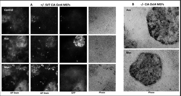
AP staining shows iPSC colonies presence:
To characterize the infected MEFs expressing GFP into growing iPSCs we used alkaline
phosphatase (AP) staining, since AP is an overexpressed enzyme in ESCs27. We observed strong,
marked stains in cells which were treated and kept under ESC conditions for 20-30 days
post-infection. The stains were concentrated in colony-like formations, indicating presence of iPSCs6.
Control
Aza
Moc
Aza
Moc
A +/- SVT CiA Oct4 MEFs B -/- CiA Oct4 MEFs
AP Stain AP Stain GFP Phase Phase
In some instances, the AP stains corresponded with GFP expressing cells but this was not
uniformly observed (Fig.5). Overall, wells with the highest AP staining also showed higher GFP
expression, even if these were not always co-localized. The method was also tried on unmodified
MEFs which did not have a GFP on one of the endogenous Oct4 alleles. The results showed highly
specific, strong and circular stains indicative of iPSC generation (Fig. 5). The staining was more
marked in non Oct4/GFP MEFs.
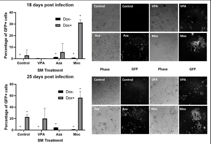
Mocetinostat is most Effective in iPSC generation:
We have also used another HDAC inhibitor, valproic acid (VPA), as a positive control due
to its reported efficacy in cell reprogramming25. Cells treated with Mocetinostat and 5-Azacytidine
Fig. 6: Flow cytometry data of GFP expression in reprogramming SVT CiA Oct 4 MEFs under different small molecule treatments at two time points during the reprogramming process. To the right are pictures of the reprogramming MEFs under different small molecules treatments corresponding to each of the two time points shown in the graphs. Flow cytometry was not performed for Aza+Dox treated cells at the 25 day timepoint due to significant cell death. Wells were allowed to grow until confluent enough to split and perform flow cytometry analysis. This was done at 33 days post infection, and Aza+Dox cells had a GFP+ percentage of 27.6. Picture to the right of 25 day timepoint graph for Aza+Dox is representative of that later timepoint as well.
Control
Control
VPA
VPA Aza
Aza
Moc
Moc
Control VPA
Aza Moc
Control VPA
Aza Moc
had GFP percentages of 1.18 and 1.48, respectively, and VPA of 11.8 percent. Importantly, an
adjustment to the Mocetinostat treatment concentration from 0.020 μM to 0.156 μM has
maximized the percentage of GFP expressing cells. This experiment has shown an increased GFP
expression in cells infected with the TetO-FUW-OSKM + FUW-M2rtTA constructs. Upon flow
cytometry analysis, reprogramming cells treated with 5-Azacytidine and Mocetinostat in a
pulsating manner show a percentage GFP expression of 27 and 31 percent on average, respectively,
while VPA shows an average of 20 percent (Fig. 6). However, the level of cell death is significantly
lower in Mocetinostat treated cells (Fig. 6).
Discussion and Conclusion
The differences observed in overall GFP expression between cells treated for one-week
post-infection, treated in a pulsating manner throughout one-month post infection or a continuous
manner suggest that a periodical treatment with the small molecules increases the efficiency of
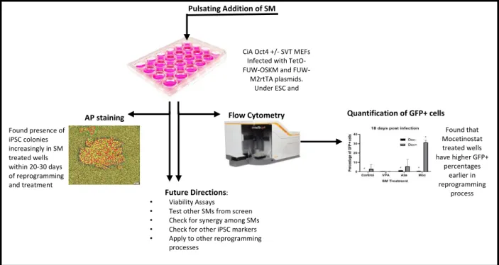
CiA Oct4 +/- SVT MEFs Infected with TetO-OSKM and
FUW-M2rtTA plasmids. Under ESC and doxycycline conditions,
Pulsating Addition of SM
AP staining
Found presence of iPSC colonies increasingly in SM treated wells within 20-30 days of reprogramming and treatment
Flow Cytometry Quantification of GFP+ cells
Found that Mocetinostat treated wells have higher GFP+
percentages earlier in reprogramming
process
Future Directions:
• Viability Assays
• Test other SMs from screen
• Check for synergy among SMs
• Check for other iPSC markers
• Apply to other reprogramming processes
GFP expression in reprogramming MEFs (Fig. 7). The continuous small molecules treatment
lowers GFP activation compared to the pulsating treatment, and the one week treated cells lose
GFP activation progressively. However, pulsating treatment allows for the maintenance of GFP
expression in small molecule treated wells over time. We hypothesize that the effects of
methylation inhibition and de-acetylation inhibition caused by the small molecules is better
maintained through the pulsating addition of the small molecules. It is likely that stopping
treatment after 1 week leads to the reversal of the small molecules’ effects over time, leading to a
decrease in endogenous Oct4 expression and thus in GFP expression. Moreover, the continuous
activation of small molecules may contribute to cell death and prevent the overall reprogramming
process.
The staining of infected MEFs with AP stain between 20-30 days post-infection yielded
positive and strong AP staining suggesting that the MEFs are reprogramming and becoming more
ESC-like (Fig. 7). Primary MEFs specially showed very marked AP positive colonies, which are
consistent with the morphology changes observed during reprogramming and characteristic of
ESCs. Interestingly, AP staining in the Oct4-GFP MEFs did not always show coincidence of AP
stain marks and GFP expressing cells. Although staining was strong and comparable to that
observed in primary MEFs, it was expected to coincide with GFP positive cells since these cells
are expressing endogenous Oct4 and driving reprogramming. Correlation was observed between
AP staining and GFP expression, but it was not exclusive to GFP expressing cells. These results
suggest that even cells which are not expressing GFP could be undergoing reprogramming since
we do not account for the three other iPSC generating factors in our assay. The literature also
reprogramming efficiency but not a marker for iPSC generation, not all GFP expressing cells will
successfully reprogram6,12,24.
Importantly, treatment with small molecules 5-Azacytidine and Mocetinostat helped
activate the Oct4 locus and drive reprogramming compared to non-treated reprogramming cells.
The reprogramming efficiency, based on quantification of GFP expressing cells using flow
cytometry, of 5-Azacytidine and Mocetinostat treated wells was of 1.48 and 1.18, respectively, in
early experiments. This is, as expected, lower than the efficiency increase caused by treatment
with VPA which leads to 11.8% iPSC generation12. However, the dose concentrations for VPA to
obtain such a result were much greater than that of Mocetinostat and 5-Azacytidine, suggesting
that these small molecules may have future applications due to higher potency for induction of
pluripotency. Interestingly, cells treated with Mocetinostat showed cell death of 11.8% whereas
those treated with 5-Azacytidine and VPA have cell death of 50%. Similarly, in the triplicate
experiments cells treated with Mocetinostat show a higher GFP expression while having
significantly less cell death than other treated wells. The percentage of GFP expressing cells is
much higher in the triplicate experiments, likely due to the use of more concentrated lentiviral
packaging plasmids during transfection as a result of better DNA isolation assays. In these later
experiments Mocetinostat shows to be fast acting, with a GFP activation of 31 percent on average
within the first 20 days of treatment. VPA and 5-Azacytidine are slower acting and reached GFP
percentages of 20 and 27 percent, respectively, later in the reprogramming process. Although the
change in GFP expression between these two sets of experiments is significant, all three treatment
conditions increased with the use of more concentrated virus to deliver the Yamanaka TFs.
Furthermore, the overall increase in GFP expression for treated wells remains higher than the
more concentrated viral constructs for infection along with the SM treatment. This is a consistent
observation in the later experiments which suggests that Mocetinostat may be of special interest
due to its high potency and low toxicity in cells during cellular reprogramming (Fig. 7).
We have found that treatment of reprogramming MEFs in a pulsating manner is more
efficient in GFP maintenance than either one time or continuous treatments. Upon AP staining, we
observed that although there is correlation between AP stain and GFP expression, the stain is not
exclusive to GFP expressing cells. This suggests cells not expressing GFP might also be
undergoing reprogramming and corroborates GFP expression suggests reprogramming but is not
a direct marker for it. Finally, pulsating treatment with 5-Azacytidine and Mocetinostat leads to
increase in reprogramming efficiency over untreated controls while showing lower cell death,
especially for Mocetinostat. Importantly, Mocetinostat is a novel small molecule which shows
potential in increasing the efficiency of cellular reprogramming at much lower doses than
traditionally used small molecules liked VPA and 5-Azacytidine.
Currently, we are using cell viability assays to corroborate that Mocetinostat not only
shows lower cell death but may also maintain cell viability during the reprogramming process.
Future steps are still needed to examine the other three identified HDACis in the process of iPSC
generation and to test combinations of the top five hits to check whether the synergism observed
in the small molecule screen is translatable. Furthermore, we will test these small molecules in
other reprogramming methods such as direct neuron transdifferentiation from MEFs. Ultimately,
the goal is to apply this work to the induction of pluripotency from human somatic cells and make
Acknowledgements:
Special thanks to Dr. Nathaniel Hathaway, Kathryn Headley and everyone at the Hathaway Lab
for teaching me patiently and for all their help in this process. Also to Dr. Scott Williams and Dr.
Amy Maddox for their help and input in this paper throughout the entire process of putting it
together. Special thanks as well to Dr. Gidi Shemer, without whose guidance and opportunity to
be a SMART scholar I would not be in this position today. Finally, to Maddie Parker for her
valuable input and help with editing.
Thanks as well to the Flow Cytometry Core University of North Carolina funded by P30
CA016086 Cancer Center Core Support Grant to the UNC Lineberger Comprehensive Cancer
Center and to S. Coquery and J. Dow for technical assistance with flow cytometry.
***This figure was made by Kathryn Headley, graduated student, as part of her small molecule
References:
1. Nishikawa S, Goldstein RA, Nierras CR. The promise of human induced pluripotent stem
cells for research and therapy. Nat Rev Mol Cell Biol. 2008;9(9):725-729.
doi:10.1038/nrm2466.
2. Singh VK, Kalsan M, Kumar N, Saini A, Chandra R. Induced pluripotent stem cells:
applications in regenerative medicine, disease modeling, and drug discovery. Front Cell
Dev Biol. 2015;3(February):2. doi:10.3389/fcell.2015.00002.
3. Mao AS, Mooney DJ. Regenerative medicine: Current therapies and future directions.
Proc Natl Acad Sci U S A. 2015. doi:10.1073/pnas.1508520112.
4. Cyranoski D. Stem cells: 5 Things to know before jumping on the iPS bandwagon.
Nature. 2008. doi:10.1038/452406a.
5. Kastenberg ZJ, Odorico JS. Alternative sources of pluripotency: science, ethics, and stem
cells. Transplant Rev. 2008. doi:10.1016/j.trre.2008.04.002.
6. Takahashi K, Yamanaka S. Induction of Pluripotent Stem Cells from Mouse Embryonic
and Adult Fibroblast Cultures by Defined Factors. Cell. 2006;126(4):663-676.
doi:10.1016/j.cell.2006.07.024.
7. Carey BW, Markoulaki S, Hanna J, et al. Reprogramming of murine and human somatic
cells using a single polycistronic vector. Proc Natl Acad Sci U S A. 2008:2-7.
doi:0811426106 [pii]\r10.1073/pnas.0811426106.
8. Li Y, Zhang Q, Yin X, et al. Generation of iPSCs from mouse fibroblasts with a single
gene, Oct4, and small molecules. Cell Res. 2011;21(1):196-204.
http://dx.doi.org/10.1038/cr.2010.142.
of progress. Nat Rev Drug Discov. 2017;16(2):115-130. doi:10.1038/nrd.2016.245.
10. Ichida JK, Blanchard J, Lam K, et al. A Small-Molecule Inhibitor of Tgf-β Signaling
Replaces Sox2 in Reprogramming by Inducing Nanog. Cell Stem Cell. 2009;5(5):491-503.
11. Li W, Zhou H, Abujarour R, et al. Generation of human-induced pluripotent stem cells in
the absence of exogenous Sox2. Stem Cells. 2009. doi:10.1002/stem.240.
12. Yuan X, Wan H, Zhao X, Zhu S, Zhou Q, Ding S. Brief report: Combined chemical
treatment enables Oct4-induced reprogramming from mouse embryonic fibroblasts. Stem
Cells. 2011. doi:10.1002/stem.594.
13. Sterneckert J, Höing S, Schöler HR. Concise review: Oct4 and more: The reprogramming
expressway. Stem Cells. 2012;30(1):15-21.
14. Hathaway NA, Bell O, Hodges C, Miller EL, Neel DS, Crabtree GR. Dynamics and
Memory of Heterochromatin in Living Cells. Cell. 2012;149(7):1447-1460.
15. Shimozaki K, Nakashima K, Niwa H, Taga T. Involvement of Oct3/4 in the enhancement
of neuronal differentiation of ES cells in neurogenesis-inducing cultures. Development.
2003;130(11):2505-2512. http://www.ncbi.nlm.nih.gov/pubmed/12702663.
16. Baranek M, Markiewicz WT, Barciszewski J. Selected small molecules as inducers of
pluripotency. Acta Biochim Pol. 2016;63(4):709-716. doi:10.18388/abp.2016_1363.
17. Radzisheuskaya A, Silva JCR. Do all roads lead to Oct4? The emerging concepts of
induced pluripotency. Trends Cell Biol. 2014;24(5):275-284.
18. Zeineddine D, Hammoud AA, Mortada M, Boeuf H. The Oct4 protein: more than a magic
stemness marker. Am J Stem Cells. 2014;3(2):74-82. www.AJSC.us.
19. Shi G, Jin Y. Role of Oct4 in maintaining and regaining stem cell pluripotency. Stem Cell
20. Kellner S, Kikyo N. Transcriptional regulation of the Oct4 gene, a master gene for
pluripotency. Histol Histopathol. 2010;25(3):405-412.
21. Pei D, Xu J, Zhuang Q, Tse H-F, Esteban M a. Induced pluripotent stem cell technology
in regenerative medicine and biology. Adv Biochem Eng Biotechnol. 2010.
doi:10.1007/10.
22. Massar M. Restricting Human Embryonic Stem Cell Research: Creating Life or
Destroying Freedom. Sch. 10(1537-405X):43-499.
23. Manuscript A, Metabolism C. c-Myc and Cancer Metabolism. 2013;18(20):5546-5553.
doi:10.1158/1078-0432.CCR-12-0977.c-Myc.
24. Zhu S, Li W, Zhou H, et al. Reprogramming of human primary somatic cells by OCT4
and chemical compounds. Cell Stem Cell. 2010. doi:10.1016/j.stem.2010.11.015.
25. Lin T, Wu S. Reprogramming with small molecules instead of exogenous transcription
factors. Stem Cells Int. 2015;2015.
26. National Center for Biotechnology Information. PubChem Compound Database;
CID=9865515, https://pubchem.ncbi.nlm.nih.gov/compound/9865515 (accessed Oct. 29,
2018).
27. Zhang H, Gayen S, Zhang H, et al. MLL1 Inhibition Reprograms Epiblast Stem Cells to
Naive Pluripotency Article MLL1 Inhibition Reprograms Epiblast Stem Cells to Naive