Assessing the Role of Imaging in Primary Breast Cancer Staging
By
Stuart-Allison M. Staley
A Master’s Paper submitted to the faculty of the University of North Carolina at Chapel Hill
in partial fulfillment of the requirements for the degree of Master of Public Health in
the Public Health Leadership Program.
Chapel Hill 2012
_________________________________ First Reader:
_________________________________ Date
TABLE OF CONTENTS
Abstract………3
Systematic Review……….4-18 Introduction………..6
Methods………6
Results………..8
Conclusion……….10
References………..12
Tables & Figures………13
Original Manuscript………..19-41 Introduction………21
Methods………..25
Results………27
Discussion………..29
Conclusion……….32
References………..33
ABSTRACT
Background: In the United States, there is no well-established staging protocol for new breast cancer patients. With various imaging modalities available, we performed the following
systematic review and original retrospective study to better characterize the utility of bone scan, liver ultrasound, chest radiograph, and computed tomography (CT) of the abdomen. For our systematic review, we determined the pooled detection rates of distant metastases using bone scan, liver ultrasound, and chest radiograph by clinical stage. In our study, we determined the utility of further imaging with abdominal CT when there is no detected disease beyond the ipsilateral axillary lymph nodes by CT of the chest.
Methods: As part of our systematic review, we searched PubMed and EMBASE databases for relevant articles using detection rate, defined as the number of patients with an abnormal test result divided by the total number of patients tested, as the primary outcome of interest for bone scan, liver ultrasound, and chest radiograph. Additionally, for our retrospective study, we reviewed medical charts for 440 patients and used cross tabulation bivariate analysis to characterize the relationship between detection of disease in the chest and disease in the abdomen.
Staging Evaluation with Bone Scan, Liver Ultrasound, and Chest Radiograph in Primary Breast Cancer Patients
By
ABSTRACT
Background: Routinely, radiological investigation with bone scan, chest radiograph, and liver ultrasound are employed as baseline staging tests. The aim of this review was to examine indications for this costly routine screening, and, thus, we posed the following question: Among women with newly diagnosed breast cancer who are otherwise asymptomatic, does evaluation with bone scanning, liver ultrasound, and chest radiograph help to determine the extent of metastatic disease?
Methods: We searched PubMed and EMBASE databases to find articles using detection rate, defined as the number of patients with an abnormal test result divided by the total number of patients tested, as the primary outcome of interest. In order to obtain overall estimates of detection rates in each test, the results from the studies were pooled and summed according to pathological stage.
Results: Eight articles out of 232 reviewed were included in the final analysis. The following pooled detection rates for bone scan were calculated according to stage: Stage I 7 of 544 (1.29%), Stage II 29 of 938 (3.09%), Stage I & II 36 of 1,482 (2.43%), and Stage III 39 of 312 (12.5%). For liver ultrasound: Stage I 1 of 213 (0.47%), Stage II 4 of 400 (1.00%), Stage I & II 5 of 613 (0.82%), and Stage III 6 of 143 (4.20%). And, for chest radiograph: Stage I 0 of 223, Stage II 2 of 473 (0.42%), Stage I & II 9 of 1,782 (0.51%), and Stage III 8 of 175 (4.57%).
INTRODUCTION
Staging is pivotal in the management of cancer and describes the extent and severity of
the patient’s disease. By classifying a person’s stage, a more accurate treatment plan and
estimate of the patient’s prognosis may be determined.1,2 Carcinoma of the breast commonly metastasizes to bone, lung, liver, and brain.1 To detect the presence of disease at these distant sites, many expensive imaging modalities with increasing sensitivity and specificity are utilized
by providers; however, as the care of cancer patients becomes more complex, the need for cost
containment is paramount to the health care system. In response, providers must find areas to
eliminate expensive staging techniques, while also protecting patient safety and providing
accurate staging and diagnosis of disease.
Routinely, radiological investigation with bone scan, chest radiograph, and liver
ultrasound are employed as baseline staging tests and are commonly referred to as “conventional
diagnostic procedures”.3,4 The aim of this review was to examine indications for this costly routine screening, and, thus, we posed the following question: Among women with newly
diagnosed breast cancer who are otherwise asymptomatic, does evaluation with bone scanning,
liver ultrasound, and chest radiograph help to determine the extent of metastatic disease?5
METHODS
Search Design & Study Criteria
PubMed and EMBASE databases were searched without language restriction or
limitations on publication date, using the terms “bone scan”, “bone neoplasms/radionuclide
Specifically, in EMBASE, the term “bone scintiscanning” was added to the search.
Bibliographies from relevant articles were also reviewed. Only retrospective case series were
included. To account for the rapid progression of the sensitivity and specificity of imaging
modalities and in order to provide more accurate results from current technology, articles were
reviewed if published after 1990. Furthermore, studies unavailable in English were excluded
from the study.
Abstracts of relevant articles were reviewed if they reported number of women with
newly diagnosed breast cancer who had disease detected by bone scan, liver ultrasound, or chest
radiograph. Studies were included only if they reported rates of positive tests by patient’s
pathological stage by the TNM staging system. These tests could be performed prior to or after
surgical intervention.
Study Evaluation
A single author evaluated the articles selected for full review. The pertinent study data
was extracted and organized into Tables 1 and 2, including setting, study method, total population, outcome measure, and total pooled results by method. The study’s quality was
assessed using a grading criteria consisting of 4 categories: (1) reporting of all disease stages, (2)
applicability to population of interest, (3) sufficient protocol detail, and (4) sufficient reporting of
results with confidence limits. Each criterion was weighted equally and assigned a grade of
good, fair, or poor (good = 2, fair = 1, poor = 0). These categorical scores were then averaged to
Data Synthesis
The primary outcome of interest was the detection rate, defined as the number of patients
with an abnormal test result divided by the total number of patients tested. In order to obtain
overall estimates of detection rates in each test, the results from the studies were pooled and
summed according to pathological stage.5
RESULTS
As seen in Figure 1, results from PubMed and EMBASE searches yielded a total of 232 articles, without duplicates. These 232 articles were screened on the basis of title and abstract
relevance, which provided 6 articles for full study review. Of the 226 articles excluded from the
review, 207 articles did not measure detection rate as their outcome, 3 articles were not provided
in English, and 15 articles were published prior to 1990.
The bibliographies of the 6 articles were reviewed for relevant titles, which yielded 5
additional articles. After review of the full manuscripts, 8 articles were included for the final
analysis. Three articles were excluded. Two studies did not provide the detection rate by
pathological stage,6,7and one study did not report detection rate as the outcome measure.8 Of the studies present in the final analysis, four studies analyzed only bone scanning; one
study analyzed only chest radiography; three studies reviewed all three imaging modalities.
Bone Scanning
As seen in Table Three, seven studies reported the detection rate of bone metastasis using routine bone scan. The calculated rate was reported by stage (Stage I, Stage II, Stage I &
metastatic disease to the bone as detected by bone scintigraphy. Similarly, only 3.09% (29/938)
of patients with Stage II tumors had detectable or possible disease spread to bone as detected by
bone scan. A pooled detection rate of only 2.43% (36/1,482) was determined for Stage I and II
cancers. Patients with Stage III tumors had a larger number of detected bone metastasis at
12.50% (39/312). In total, across all three stages, bone scan detected disease spread in 4.18%
(75/1,794) of new breast cancer patients.
Liver Ultrasound
As seen in Table Four, three studies reported the detection rate of liver metastasis using routine liver ultrasound. The calculated rate was reported by stage (Stage I, Stage II, Stage I &
II, Stage III) for each study. The pooled rates for all studies are provided by stage as well. For
patients with Stage I disease, only 0.47% (1/213) were found to have metastatic or possible
metastatic disease to the liver as detected by ultrasound. Similarly, only 1.00% (4/400) of
patients with Stage II tumors had detectable or possible disease spread to the liver as detected by
ultrasound. A pooled detection rate of only 0.82% (5/613) was determined for Stage I and II
cancers. Patients with Stage III tumors had only 4.20% (6/143) of patients with detected liver
metastasis. In total, across all three stages, liver ultrasound detected disease spread in 1.34%
(11/822) of new breast cancer patients.
Chest Radiograph
As seen in Table Five, four studies reported the detection rate of pulmonary metastasis using routine chest X-ray. The calculated rate was reported by stage (Stage I, Stage II, Stage I &
patients with Stage II tumors had detectable or possible disease spread to the lung as detected by
chest X-ray. A pooled detection rate of only 0.51% (9/1,782) was determined for Stage I and II
cancers. Patients with Stage III tumors had a higher percentage of patients with potential lung
metastasis at 4.57% (8/175). In total, across all three stages, a routine chest radiograph detected
possible disease spread to the lung in 0.87% (17/1,957) of new breast cancer patients.
CONCLUSION
Many previous studies have evaluated the use of CDPs (bone scanning, liver ultrasound,
and chest radiograph) in primary breast cancer staging. With all methods, the detections rates
increased with tumor size; however, the overall detection rates remain low for all three
modalities, particularly in asymptomatic patients, which questions the utility of these imaging
exams. Moreover, these techniques do not include many of the other common sites of
metastasis. Many studies have recommended limited use of CDPs, particularly in patients with
smaller tumor sizes.5, 6, 8, 13, 15
Future Research
The strength of this study is mainly a result of the number of patients studied – 1,794 for
bone scan, 822 for liver ultrasound, and 1,957 for chest radiograph – and the consistently low
detection rates seen for each method; however, further research to determine the false negative
and false positive rates are needed to fully ascertain the usefulness of these methods as baseline
staging exams. Moreover, the differences in rates of detection in patients who are clinically
symptomatic versus asymptomatic would be helpful in determining if particular subgroups of
Recommendations
Based on this data, the detection rates in patients with Stage I & Stage II tumors are
incredibly low, with 0.50% to 3.00% detection rates among the three methods. With such a low
number, we question if there is significant utility in performing any CDP on patients with small
tumors, T1 and T2, who are clinically asymptomatic. Further research can more clearly answer
this question, but the consensus of this research group is that patients with clinically early
cancers do not benefit from bone scanning, chest radiograph, or liver ultrasound. It is our
recommendation that asymptomatic patients proceed with only a screening chest CT scan.
Further evaluation with abdominal and pelvic CT scans should only be performed in the presence
of disease spread beyond the axillary lymph nodes or if the patients presents with specific
REFERENCES
1. American Cancer Society. “Breast Cancer: Facts and Figures 2011-2012”. American Cancer Society. Atlanta, Georgia. Last updated: 2011. Accessed on: February 28, 2012. Found at:
http://www.cancer.org/acs/groups/content/@epidemiologysurveilance/documents/document/acspc-030975.pdf.
2. The National Cancer Institute. “Cancer Staging”. The National Cancer Institute. Accessed on: February 28, 2012. Found at: http://www.cancer.gov/cancertopics/factsheet/detection/staging.
3. Barry MC, Thornton F, Murphy M, Younis F, Watson RG. The value of metastatic screening in early primary breast cancer. Ir J Med Sci. 168: 248-250, 1999.
4. Samant R and Ganguly P. Staging investigations in patients with breast cancer: The role of bone scans and liver imaging. Arch Surg 134: 551-553, 1999.
5. Myers RE, Johnston M, Pritchard K, Levine M, Oliver T, and the Breast Cancer Disease Site Group of the Cancer Care Ontario Practice Guidelines Initiative. Baseline staging tests in primary breast cancer: A practice guideline. Canadian Medical Association Journal 164(10): 1439-1444, 2001. 6. Gerber B, Seitz E, Muller H, Krause A, Reimer T, Kundt G, Friese K. Perioperative screening for
metastatic disease is not indicated in patients with primary breast cancer and no clinical signs of tumor spread. Breast Cancer Research and Treatment. 82:29-37, 2003.
7. Muller D, Kohler G, Ohlinger R. Staging procedures in primary breast cancer. Anticancer Research. 28: 2397-2400, 2008.
8. Hadley D, Fowble B, Torosian M. Evidence for selective use of bone scans in early stage breast cancer. Oncology Reports. 5: 991-993, 1998.
9. Ahmed A, Glynne-Jones R, Ell PJ. Skeletal scintigraphy in carcinoma of the breast – a ten year retrospective study of 389 patients. Nucl Med Commun 11: 421-426, 1990.
10. Brar IS, Sisley JF, Johnson RI. Value of preoperative bone and liver scans and alkaline phosphatase in the evaluation of breast cancer patients. Am J Surg 165: 221-223, 1991.
11. Glynne-Jones R, Young T, Ahmed A, Ell PJ, Berry RJ. How far investigations for occult metastases in breast cancer aid the clinician. Clin Oncol 3(2): 65-72, 1991.
12. Kennedy H, Kennedy N, Barclay M, Horobin M. Cost efficiency of bones scans in breast cancer. Clin Oncol 3: 73-77, 1991.
13. Yeh KA, Fotunato L, Ridge JA, Hoffman JP, Eisenberg BL, Sigurdson ER. Routine bone scanning in patients with T1 and T2 breast cancer: A waste of money. Ann Surg Oncol 2: 319-324, 1995.
14. Barry MC, Thornton F, Murphy M, Younis F, Watson RG. The value of metastatic screening in early primary breast cancer. Ir J Med Sci 168(4): 248-250, 1999.
15. Chen EA, Carlson GA, Coughlin BF, Reed WP, Garb JL, Frank JL. Routine chest roentgenography is unnecessary in the work-up of stage I and II breast cancer. J Clin Oncol 18(20): 3503-3506, 2000. 16. Schneider C, Fehr MK, Steiner RA, Hagen D, Haller U, Fink D. Frequency and distribution pattern of
distant metastases in breast cancer patients at the time of primary presentation. Arch Gynecol Obstet
TABLES & FIGURES
Figure One: Literature Search Flow Diagram
Literature Search:
Databases: PubMed & EMBASE
Limitations: None
Literature Search:
Databases: PubMed & EMBASE
Limitations: None
Search Results: (n = 232)
PubMed: 223
EMBASE: 9 Duplicates: 0
Articles screened on basis of title and abstract (n = 6 )
Excluded: (n = 226)
Different outcome measure: 208
Articles not provided in English: 3
Articles published before 1990: 15
Included: 11
Articles provided from screened bibliographies (n = 5)
Manuscript review and application
of inclusion criteria Excluded: (n = 3)
Did not meet inclusion criteria: Gerber, et. al. (2003)6
Hadley, et. al. (1998)8 Muller, et. al. (2008)7
Included: 8
Liver U/S: Glynne-Jones, et. al. (1991)11 Barry, et. al. (1999)14 Schneider, et. al. (2002)16 Bone Scan:
Ahmed, et. al. (1990)9 Brar, et. al. (1991)10
Glynne- Jones, et. al. (1991)11 Kennedy, et. al. (1991)12 Yeh, et. al. (1995) 13 Barry, et. al. (1999)14 Schneider, et. al. (2002)16
CXR:
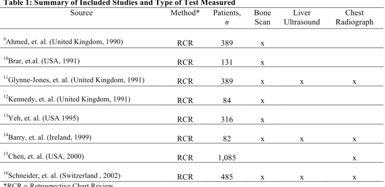
Table 1: Summary of Included Studies and Type of Test Measured
Source Method* Patients,
n
Bone Scan
Liver Ultrasound
Chest Radiograph
9Ahmed, et. al. (United Kingdom, 1990)
RCR 389 x
10Brar, et.al. (USA, 1991)
RCR 131 x
11Glynne-Jones, et. al. (United Kingdom, 1991)
RCR 389 x x x
12Kennedy, et. al. (United Kingdom, 1991)
RCR 84 x
13Yeh, et. al. (USA 1995)
RCR 316 x
14Barry, et. al. (Ireland, 1999)
RCR 82 x x x
15Chen, et. al. (USA, 2000)
RCR 1,085 x
16Schneider, et. al. (Switzerland , 2002)
RCR 485 x x x
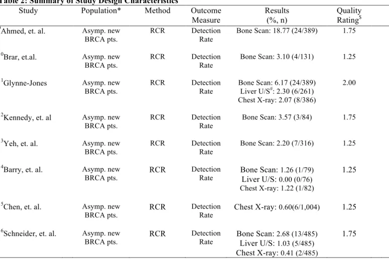
Table 2: Summary of Study Design Characteristics
Study Population* Method Outcome Measure
Results (%, n)
Quality Rating$ 9Ahmed, et. al. Asymp. new
BRCA pts.
RCR Detection
Rate
Bone Scan: 18.77 (24/389) 1.75
10Brar, et.al. Asymp. new BRCA pts.
RCR Detection
Rate
Bone Scan: 3.10 (4/131) 1.25
11Glynne-Jones Asymp. new BRCA pts.
RCR Detection
Rate
Bone Scan: 6.17 (24/389) Liver U/S#: 2.30 (6/261) Chest X-ray: 2.07 (8/386)
2.00
12Kennedy, et. al Asymp. new BRCA pts.
RCR Detection
Rate
Bone Scan: 3.57 (3/84) 1.75
13Yeh, et. al. Asymp. new BRCA pts.
RCR Detection
Rate
Bone Scan: 2.20 (7/316) 1.25
14Barry, et. al. Asymp. new
BRCA pts. RCR
Detection
Rate Bone Scan: Liver U/S: 0.00 (0/76) 1.26 (1/79) Chest X-ray: 1.22 (1/82)
1.25
15Chen, et. al. Asymp. new
BRCA pts. RCR
Detection
Rate Chest X-ray: 0.60(6/1,004) 1.25 16Schneider, et. al. Asymp. new
BRCA pts. RCR
Detection
Rate Bone Scan: Liver U/S: 2.68 (13/485) 1.03 (5/485)
Chest X-ray: 0.41 (2/485)
1.75
*Asymptomatic, new breast cancer patients #U/S = Ultrasound
$The study’s quality was assessed using a grading criteria consisting of 4 categories: (1) reporting of all disease
Table 3: Bone Scan Results by Stage of Breast Cancer
Cancer Stage; % (and no.) of patients with abnormal scan results
Study Year Stage I Stage II Stage I & II Stage III Total
9Ahmed, et. al.
1990 2.50 (2/80) 4.00 (9/226) 3.59 (11/306) 15.66 (13/83) 18.77 (24/389)
10Brar, et. al. 1991 0 (0/21) 3.00 (2/67) 2.27 (2/88) 4.70 (2/43) 3.10 (4/131)
11Glynne- Jones 1991 1.50 (1/67) 4.20 (10/240) 3.58 (11/307) 16.00 (13/82) 6.17 (24/389)
12Kennedy, et. al 1991 0 (0/13) 1.67 (1/60) 1.37 (1/73) 18.18 (2/11) 3.57 (3/84)
13Yeh, et. al. 1995 1.00 (2/204) 4.50 (5/112) 2.20 (7/316) --- 2.20 (7/316)
14Barry, et. al. 1999 --- --- 1.26 (1/79) --- 1.26 (1/79)
16Schneider, et. al. 2002 1.30 (2/159) 0.80 (2/233) 1.02 (4/392) 9.70 (9/93) 2.68 (13/485)
Table 4: Liver Ultrasonography Results by Stage of Breast Cancer
Cancer Stage; % (and no.) of patients with abnormal results
Study Year Stage I Stage II Stage I & II Stage III Total
11Glynne-Jones 1991 1.80 (1/54) 1.80 (3/167) 1.80 (4/221) 4.00 (2/50) 2.30 (6/261)
14Barry, et. al. 1999 --- --- 0.00 (0/76) --- 0.00 (0/76)
16Schneider, et. al. 2002 0 (0/159) 0.40 (1/233) 0.26 (1/392) 4.30 (4/93) 1.03 (5/485)
Table 5: Chest Radiography Results by Stage of Breast Cancer
Cancer Stage; % (and no.) of patients with abnormal results
Study Year Stage I Stage II Stage I & II Stage III Total
11Glynne-Jones 1991 0 (0/64) 0.80(2/240) 0.65 (2/304) 7.30 (6/82) 2.07 (8/386)
14Barry, et. al. 1999 --- --- 1.22 (1/82) --- 1.22 (1/82)
15Chen, et. al. 2000 --- --- 0.60 (6/1,004) --- 0.60(6/1,004)
16Schneider, et. al. 2002 0 (0/159) 0 (0/233) 0 (0/392) 2.20 (2/93) 0.41 (2/485)
Assessing the Role of Computed Tomography of the Abdomen in the Staging Evaluation of Primary Breast Cancer
By
ABSTRACT
Objective: As there is currently no well-established staging protocol for primary breast cancer, we sought to evaluate the role of computed tomography (CT), a commonly used imaging modality. The purpose of this study was to determine the utility of routine abdominal CT in the staging evaluation of women with newly diagnosed primary breast cancer given no detectable disease beyond the ipsilateral axillary nodes on chest CT.
Methods: The chest and abdominal CT scans from 440 patients over a 10-year period were reviewed. The presence of definite or possible metastatic disease in the axillary nodes, chest wall, internal mammary nodes, mediastinal nodes, lungs, liver and adrenals were recorded for each patient. Cross tabulation bivariate analysis as well as a chi-square test were performed to characterize the relationship between detection of disease in the chest and disease in the abdomen.
Results: Of the 440 patients reviewed, the following were found to have detectable metastatic disease by CT scan: axillary nodes 258 of 440 (56.5%), chest wall 40 of 440 (9.1%), internal mammary nodes 8 of 440 (1.8%), mediastinal nodes 29 of 440 (6.6%), lung 25 of 440 (5.7%), liver 12 of 437 (2.7%), and adrenals 8 of 440 (1.8%). In total, 81 patients had disease detectable in the chest beyond the ipsilateral axillary nodes (ie, chest wall, internal mammary nodes,
mediastinal nodes, and lung), and only 12 patients had detectable disease spread in the abdomen (ie, liver and adrenals). Of the 359 patients who had a negative chest CT, only 1 patient had detectable or possible metastatic disease spread on abdominal CT, resulting in a 99.7% negative predictive value (p < 0.001).
INTRODUCTION
Primary Breast Cancer Staging in the United States
Breast cancer is the most frequent malignant tumor in women of the Western countries.1 In 2011, an estimated 230,000 women were diagnosed with a new invasive breast cancer in the
United States.1 These patients will receive some form of staging evaluation at the time of their diagnosis. Staging describes the extent and severity of the patient’s disease and is pivotal in the
management of cancer. A patient’s specific stage is based on the invasiveness of the cancer, the
size of the tumor, the number of lymph nodes involved, and presence of distant metastasis.2 By classifying a person’s stage, a more accurate treatment plan and estimate of the patient’s
prognosis may be determined.3
Several imaging techniques are among the various tests that are used during a staging
evaluation. Imaging technology is advancing at a rapid pace, with tests available that are
increasingly more sensitive and specific in detecting morphologic as well as functional changes
in anatomy.4 With these advances, however, comes the added expense to the medical system as well as to the patient, additional time consumption, and emotional toll. In the U.S., there is no
clearly defined and accepted protocol for staging of primary breast cancers. Positron emission
tomography (PET) and computed tomography (CT) are commonly used in addition to less
advanced techniques such as ultrasound and radiography.4,5 Moreover, to our knowledge, no variations in imaging protocols exist based on the patient’s initial tumor size and axillary lymph
node metastasis.6
factors, but most particularly, the presence and extent of disease spread to the axilla.7 Moreover, these findings have a significant impact on determining the therapeutic plan. The most common
sites for distant metastasis include lung, liver, brain, bone, and adrenals.2 Effective staging evaluation must utilize imaging or laboratory techniques that can assess these locations in
totality.
Conventional Diagnostic Procedures (CDPs)
The conventional diagnostic procedures (CDPs) include chest radiograph
(posterior/anterior and lateral), ultrasound of the liver, and bone scan. In many institutions, these
modalities are commonly used to determine presence of metastases in the lung, liver, and bone,
respectively. The documented cumulative sensitivity of these tests may range from 36% to 51%,
with a greater specificity ranging from 81% to 95%.5 One possible explanation for these suboptimal results is that metastases may be too small to detect with these methods. Moreover,
these conventional techniques do not examine other common sites of disease spread, including
other thoracic lymph nodes or the adrenal glands.8 However, one study estimated the positive predictive value of CDPs to be as high as 91%, but a negative predictive value of 53%.5
Because CDPs are often inconclusive with low sensitivities, additional imaging may be
needed to achieve a final diagnosis. To further assess suspicious findings, providers may pursue
computed tomography (CT), magnetic resonance imaging (MRI), positron emission tomography
(PET), and/or bone scanning. With this additional imaging comes added financial cost, radiation
exposure, and an increase in a patient’s anxiety. Regardless, CDPs continue to be mainstays in
staging options as other imaging modalities are very costly, with longer scanning times and may
Positron Emission Tomography (PET)
Positron emission tomography (PET) is a more advanced form of imaging that has been
shown to be useful in detecting many tumors as well as distant metastases. It is well-established
in radiologic research that there is increased uptake of 2-[18F]fluoro-2-deoxy-D-glucose (FDG) in numerous malignancies.5 This increased uptake is evidence for enhanced glycolytic rate compared to the surrounding benign tissue. As a result, FDG PET is another method used for
whole-body staging evaluation.5
Few studies report data concerning use of FDG PET in detection of distant disease
spread; however, results from one prospective study indicate that FDG PET has a superior
sensitivity to CDPs (78.6%-100%) as well as higher specificity (89.4%-100%).5 Compared to CDPs, FDG PET has a comparable positive predictive value of 93% and a higher negative
predictive value (83% vs. 53%).5 Although superior in disease detection, FDG PET is a costly method with long scanning times. Furthermore, depending on the location, PET may not be
widely available to all patients.5,9
Generally, based on few studies reviewing PET for whole-body staging, FDG PET is not
recommended in patients with small primary tumors on imaging (defined as < 3cm) and negative
lymph nodes, as determined by surgical dissection or sentinel lymph node biopsy.5,9 In contrast, it is recommended for those with locally advanced disease, extended lymph node metastases and
those scheduled to undergo intense chemotherapy. Furthermore, scanning is recommended in
cases of uncertain radiographic results or rising tumor markers.5 In spite of these
The Role of Computed Tomography (CT) in Breast Cancer Staging
The clinical experience of members of our multidisciplinary breast oncology program has
been that CT is a commonly utilized modality at the University of North Carolina Hospitals
(UNCH) as well as referring institutions for breast cancer staging. Computed tomography is
used to evaluate the chest, abdomen, and pelvis. With CT of the chest, the following structures
may be evaluated for disease spread: axillary lymph nodes, chest wall, mediastinal nodes,
internal mammary nodes, and lung. An abdominal CT will allow evaluation of the liver and
adrenal glands, and the pelvic CT will allow visualizaiton of the ovaries, endometrium, and
pelvic bone.
In 2000, retrospective research from authors at Memorial Sloan-Kettering reported that
patient treatment was not substantially altered based on findings from pelvic CT scans.10
Moreover, findings were reported to lead to additional examinations and procedures that yielded
normal, benign, or indeterminate results that were not relevant to the patient’s cancer therapy.
Specifically, out of 2426 new primary breast cancer patients, only 17 patients (0.7%) were found
to have metastatic disease isolated to the pelvis. Of the 17 patients, eleven had metastasis
present only in the pelvic bone, which was simultaneously detected by concomitant bone
scanning. Pelvic CT only contributed relevant information for six patients, who had disease
present in the adnexa and/or endometrium. As a result, routine scanning of the pelvis by CT was
non-contributory in nearly all new cancer patients and, therefore, was not recommended based
on these findings.10
Likewise, it has been our anecdotal experience that evaluation of the abdomen yields no
further information when no metastases are detected in the chest beyond the ipsilateral axillary
disease beyond the ipsilateral axilla in the chest, then CT will not detect the spread of disease in
the abdomen. In other words, if a negative CT scan of the patient’s chest yields no detectable
disease beyond the axilla, then further CT imaging of the abdomen is of no additional benefit to
the patient and should not be performed.
METHODS
Patient Selection and Data Collection
With Internal Review Board (IRB) approval, we performed a retrospective case series of
all patients seen in the multidisciplinary breast oncology program at the University of North
Carolina Hospital (UNCH) between 1998 and 2011. We reviewed clinical records in order to
identify all patients referred for diagnosis of suspected breast cancer or treatment of a newly
diagnosed primary breast cancer by fine needle aspiration, core biopsy, or excisional biopsy.
Male patients, patients with a previous diagnosis of breast cancer, those with a diagnosis of
benign breast disease, as well as those with a metastatic breast carcinoma from another primary
cancer were excluded. Patients referred after surgical intervention were also excluded. There
was no age exclusion.
Patients with a CT scan of the chest and abdomen within 30 days of diagnosis were
included in the review. Patients evaluated with a CT chest or abdomen greater than 30 days after
diagnosis were excluded. Those referred with imaging studies from outside institutions that
could not be reviewed were not included in the study. Likewise, patients imaged at UNCH
either at UNCH or referring institutions, were excluded. This provided a final study group of
440 subjects.
CT scans were all performed on helical scanners and reconstructed with 5mm or 8mm
slice thickness and table incrementation. Between 1999 and 2002, patients were imaged on a
Siemens® Plus 4 scanner, which has a single detector with helical acquisition. After 2002, all
patients were imaged on a multidetector scanner, also with helical acquisition. All CT scans
were reviewed by a board certified chest radiologist with fellowship training in body computed
tomography. Scans of the chest were reviewed for the presence, questionable presence, or
absence of axillary, mediastinal, and internal mammary adenopathy; chest wall invasion; and
pulmonary metastases. Scans of the abdomen were reviewed for presence, possible presence, or
absence of hepatic and adrenal metastases. Information from patient medical records included
initial clinical stage, final pathologic stage post-operatively (if available), results of liver function
tests (if determined), and findings from bone scan (if performed).
Data Analysis
We used descriptive statistics to tabulate patient staging distribution and the presence of
disease in the chest and abdomen as detected by CT scan. The total number of patients included
in the final analysis were calculated and categorized by their clinical stage. Within each clinical
stage, the number and percentage of patients with metastases to the axillary nodes, chest wall,
internal mammary nodes, lung, mediastinal nodes, liver, and adrenal glands were also calculated.
Additionally, for each clinical stage, the total number and percentage of patients who had
detected presence of disease in either the chest or the abdomen were summed. Cross tabulation
the chest and detection of disease in the abdomen. The following contingency tables provide
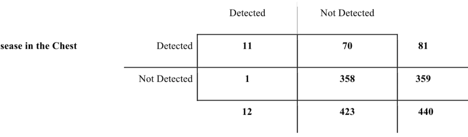
matrix displays of each variable based on location of disease. For contingency Table Two, a chi-square test was performed as a test for statistical significance.
RESULTS
Presence of Metastases by Anatomical Sites and Clinical Stage
For analysis, 440 patient scans were reviewed. Each patient received a chest CT and an
abdominal CT. As seen in Table One, most patients were classified as stage IIB (129 of 440). The distribution by stage was as follows: Stage IIIA (90), Stage IIA (83), Stage IIIB (63), Stage I
(40), and Stage IV (33). Two patients diagnosed with in situ carcinoma by core biopsy
underwent staging evaluation by CT.
In total, 258 (56.46%) patients had disease spread to the axillary lymph nodes as detected
by CT chest. Of note, axillary lymph node data was unavailable for 31 patients, as their CT
evaluation was performed following surgical dissection. By stage, data was missing for the
following: in situ (0), Stage I (3), Stage IIA (10), Stage IIB (14), Stage IIIA (2), Stage IIIB (2),
Stage IV (0). These 31 patients were subtracted from the study population when calculating the
percentage of metastases by stage and in total.
For other sites beyond the axillary lymph nodes, disease spread was less frequent: chest
wall 40 (9.10%), internal mammary nodes 8 (1.82%), mediastinal nodes 29 (6.59%), lung 25
(5.68%), liver 12 (2.73%), and adrenals 8 (1.82%). For three patients, liver data was unavailable
patients were subtracted from the study population when calculating the percentage of metastases
by stage and in total.
Comparison of Disease Detection in the Chest versus Abdomen
As seen in Table Two, 359 of the 440 patients scanned did not have disease detected in structures beyond the axillary lymph nodes (ie, chest wall, internal mammary nodes, mediastinal
nodes, lung) based on CT imaging. Of these 359 patients without detectable disease in the chest,
only 1 patient was found to have disease spread to the abdomen. This patient had metastatic
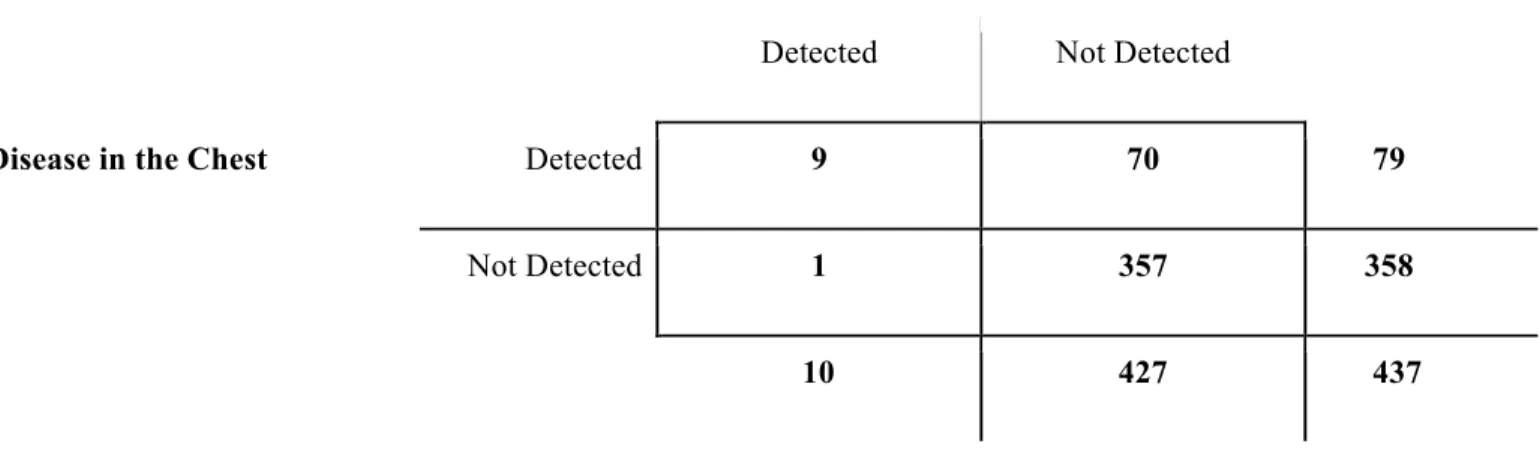
disease spread to the liver, but not detectable disease to the adrenals as noted in Tables Three and Four. Moreover, PET/CT scanning of this patient found detectable disease spread in the supraclavicular, which was not present on chest CT.
From these data, a 99.7% negative predictive value was calculated. In other words, of
those who did not have disease detected beyond the axillary lymph nodes in the chest, 99.7% did
not have detectable disease in the liver or adrenals (p<0.001).
Comparison of Disease Detection by Individual Chest Structures versus Abdomen
As additional information, Tables Five through Eight display the contingency tables for each individual chest structure and disease spread to the abdomen. Of note, out of the 12
patients with detected abdominal metastases, 8 had detectable lung metastasis on CT scan. The
other chest structures had detectable disease with concomitant abdominal metastasis seen in the
following number of cases: chest wall invasion (4), mediastinal nodes (4), and internal mammary
DISCUSSION
Once the initial diagnosis of invasive breast cancer is made, the extent of disease spread
is assessed in order to inform appropriate therapy, patient prognosis, and formally stage the
tumor. This assessment includes a number of imaging modalities, such as chest radiograph, liver
ultrasound, bone scintigraphy, MRI, PET, and CT scan of the chest, abdomen, and pelvis.
Results from these various tests may also inform further imaging to reach a final diagnosis. To
our knowledge, the routine use of many of these staging techniques is not standardized with no
well-established protocol.10 Our goal was to assess the role of abdominal computed tomography in staging primary breast cancer patients.
Our study showed that pursuing a CT abdomen provides little to no additional
information in light of a negative chest CT scan. Among the 359 patients who had no detectable
disease spread to the chest wall, lung, mediastinal or internal mammary lymph nodes, only 1 had
detectable disease to the abdomen.
Harms and Costs
With the benefits of additional knowledge achieved with CT, there are still many risks
with ordering this test. These risks are further reasoning for why standard staging protocol is
necessary for quality improvement, cost-effective medicine, and reducing patient exposure to
potentially harmful testing. First, although there is some data that suggests hormesis at low
levels of exposure, the generally accepted theory is the Linear No Threshold (LNT) theory based
based on the information gained from the test.11 Furthermore, without a standardized protocol, patients risk multiple forms of testing that may lead to additional follow-up imaging. This
cumulative exposure may sum to a greater radiation risk for breast cancer patients.10,12
Secondly, in order to appropriately image liver metastases, patients will receive contrast
material intravenously prior to CT scanning. In some cases, contrast material may cause an
allergic reaction or nephrotoxicity.12 Third, the financial cost of additional scanning, travel time, and work time lost are growing expenses to the U.S. health care system and the individual
patient. Even though pricing for scans are variable between insurance providers and hospital
systems, the most current hospital charges at UNC for a chest CT with contrast is $2890 with
professional fee of $155, totaling over $3000 for one scan. An abdominal and pelvic CT scan
totals to $2756 with an additional professional fee of $281. In this study, if the 358 patients with
negative scans had not undergone abdominal CT in light of their negative chest CT, the total
savings amounts to $1,087,246. The cumulative cost and time associated with pursuing
abdominal CT scans in large numbers of patients are substantial, as exemplified by this value.
Study Limitations
This study was a retrospective review of patient medical records. Several factors may
have biased our results. Primarily, it is inherently difficult to fully determine retrospectively why
a CT may have been ordered for a certain patient; however, to counteract this potential bias, only
patients with CT scans performed within one month of diagnosis and listed as evaluations due to
breast cancer diagnosis were included. It is our belief that the included subjects were scanned as
part of routine cancer evaluation as opposed to concurrent medical problems or preoperative
Additionally, CT scans performed at other institutions and reviewed by outside
physicians were excluded from the study. This limited a large number of imaging studies that
could be added to the analysis. Nevertheless, our sample size was significant, representing
patients seen at UNCH over a 13-year time period, various presenting stages and
symptomatology.
As this study is retrospective, assessing the clinical impact of the test result, even when
negative, cannot be fully evaluated. Furthermore, the effect of routine abdominal CT scanning
on patient survival cannot be assessed through this database.
Future Research
The need for routine imaging with CT chest, abdomen, and pelvis should be reconsidered
when developing staging protocol based on our study results. A prospective study including a
cost-effectiveness analysis as well as well-documented physical examination findings is needed
to determine if a particular subset of new cancer patients could benefit from CT scanning beyond
the chest. Furthermore, additional information concerning routine use of CT pelvis can inform
future protocol.
Recommendations for CT Imaging and Staging
Based on our study results and those of Drotman et. al.10 in 2000, we believe that routine CT imaging protocol does not require scanning of the chest, abdomen, and pelvis for all new
routine CT of the pelvis has shown to be of benefit to less than 1% of new breast cancer
patients.10 The results from these CT scans are rarely relevant to cancer therapy and, therefore, of no benefit to patients. In fact, the exposure to and cost of the testing may be greater potential
harm to women.
CONCLUSION
The routine use of abdominal CT in women with newly diagnosed primary breast cancer
and no detectable disease beyond the ipsilateral axillary nodes on staging chest CT scan has little
value with a 99.7% negative predictive value. Based on this information, we recommend that if
a negative CT scan of the patient’s chest yields no detectable disease beyond the axillary nodes,
then further CT imaging of the abdomen is of no additional benefit to the patient and should not
be performed.
REFERENCES
1. American Cancer Society. Breast Cancer: Facts and Figures, 2011-2012. American Cancer Society. 2011. Accessed on: January 2012. Available at:
http://www.cancer.org.libproxy.lib.unc.edu/acs/groups/content/@epidemiologysurveilance/document s/document/acspc-030975.pdf
2. American Cancer Society. How Is Breast Cancer Staged? American Cancer Society. 2011. Accessed on: January 2012. Available at:
http://www.cancer.org.libproxy.lib.unc.edu/Cancer/BreastCancer/DetailedGuide/breast-cancer-staging
3. The National Cancer Institute. Cancer Staging. The National Cancer Institute. 2011. Accessed on: January 2012. Available at:
http://www.cancer.gov.libproxy.lib.unc.edu/cancertopics/factsheet/detection/staging 4. Blake, Michael. Imaging in Oncology. Springer. New York, New York: 2008
5. Dose J, et. al. Comparison of fluorodeoxyglucose positron emission tomography and conventional diagnostic procedures for the detection of distant metastases in breast cancer patients. Nucl Med Commun. 23(9): 857-864, 2002
6. Based on systematic literature review of PubMed and EMBASE performed in conjunction with assistance from science information specialists at the UNC Health Sciences Library
7. Smith RA, Guisti R. The epidemiology of breast cancer. In: Basset LW, Jackson VP, eds. Diagnosis of diseases of the breast. Philadelphia: Saunders, 1996: 293-316
8. Koolen BB, et. al. 18F-FDG PET/CT as a staging procedure in primary stage II and III breast cancer: comparison with conventional imaging techniques. Breast Cancer Res Treat. 131(1): 117-126, 2012. 9. Benard F, Turcotte E. Imaging in breast cancer: Single-photon computed tomography and
positron-emission tomography. Breast Cancer Res. 7(4):153-162, 2005.
10. Drotman MB, Machnicki SC, Schwartz LH, Winston CB, Yoo HH, Panicek DM. Breast cancer: Assessing the use of routine pelvic CT in patient evaluation. AJR Am J Roentgenol. 176(6): 1433-1436, 2001
11. Hiserodt E. Underexposed: What id Radiation is Actually Good for You? Laissez Faire Books. Little Rock, Arkansas: 2005.
12. Mayo Clinic. “Overview of CT Scanning”. Mayo Clinic. Updated: May 24, 2012. Accessed on: June 12, 2012. Available at:
TABLES
Table One: Presence of Metastasis as Detected by CT Scan by Pre-Operative Clinical Stage (n, %)
Clinical Stage (n)
Axillary Nodes*
Chest Wall Internal
Mammary Nodes
Lung Mediastinal
Nodes
Liver** Adrenal
In Situ (2) 0 (0) 0 (0) 0 (0) 0 (0) 0 (0) 0 (0) 0 (0)
I (40) 9 (24.32) 1 (2.50) 0 (0) 0 (0) 2 (5.00) 0 (0) 0 (0)
IIA (83) 27 (36.99) 3 (3.61) 1 (1.20) 4 (4.82) 1 (1.20) 2 (2.41) 1 (1.20)
IIB (129) 67 (58.26) 8 (6.20) 1 (0.78) 2 (1.55) 6 (4.65) 2 (1.56) 2 (1.55)
IIIA (90) 75 (85.23) 4 (4.44) 1 (1.11) 3 (3.33) 4 (4.44) 0 (0) 1 (1.11)
IIIB (63) 51 (83.61) 11 (17.46) 4 (6.35) 4 (6.35) 4 (6.35) 1 (1.61) 0 (0)
IV (33) 29 (87.88) 13 (39.39) 1 (3.03) 12 (36.36) 12 (36.36) 7 (21.21) 4 (12.12)
Total (440) 258 (56.46) 40 (9.10) 8 (1.82) 25 (5.68) 29 (6.59) 12 (2.73) 8 (1.82)
*Axillary Node data was missing for the following number of patients by stage: in situ (0), Stage I (3), Stage IIA (10), Stage IIB (14), Stage IIIA (2), Stage IIIB (2), Stage IV (0). These patients were subtracted from the study population when calculating the percentage of metastases by stage, respectively.
**Liver data was unavailable for three patients due to lack of contrast for imaging: in situ (0), Stage 1 (0), Stage IIA (0), Stage IIB (1), Stage IIIA (1), Stage IIIB (1), Stage IV (0). These patients were subtracted from the study population when calculating the percentage of metastases by stage, respectively.
Table Two: Comparison of Disease Detection in the Chest versus Disease Detection in the Abdomen (n)$,#
Disease in the Abdomen
Detected Not Detected
Disease in the Chest Detected 11 70 81
Not Detected 1 358 359
12 423 440
____________________________________________________
$Detection of disease in the chest is defined as disease detected beyond the axillary nodes.
#Positive detection of disease in the chest was defined as disease detected beyond the ipsilateral axillary nodes and present in the
chest wall, internal mammary nodes, lung, and/or mediastinal nodes. Positive detection of disease in the abdomen was defined as disease detected in the liver and/or adrenals.
Table Three: Comparison of Disease Detection in the Chest versus Disease Detection in the Liver (n)%
Disease in the Liver
Detected Not Detected
Disease in the Chest Detected 9 70 79
Not Detected 1 357 358
10 427 437
_______________________________________
%Liver data was unavailable for three patients due to lack of contrast for imaging. Their results are not included in the above
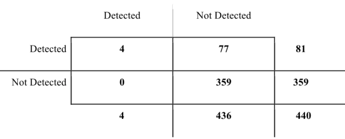
Table Four: Comparison of Disease Detection in the Chest versus Disease Detection in the Adrenals (n)
Disease in the Adrenals
Detected Not Detected
Disease in the Chest Detected 4 77 81
Not Detected 0 359 359
Table Five: Comparison of Disease Detection in the Chest Wall versus Disease Detection in the Abdomen (n)
Disease in the Abdomen
Detected Not Detected
Chest Wall Invasion Detected 4 36 40
Not Detected 8 392 400
Table Six: Comparison of Disease Detection in the Internal Mammary (IM) Nodes vs. Disease Detection in the Abdomen (n)
Disease in the Abdomen
Detected Not Detected
Disease in the IM Nodes Detected 2 6 8
Not Detected 10 422 432
Table Seven: Comparison of Disease Detection in the Lungs vs. Disease Detection in the Abdomen (n)
Disease in the Abdomen
Detected Not Detected
Disease in the Lungs Detected 8 17 25
Not Detected 4 411 415
Table Eight: Comparison of Disease Detection in the Mediastinal (MD) Nodes vs. Disease Detection in the Abdomen (n)
Disease in the Abdomen
Detected Not Detected
Disease in MD Nodes Detected 4 25 29
Not Detected 8 403 411