Introduction
Cystic fibrosis (CF) is a genetic disease characterized by hyperosmolar and abnormally viscous mucus in the lungs (Oliver et al., 2000). The defect is caused by a mutation in the cystic fibrosis transmembrane conductance regulator (CFTR) (Therien et al., 2001). The lungs traditional bacterial defenses, mucociliary clearance, polymorphonuclear neutrophil phagocytosis, and local cationic peptide release are all considerably impaired in the CF lung (Oliver et al., 2000). It is the impaired immune defense resulting from CFTR mutation that is responsible for CF and the host of bacterial infections that follow, including chronic colonization by Pseudomonas aeruginosa.
P. aeruginosa is an opportunistic bacterial pathogen capable of infecting a variety of eukaryotes, including humans (Fulcher et al., 2010; Bertrand et al., 2010). With a pervasive prevalence in water and soil, P. aeruginosa stands as a considerable threat to immunocompromised patients (Mandell et al., 2005). The CDC estimates that over 50,000 new cases of P. aeruginosa infection occur annually in the United States, with an increasing percentage of these cases being multi-drug resistant infections (CDC, 2014). Of particular importance is the nearly universal infection of cystic fibrosis patients with P. aeruginosa (Smith et al., 1996; Goldman et al., 1997).
(Strom and Lory, 1993). Because virtually all CF patients are infected with P. aeruginosa, it is presumed that the CF lung environment is an exceptional niche for P. aeruginosa adhesion and subsequent colonization.
Previous studies have shown that TFP proteins are part of a suite of virulence factors that are regulated by second messengers (Beatson et al., 2002; Wolfgang et al. 2003). The second messenger molecule adenosine 3’5’-cyclic monophosphate (cAMP) has been shown to play a critical role in regulating virulence. Synthesized primarily by adenylate cyclase enzyme CyaB1, cAMP acts as an allosteric regulator of the transcription factor Vfr (Virulence factor regulator) (Fulcher et al., 2010). Together, cAMP and Vfr are considered global regulators of virulent gene expression in P. aeruginosa (Suh et al., 2002), controlling multiple systems including TFP regulation (Beatson et al., 2002; Wolfgang et al., 2003). In summation, TFP production and function, key components of P. aeruginosa infection, are dependent on intracellular cAMP and Vfr levels. We focus on cAMP production and its relationship to the TFP proteins PilG and PilH. PilG and PilH are two CheY-like response regulators that are part of the Chp chemosensory system (Fultcher et al., 2010). The Chp chemosensory system has been shown to both positively and negatively regulate CyaB-dependent cAMP levels (Fulcher et al., 2010). PilG deletion mutants show a severe drop in cAMP (~80% drop from WT levels) while pilH deletion mutants show a roughly three-fold increase in cAMP (Fulcher et al., 2010).
The objective of this study is to develop fluorescent fusion proteins to investigate the localization patterns and molecular targets of PilG and PilH in P. aeruginosa. It is because of PilG and PilH’s tremendous effect on intracellular cAMP and, thus, on TFP production, function,
and overall virulence that we have chosen to study this system. The mechanisms by which PilG and PilH function are unclear; however, CheY-like proteins such as PilG and PilH are typically regulated through phosphorylation (Bertrand et al., 2009). It has been proposed that PilG modulates pilus extension and PilH allosterically modulates pilus retraction (Bertrand et al., 2009). Because of function and preliminary testing, it is presumed that PilG localizes to the leading pole of the cell and that PilH is a diffuse intracellular protein. Our plan was to test protein localization using GFP fluorescent tagging. To uncover potential PilG and PilH targets, we sought to screen mutant strains and look for changes in localization.
Materials and Methods
All plasmids for this study were isolated using Promega Wizard Plus SV Miniprep DNA Purification System. PCR amplicons were purified using Qiagen QIAquick PCR Purification Kit. Cloning was performed using Invitrogen Gateway LR and BP clonase II. Refer to the appendix for a summary of plasmids, primers, and strains used in this study.
Construction of a stable, monomeric GFP. Plasmid pRGFP was isolated from Escherichia coli strain DH5α as a source of GFPmut3. 2 The plasmid was used as a template for splice by overlap extension mutagenesis. Primers 1 and 2 were used to amplify a portion of GFP; primers 3 and 4 were used to amplify the remaining portion of GFP by PCR.3 Primers 2 and 3 introduced a point mutation (L221K) in the amplified PCR products. The introduced point mutation was shown to effectively eliminate any potential for GFP dimerization and
2 GFPmut3 is an isoform of GFP optimized for expression in bacteria. All GFP used in this study are mut3 and will henceforth be referred to as GFP.
unpredictable localization at high concentrations (Zacharias et al., 2002). The mutation is referred to as monomericGFP, or mGFP.
The two GFP PCR products with the introduced point mutation (L221K) were spliced together and amplified using primers 5 and 6. This amplification product was cloned into pDONR201 plasmid by way of BP Gateway Cloning. Following cloning, E. coli strain DH5α was transformed with pDONR201-mGFP. Plasmid pDONR201-mGFP was isolated and sequence confirmed using primers 11 and 12.
pilGmGFP and pilHmGFP construction via splice by overlap extension. In pilGmGFP construction, mGFP was amplified from pDONR201-mGFP by PCR with primers 4 and 9. Plasmid pMMBV1-pilG was used to amplify pilG via PCR using primers 7 and 8. Primers 8 and 9 were designed with an overlapping region encoding SGGGG as a strong, flexible linker between PilG and mGFP. Primers were designed to attach mGFP to the carboxyl-terminus of PilG.
Constructing pilHmGFP was performed using the same methods as pilGmGFP. PCR amplification of mGFP was done on pDONR201-mGFP using primers 4 and 13. Plasmid pMMBV1-pilH was used to amplify pilH via PCR using primers 14 and 15. As in pilGmGFP construction, primers 13 and 14 were designed with an overlapping region encoding SGGGG as a flexible linker between PilH and mGFP. Primers were designed to attach mGFP to the carboxyl-terminus of PilH.
transformed with pDONR201-pilGmGFP and pDONR201-pilHmGFP, independently. Both plasmids were isolated from DH5α and sequence confirmed using primers 11 and 12.
Cloning into inducible plasmids and mating with P. aeruginosa. pilHmGFP,
pilGmGFP, and mGFP were cloned from their pDONR201 plasmids into two plasmids, pMMBV1GW and pMMBGW, which have different degrees of inducible expression in P. aeruginosa. The destination pMMB- plasmids were linearized with restriction enzyme BspEI (New England Biolabs Inc.). pilGmGFP, pilHmGFP, and mGFP were inserted into pMMB- plasmids using LR Gateway Cloning. Following cloning, E. coli strain DH5α was transformed. pMMB- plasmids containing pilGmGFP and pilHmGFP constructs were individually mated into P. aeruginosa PAK strains with chromosomal pilG or pilH deletions such that the introduced plasmids contained the sole copy of pilG or pilH. The PAK strains used also contain a chromosomal reporter gene for measuring virulence factor expression. A helper E. coli strain pRK2013 was used to assist in a tri-parental mating. At this time plasmid pMMB67EH was mated into PAK strains as an empty vector control for future experiments.
Β-galactosidase assays. The PAK strains used in this study contained a background chromosomal insertion, lacP1ΔlacI-lacZ, that enables indirect measurement of cAMP levels by measuring B-galactosidase activity. In E. coli, CRP binds to the P1 promoter region of the lac operon and facilitates transcription of lacZ in the presence of cAMP. Similarly, in P. aeruginosa, Vfr functions as a CRP homolog that interacts with the P1 promoter and, in the presence of cAMP, facilitates the transcription of lacZ (Fulcher et al., 2010). In summation, PilG and PilH regulate cAMP levels positively and negatively, respectively. cAMP acts as a cofactor for the transcription of lacZ, which codes for B-galactosidase. Therefore, B-galactosidase activity reflects cAMP levels, and can be used to surmise the functionality of fusion proteins PilGmGFP and PilHmGFP. The assumption being made is that if PilGmGFP or PilHmGFP are able to regulate cAMP in a wild type fashion, then the fluorescent tags are not inhibiting their function.
P. aeruginosa strains with pMMB- pilGmGFP inserts were streaked for isolation and cultured overnight on LB plates with carbomycin (150 ug/ml). Single colonies were then picked and inoculated in LB broth with carbomycin (150ug/ml) overnight at 37° C and 300 rpm. Overnight inoculations were diluted 1:100 in LB broth with low dose antibiotic (carbomycin 50 ug/ml) and a gradient of IPTG4 concentrations. An IPTG gradient was used to determine the induction concentration that produced wild type expression levels of pilGmGFP. Samples were grown to OD600 ≈ 0.5 and placed on ice to arrest growth. At this point, 1 ml samples were taken for immunoblotting.
B-galactosidase assays were conducted with three technical replicates per sample. To 0.05 ml of sample, 0.95 ml of Z buffer + Β-mercaptoethanol (1000Z-Buffer:3.58B-ME) were added and vortexed briefly. To each sample, 15 μl of 0.1% SDS and 15 μl of chloroform were added to
lyse cells, with brief vortexing between each addition. Samples were incubated at 37° C for 5 minutes. To develop samples, 0.2 ml of developing solution (4 mg ONPG/ml Z-buffer + Β-ME) were added. Samples were vortexed briefly and returned to a 37° C water bath for timed developing. Developing was stopped by the addition of 0.5 ml 1M Na2CO3 and brief vortexing after the sample had turned a straw-yellow color, or if an hour had passed. Samples OD420 was then measured and Miller Units5 calculated.
Immunoblotting. Aliquots of 1 ml were taken from B-galactosidase assay samples of P. aeruginosa with pMMB- pilGmGFP inserts grown to OD600 ≈ 0.5. Aliquots were pelleted, supernatant removed, and the pellet re-suspended in 50 μl of loading buffer (950SDS-Page 2x Loading Buffer:50B-ME). Samples were then boiled before being loaded onto 12% acrylamide SDS-PAGE
gels. Gels were run using10 μl of sample, separated at 150V for roughly 1 hour. Proteins were subsequently transferred to nitrocellulose at 100V for 1 hour. PilG rabbit antibodies (1:10,000) and 800 nm anti-rabbit secondary antibodies (1:10,000) were used to develop blots.
Results
The objective of this study was to construct PilG and PilH fluorescent fusion proteins to study their localization patterns in P. aeruginosa. Sequencing showed success in point mutating GFP (L221K), creating a monomeric copy of GFP that does not dimerize or localize at high concentrations (Zacharias et al., 2002). PilGmGFP and pilHmGFP fusions were sequence confirmed to ensure no mutations had occurred during the splicing process. By observing fluorescence in E. coli expressing PilHmGFP, we surmised that the fluorescent tag was
functional (figure 1). Conducting B-galactosidase assays and immunoblotting tested PilGmGFP function and expression in P. aeruginosa. The mechanisms underlying the B-galactosidase assay can be found in the methods. In general, we compared trends in B-galactosidase activity between mutants and wild type controls.
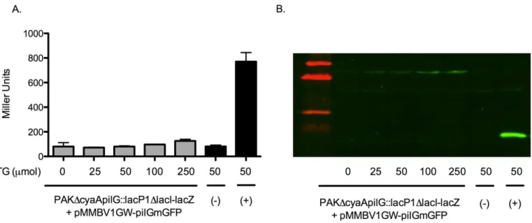
B-galactosidase assay and immunoblotting for pMMBV1GW-pilGmGFP complementation
Figure 2A. B-galactosidase activity across an IPTG induction gradient to test PilGmGFP functionality. The (+) label refers to a pseudo-WT positive control and (-) refers to a ΔpilG negative control (appendix). Error bars represent SD between three technical replicates. Figure 2B represents an immunoblot of the same samples depicted in 2A. Bands at ~38 kDa represent PilGmGFP and bands at ~14 kDa represent PilG.
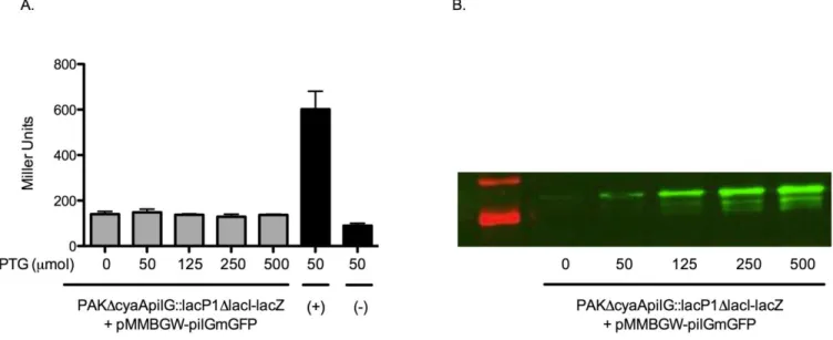
pMMBGW. Fluorescent constructs were cloned into pMMBGW; B-galactosidase assays and immunoblotting were repeated.
B-galactosidase assay and immunoblotting for pMMBGW-pilGmGFP complementation
Figure 3A. B-galactosidase activity across an IPTG induction gradient to test PilGmGFP functionality. The (+) label refers to a pseudo-WT positive control and (-) refers to a ΔpilG negative control (appendix). Error bars represent SD between three technical replicates. Figure 3B represents an immunoblot of the same samples depicted in 3A. The band at ~38 kDa represents PilGmGFP.
Complementation data
Figure 4. This figure is borrowed from previous studies conducted in our laboratory (Fulcher et al., 2010). Relative reporter activity is reported as the B-galactosidase activity of an individual mutant strain divided by that of the ΔcyaA mutant parent stain harboring pMMB67EH empty vector (Fulcher et al., 2010). V indicates deletion mutants and C indicates deletion mutants rescued with pMMB based plasmids.
Figure 4 from a previous study was included here to demonstrate that ΔpilG mutants could be rescued with pMMB based complementary plasmids (Fulcher et al., 2010). These data indicate that, in this study, failure to rescue mutants by complementation is a result of fluorescent tagging. Because PilG protein function is severely compromised localization data were not collected.
Discussion
The results of this study provide insight into an artificial means of tracking protein localization patterns in P. aeruginoas. Fundamental to experimental design is the caution taken to preserve the fidelity of the naturally functioning system. Thus, careful procedures were enacted
Rela
tive
Re
p
o
rte
r Ac
in this work to determine if our means of testing protein localization were inherently disruptive to native protein function.
Sequencing results indicated successful creation of a monomeric version of GFP that does not dimerize or localize at high concentrations (Zacharias et al., 2002). Sequencing also indicated successful splicing of mGFP to pilG and pilH. Confocal microscopy was utilized to determine that mGFP was functional when fused to PilH. These preliminary findings prompted the use of B-galactosidase assays and immunoblotting to measure intracellular cAMP levels and pilGmGFP expression, respectively. In wild type systems, PilG positively regulates cAMP levels. In contrast, our results show that PilGmGFP has no capacity to regulate cAMP levels at any expression threshold. It was decided that fusing mGFP to the carboxyl-terminus of PilG was too disruptive to native protein function for a faithful interpretation of localization patterns. For these reasons, PilGmGFP localization data were not collected.
PilHmGFP function and expression were not sampled due to a high degree of homogeneity between pilG and pilH. Because of biochemical and primary sequence similarities between the PilG and PilH, it is presumed that pilHmGFP would be impaired similarly to PilGmGFP. Future studies can further investigate this presumption by employing B-galactosidase assays and immunoblotting for PilHmGFP.
fused to the amino-termini. Amino tagging may reduce or eliminate PilGmGFP loss of function observed here, serving as a more faithful representation of the natural system. Additionally, it was observed that the rabbit PilG antibody has affinity for PilGmGFP and does not target PilH despite the high degree of homogeneity between the two proteins. These observations demonstrate the possibility of conducting PilG and PilH localization studies on fixed cells using antibodies should amino-terminal fluorescent tagging prove unsuccessful.
In conclusion, this study fell short of its ultimate goal of tracking PilG and PilH localization patterns in P. aeruginosa. We were successful in constructing PilGmGFP and PilHmGFP fusion proteins; however, unexpected PilGmGFP loss of function attributed to the fluorescent tag has led to redesigning our approach to study localization. Our current investigation into amino-terminal tagging may prove to remedy the protein loss of function observed here.
Acknowledgements
Appendix
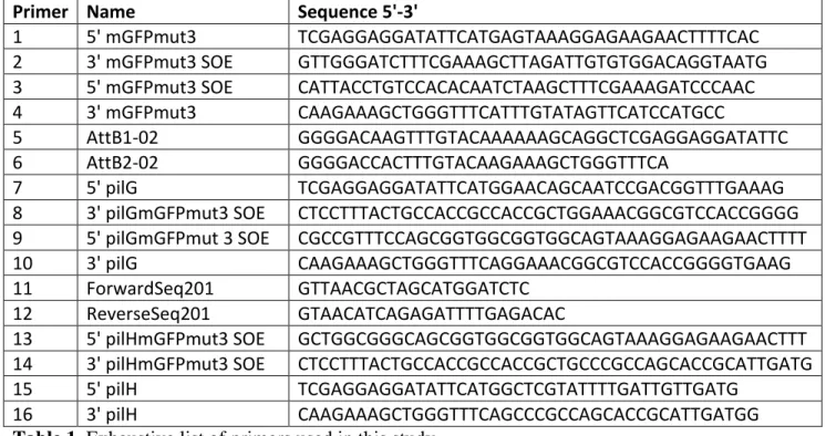
Primer Name Sequence 5'-3'
1 5' mGFPmut3 TCGAGGAGGATATTCATGAGTAAAGGAGAAGAACTTTTCAC
2 3' mGFPmut3 SOE GTTGGGATCTTTCGAAAGCTTAGATTGTGTGGACAGGTAATG 3 5' mGFPmut3 SOE CATTACCTGTCCACACAATCTAAGCTTTCGAAAGATCCCAAC
4 3' mGFPmut3 CAAGAAAGCTGGGTTTCATTTGTATAGTTCATCCATGCC
5 AttB1-02 GGGGACAAGTTTGTACAAAAAAGCAGGCTCGAGGAGGATATTC
6 AttB2-02 GGGGACCACTTTGTACAAGAAAGCTGGGTTTCA
7 5' pilG TCGAGGAGGATATTCATGGAACAGCAATCCGACGGTTTGAAAG
8 3' pilGmGFPmut3 SOE CTCCTTTACTGCCACCGCCACCGCTGGAAACGGCGTCCACCGGGG 9 5' pilGmGFPmut 3 SOE CGCCGTTTCCAGCGGTGGCGGTGGCAGTAAAGGAGAAGAACTTTT
10 3' pilG CAAGAAAGCTGGGTTTCAGGAAACGGCGTCCACCGGGGTGAAG
11 ForwardSeq201 GTTAACGCTAGCATGGATCTC
12 ReverseSeq201 GTAACATCAGAGATTTTGAGACAC
13 5' pilHmGFPmut3 SOE GCTGGCGGGCAGCGGTGGCGGTGGCAGTAAAGGAGAAGAACTTT 14 3' pilHmGFPmut3 SOE CTCCTTTACTGCCACCGCCACCGCTGCCCGCCAGCACCGCATTGATG
15 5' pilH TCGAGGAGGATATTCATGGCTCGTATTTTGATTGTTGATG
16 3' pilH CAAGAAAGCTGGGTTTCAGCCCGCCAGCACCGCATTGATGG
Table 1. Exhaustive list of primers used in this study.
Strain/plasmid Description
DH5α pMMBV1-pilG Initial source of pilG DH5α pMMBV1-pilH Initial source of pilH DH5α pRGFP Initial source of GFPmut3
DH5α pMMBV1GW Initial source of plasmids used for cloning
DH5α pMMBGW
DH5α HB101 pMMB67EH Empty vector control plasmid
Table 2. A list of initial plasmids used to construct inserts for this study. Constructs Created or Utilized
E. coli DH5α P. aeruginosa
pDONR201-mGFPmut3 In PAKΔcyaA::lacP1ΔlacI-lacZ Background: pDONR201-pilGmGFPmut3 pMMBV1GW-mGFPmut3
pDONR201-pilHmGFPmut3 ΔpilG + pMMBV1GW-pilGmGFPmut3 pMMBV1GW-mGFPmut3 ΔpilG + pMMBGW-pilGmGFPmut3 pMMBV1GW-pilGmGFPmut3 ΔpilH + pMMBV1GW-pilHmGFPmut3 pMMBV1GW-pilHmGFPmut3 ΔpilH + pMMBGW-pilHmGFPmut3 pMMBGW-mGFPmut3 pMMB67EH (positive control)
pMMBGW-pilGmGFPmut3 ΔpilG + pMMB67EH (negative control) pMMBGW-pilHmGFPmut3
References
1) Beatson, S.A., Whitchurch, C.B., Sargent, J.L., Levesque, R.C., Mattick, J.S. (2002) Differential regulation of twitching motility and elastase production by Vfr in
Pseudomonas aeruginosa. J. Bacteriology 184: 3605-3613
2) Bertrand, J.J., West, J.T., Engel, J.N. (2010) Genetic analysis of the regulation of type IV pilus function by the Chp chemosensory system of Pseudomonas aeruginosa. Journal of Bacteriology 192: 994-1010
3) CDC, (2014) Pseudomonas aeruginosa in Healthcare Settings.
http://www.cdc.gov/hai/organisms/pseudomonas.html
4) Dasgupta, N., Ferrel, E.P., Kanack, K.J., West, S.E., ramphal, R. (2002) fleQ, the gene encoding the major flagellar regulator of Pseudomonas aeruginosa, is sigma70 dependent and is downregulated by Vfr, a homolog of Escherichia coli cyclic AMP receptor protein. J. Bacteriology 184: 5240-5250
5) Ferrel, E., Carty, N.L., Colmer-Hamood, A.N., and West, S.E. (2008) Regulation of Pseudomonas aeruginosa ptxR by Vfr. Microbiology 154: 431-439
6) Fulcher, N.B., Holliday, P.M., Klem, E., Cann, M.J., Wolfgang, M.C. (2010)
Pseudomonas aeruginosa Chp chemosensory system regulates intracellular cAMP levels by modulating adenylate cyclase activity. Microbiology 76: 889-904
7) Goldman, M.J., Anderson, G.M., Stolzenberg, E.D., Kari, U.P., Zasloff, M., Wilson, J.M. (1997) Human b-defensin-1 is a salt-sensitive antibiotic in lung that is inactivated in cystic fibrosis. Cell 88:553–560.
9) Oliver, A., Cantón, R., Campo, P., Baquero, F., Blázquez, J. (2000) High frequency of hypermutable Pseudomonas aeruginosa in cystic fibrosis lung infection. Science 288:
1251-1253
10) Smith, J. J., Travis, S.M., Greenberg, E.P., Welsh, M.J. (1996) Cystic fibrosis airway epithelia fail to kill bacteria because of abnormal airway surface fluid. Cell 85: 229–236
11) Strom, M.S., Lory, S. (1993) Structure-function and biogenesis of the type IV pili. Dept. of Microbiology, University of Washington, Seattle, Washington
12) Suh S, Runyen-Janecky L, Maleniak T, Hager P, MacGregor C, Zielinski-Mozny N,
Phibbs P, West S. (2002) Effect of vfr mutation on global gene expression and catabolite repression control of Pseudomonas aeruginosa. Microbiology 148:1561-1569
13) Therien, A.G., Fiona, E.M., Deber, G. & C.M. (2001) Interhelical hydrogen bonds in the CFTR membrane domain. Nature Structural Biology 8: 597-601
14) West, S.E., Sample, A.K., Runyen-Janecky, L.J. (1994) The vfr gene product, required for Pseudomonas aeruginosa exotoxin A and protease production, belongs to the cyclic AMP receptor protein family. J. Bacteriology 176: 7532-7542
15) Wolfgang, M.C., Lee, V.T., Gilmore, M.E., Lory, S. (2003) Coordinate regulation of bacterial virulence genes by a novel adenylate cyclase-dependent signaling pathway. Dev Cell 4: 253-263