ARTIGO ORIGINAL
ABSTRACT
Introduction: In prostate cancer, after therapy with curative intent, biochemical recurrence frequently occurs. The purpose of this study was to evaluate the impact of PET/CT with 18F-Fluorocholine in restaging these patients and in their orientation, and to analyze the effect of the risk stratification, the values of PSA and the hormone suppression therapy, in the technique sensitivity.
Material and Methods: Retrospective analysis of 107 patients with prostate carcinoma in biochemical recurrence who underwent PET/ CT with 18F-Fluorocholine in our hospital, between December 2009 and May 2014.
Results: The overall sensitivity was 63.2% and 80.0% when PSA > 2 ng/mL. It was possible to identify distant disease in 28% of the patients. The sensitivity increased from 40.0%, in patients with low and intermediate risk, to 55.2% in high-risk patients. Without hormonal suppression therapy, the sensitivity was 61.8%, while in the group under this therapy, was 67.7%.
Discussion: PET/CT with 18F-Fluorocholine provided important information even in patients with low levels of PSA, however, with significantly increased sensitivity in patients with PSA > 2 ng/mL. Sensitivity was higher in high-risk patients compared with low and intermediate risk patients, however, without a statistically significant difference. The hormone suppression therapy does not appear to influence uptake of 18F-Fluorocholine in patients resistant to castration.
Conclusions: In this study, PET/CT with 18F-Fluorocholine showed good results in restaging patients with prostate cancer biochemical recurrence, distinguishing between loco regional and systemic disease, information with important consequences in defining the therapeutic strategy.
Keywords: Fluorocholine; Positron-Emission Tomography; Prostatic Neoplasms; Radiopharmaceuticals.
PET/CT with 18F-Fluorocholine in Patients with Prostatic
Cancer in Biochemical Recurrence
PET/CT com Fluorocolina-F18 em Doentes com
Carcinoma da Próstata em Recidiva Bioquímica
1. Serviço de Medicina Nuclear. Centro Hospitalar e Universitário de Coimbra. Coimbra. Portugal. 2. Serviço de Urologia. Centro Hospitalar e Universitário de Coimbra. Coimbra Portugal.
Autor correspondente: Paula Alexandra Amado Lapa Matos dos Santos. [email protected] Recebido: 05 de outubro de 2015 - Aceite: 25 de Novembro de 2015 | Copyright © Ordem dos Médicos 2016
Paula LAPA1, Rodolfo SILVA1, Tiago SARAIVA1, Arnaldo FIGUEIREDO2, Rui FERREIRA1, Gracinda COSTA1,
João Pedroso LIMA1
Acta Med Port 2016 Mar;29(3):182-192 ▪ http://dx.doi.org/10.20344/amp.7056
RESUMO
Introdução: No carcinoma da próstata, é frequente, após terapêutica com intuito curativo, ocorrer recidiva bioquímica. O objectivo deste trabalho foi avaliar o impacto da PET/CT com Fluorocolina-F18 no restadiamento e orientação destes doentes e analisar a in-fluência, da estratificação de risco, dos valores do PSA e da terapêutica de supressão hormonal, na sensibilidade da técnica.
Material e Métodos: Análise retrospectiva de 107 doentes com carcinoma da próstata em recidiva bioquímica que realizaram PET/CT com Fluorocolina-F18 no nosso hospital, entre dezembro de 2009 e maio de 2014.
Resultados: A sensibilidade global foi de 63,2% sendo 80,0% quando PSA > 2 ng/mL. Foi possível identificar doença à distância em 28% dos doentes. A sensibilidade aumentou de 40,0% em doentes de risco baixo e intermédio para 55,2% em doentes de alto risco. Sem terapêutica de supressão hormonal, a sensibilidade foi de 61,8% enquanto no grupo sob essa terapêutica, foi de 67,7%.
Discussão: A PET/CT com Fluorocolina-F18 forneceu informações relevantes, mesmo em doentes com baixos valores do PSA, con-tudo, com incremento significativo da sensibilidade nos doentes com PSA >2 ng/mL. A sensibilidade foi superior nos doentes de alto risco comparativamente com os de risco baixo e intermédio, contudo, sem uma diferença estatisticamente significativa. A terapêutica de supressão hormonal parece não influenciar a captação de Fluorocolina-F18 nos doentes resistentes à castração.
Conclusões: Neste estudo, a PET/CT com Fluorocolina-F18 apresentou bons resultados no restadiamento de doentes com carci-noma da próstata em recidiva bioquímica, distinguindo entre doença loco-regional e sistémica, informação com importantes conse-quências na definição da estratégia terapêutica.
Palavras-chave: Fluorocolina; Neoplasias da Próstata; Radiofármacos; Tomografia por Emissão de Positrões.
INTRODUCTION
Prostate cancer (PCa) is the most common cancer in males and the second leading cause of cancer death in Europe, with an increasing incidence over the last few
decades.1 Patient’s age is a well-recognized risk factor
for PCa and therefore with increasing life expectancy it is estimated to become an ever more relevant public health
concern.2
Follow-up upon therapy with a curative intent is mainly based on serum prostate specific antigen (PSA) levels and
biochemical recurrence is a common event.3
Patients with locally recurrent cancer have an indication for salvage therapy. Hormonal therapy (HT) is the most commonly used palliative therapy in patients
with metastatic cancer.4 However, with the introduction of
tailored therapies new therapies are available and therefore optimized resources are required with an early and accurate identification of the affected locations.
ARTIGO ORIGINAL
re-staging as well as in therapeutic decision. Relevant advances have also occurred in imaging techniques, allowing for a better assessment of patients with PCa, adding to the optimization of therapy. The 18F-fluorocholine (18F-FCH) positron emission tomography/computed tomography (PET/CT) assumed a relevant role in PCa assessment and showed promising results, namely in patients with recurrent
disease.5-16 This is a non-invasive anatomo-functional
tomographic imaging technique providing whole-body and multi-organ information, with high accuracy for the identification of local disease, as well as local and distant lymph node involvement, including with bone involvement. It has emerged allowing for the identification of lesions, regardless of any dimensional criteria, leading to an earlier and more sensitive detection, when compared to other imaging techniques, such as CT scan and MRI (magnetic
resonance imaging).17,18
Choline plays a role in biosynthesis of phospholipid components of cell membrane and enters the cell through a specific transporter and is used in the synthesis of phosphatidylcholine. Phosphorylation of choline into phosphocholine is the first step and is catalyzed by the
enzyme choline kinase19. 11C- or 18F-labelled choline are
therefore considered as useful radiotracers for PCa cells. Both radiotracers (11C-choline and 18F-FCH) provide similar information. Nevertheless, 18F longer half-life time
(110 versus 20 minutes for 11-C) allows for a wider use and
in locations away from production sites (cyclotrons).20
Different sensitivities and specificities, positive and negative predictive values, as well as diagnostic accuracy have been described in literature. Despite this disparity, 18F-FCH PET/CT is probably the best-performing imaging method for staging and re-staging of PCa, for
the identification of lymph node involvement21 as well as
the detection of intramedullary bone involvement.22 It is
also useful in the identification of dormancy in PCa, with a 74% success rate, as well as in the differentiation between
local and systemic disease.23 However, issues such as its
impact on the approach to the patient with PCa, as well as the influence of PSA levels and HT on information provided need further clarification; optimal trigger PSA level for the
use of this technique is also controversial.7,12-14,24,25
Different studies have found a good positive correlation between 18F-FCH PET/CT sensitivity and PSA level in recurrent disease. Other variables were also suggested as positive predictive factors, namely patient’s age, the stage of disease, Gleason score and the presence of previous
biochemical recurrence.26,27
The influence of HT on 18F-FCH uptake was assessed in only a few studies and in a limited number of patients. However, current scientific evidence seems to indicate that 18F-FCH uptake is not significantly influenced by HT in
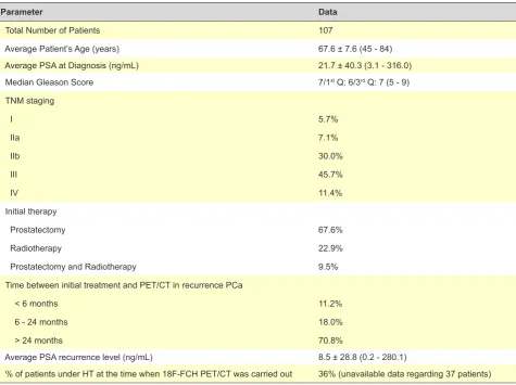
Table 1 – Characteristics of our group of patients
Parameter Data
Total Number of Patients 107
Average Patient’s Age (years) 67.6 ± 7.6 (45 - 84)
Average PSA at Diagnosis (ng/mL) 21.7 ± 40.3 (3.1 - 316.0)
Median Gleason Score 7/1st Q: 6/3rd Q: 7 (5 - 9)
TNM staging
I 5.7%
IIa 7.1%
IIb 30.0%
III 45.7%
IV 11.4%
Initial therapy
Prostatectomy 67.6%
Radiotherapy 22.9%
Prostatectomy and Radiotherapy 9.5%
Time between initial treatment and PET/CT in recurrence PCa
< 6 months 11.2%
6 - 24 months 18.0%
> 24 months 70.8%
Average PSA recurrence level (ng/mL) 8.5 ± 28.8 (0.2 - 280.1)
ARTIGO ORIGINAL patients with castration-resistant disease, although it may be significantly attenuated in hormone-sensitive patients.28,29
OBJECTIVE
Our study aimed to assess the impact of 18F-FCH PET/ CT in re-staging and therapy orientation of patients with biochemically recurrent PCa.
We also aimed to assess the influence of different factors in 18F-FCH PET/CT sensitivity, namely risk stratification, as well as PSA levels and HT.
MATERIAL AND METHODS Population
In total, 107 patients (aged 67.6 ± 7.6, 45-84) with PCa from our hospital, submitted to 18F-FCH PET/CT imaging between Dec 2009 and May 2014 due to biochemical recurrence upon therapy with a curative intent were included in the study.
Initial therapy with a curative intent involved prostatectomy or radiotherapy (RT) in 75.7% (81/107) and 33.6% (36/107) of the patients, respectively (10 patients underwent prostatectomy and consolidation RT).
The presence of two consecutive PSA levels >0.2 ng/ mL obtained upon prostatectomy or Biochemical recurrence was considered when two PSA consecutive values over 0.2 ng/mL were obtained upon prostatectomy or an increase >2
ng/mL compared to the PSA nadir upon RT.1,30,31
At the time when the 18F-FCH PET/CT was performed, 36.0% (31/86) of the patients were under HT (this information was not available for 21 patients). Patients on HT with PSA values in progression were considered as castration-resistant.
The characteristics of our group of patients are shown in Table 1.
Imaging protocol for 18F-FCH PET/CT
Patients who were referred for 18F-FCH PET/CT were previously informed as regards the aims of the scan and the whole procedure and formally accepted it through completion of a written informed consent.
The acquisition protocol involved the intravenous administration of 3MBq/kg of 18F-FCH, with immediate acquisition of a 5-min pelvic dynamic study. This initial acquisition aimed to allow for the study of the pelvic region before the arrival of the radiotracer to the bladder due to its normal urinary excretion, which, due to its proximity, would prevent the assessment of prostate and periprostatic region. Thirty minutes upon the administration of the radiotracer, a whole-body acquisition study was subsequently obtained, in order to search for the presence of any metastatic involvement. Patients assumed the dorsal decubitus position with the arms above the head within a GE Discovery ST PET/CT scanner. CT attenuation and anatomical mapping acquisition parameters were as follows: 120 kV, smart mA: 35 noise index with current values between 10-200mA, pitch 1.5:1, rotation 0.5 s and a 3.75 mm slice thickness. PET emission study was obtained in 3D mode, with a 70
cm Field Of View (FOV) diameter and 3-min acquisition time whole-body studies were acquired per table position. Data were collected in list mode and rebuilt using a 3D Ordered Subset Expectation Maximization (OSEM) iterative reconstruction algorithm, with 20-subset per two iterations, 128 x 128 matrix and one 5-mm Full Width at Half Maximum (FWHM) post-reconstruction filtering.
Image interpretation
Consensus interpretations by two specialists in Nuclear Medicine were obtained and patient’s clinical history as well as the available laboratory and imaging data were previously provided.
A semi-quantitative analysis was carried out and the
value of the Maximum Standard Uptake Value (SUVmax)
was calculated for each lesion, based on the creation of a volume of interest completely and solely involving the
lesion. SUVmax was used as an indicator of the radiotracer’s
uptake intensity by lesions.
18F-FCH uptake by the prostate and prostate bed was considered as abnormal when uptake intensity was above the background activity.
Lymph nodes with increased 18F-FCH uptake were considered as lymph node metastases, even without any anatomical criteria as lymphadenopathies. Lymph nodes with anatomical criteria as lymphadenopathies and no 18F-FCH uptake were not considered as metastases. Inguinal lymph nodes with mild 18F-FCH uptake and no suspicious morphological criteria were considered as reactive or inflammatory. This interpretation was carried out according to different criteria described in several published
studies.7,10,15
Skeletal areas of abnormally increased 18F-FCH uptake were considered as malignant according to its uptake intensity, anatomical location and morphological characteristics (tomodensitometric characteristics). These data were also correlated to those obtained in additional
tests, such as the 18F-Sodium Fluoride (18F-NaF) bone
PET/CT scan, the bone scintigraphy or MRI, whenever available. Discrepancies between 18F-FCH uptake and CT scan morphological characteristics found in PET/CT lead to an additional study through clinical follow-up and, in 16 patients, through a control 18F-FCH PET/CT. In these patients, lesions showing persistent 18F-FCH uptake or with a clinical or laboratory evidence of disease progression were considered as malignant.
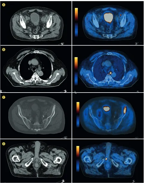
The tests of two patients with biochemical recurrence upon RT and prostatectomy are shown in Figure 1-2 and in Figure 3, respectively.
Recurrence diagnosis was histopathologically
ARTIGO ORIGINAL
as malignant, according to the abovementioned criteria. Clinical and laboratory follow-up, as well as the results obtained from several control imaging tests have also contributed to the assignment of any pathological meaning. Average follow-up time of our patients was 18.7 ± 13.3 months.
Statistical analysis
SPSS (version 20) software was used in statistical analysis. Univariable studies were carried out for the descriptive and frequency analysis. Mann-Whitney’s U-test and Chi-square test were used in comparison of quantitative and qualitative variables between groups, respectively. Correlation between quantitative variables was assessed using Spearman’s coefficient. Logistic regression test was carried out for assessing the influence of the different variables in the sensitivity of 18F-FCH PET/CT scan.
Statistical significance was met for a value of p below 0.05
for all tests.
RESULTS
The 18F-FCH PET/CT scan was positive in 63.6% (68/107) of the patients. Prostatic/prostate bed recurrence was found in 38.2% of these patients (26/68) with absence of disease at any other location; isolated lymph node involvement was found in 20.6% (14/68) and isolated bone involvement in 8.8% (6/68) of the patients. Regardless of the presence of any other spread, 60.3% (41/68) of the patients presented with prostatic recurrence, 51.5% (35/68) with lymph node involvement and 26.5% (18/68) with bone involvement.
The presence of local spread of the disease was found in 55.9% (38/68) of the patients, corresponding to prostatic or prostate bed recurrence and/or to pelvic lymph node involvement and 44.1% (30/68) presented with distant spread, corresponding to extra-pelvic lymph node and/or bone involvement.
A false positive result corresponding to prostatitis was found in one of the eight patients from whom we were provided with histology results. In the remaining seven
ARTIGO ORIGINAL
Figure 2 - 18F-FCH PET/CT imaging regarding the same patient as in Fig. 1, showing the presence of lymphadenopathies with abnormally increased prostatic 18F-FCH uptake, suggesting the presence of local-regional (A) and distant (B) lymph node metastatic spreading as well as bone involvement (C), in addition to the involvement of the right corpus cavernosum (D).
A
B
C
D
patients the disease was confirmed in the prostatectomy sample, corresponding to true positive results. No lymph node involvement (19 lymph nodes in total) was found in
ARTIGO ORIGINAL
Figure 3 - 18F-FCH PET/CT imaging from patient with prostate cancer who underwent prostatectomy (current PSA level - 8 ng/mL). The presence of one right pararectal lymphadenopathy with abnormally increased uptake of the radiotracer suggesting the presence of lymph node involvement, showing no other suspicious activity at the prostatic bed or at any other location.
A single lesion was detected by the 18F-FCH PET/CT in 54.4% (37/68) of the patients (70.3% with local recurrence, 21.6% with a single lymph node metastasis and 8.1% with a single bone metastasis).
Positive 18F-FCH PET/CT scan and PSA levels
Globally, an average 8.2 ± 29.1 ng/mL(0.2 – 280.1) PSA level was found. An average 2.0 ± 3.5 ng/mL (0.2 - 20) PSA level was found in patients with negative 18F-FCH PET/ CT, when compared to 11.8 ± 36.2 ng/mL (0.3 – 280.1)
in patients with positive 18F-FCH PET/CT (p < 0.05). An
average 5.6 ± 6.7 ng/mL (0.4 – 29.1) PSA level was found in patients with local recurrence only and no disease in any other location, 4.5 ± 3.3 ng/mL (0.7 - 14) in patients with lymph node involvement only and 7.3 ± 12.9 ng/mL (1.2
– 33.5) in patients with bone involvement only (p > 0.05).
An average 7.6 ± 33.9 ng/mL (0.3 – 280.1) PSA level was found in patients with local and regional disease, while a 9.4 ± 12.4 ng/mL (0.4 - 50) level was found in patients with
distant spread disease (p < 0.05). An average 36.8 ± 86.4
ng/mL (1.1 – 280.1) PSA level was found in patients with local recurrence and lymph node involvement and 0.3 ng/ mL (one patient only) in patients with local recurrence and bone involvement; an average 12.7 ± 3.1 ng/mL (9.0 – 16.5) PSA level was found in patients with local recurrence and with both lymph node and bone involvement. These data are shown in Table 2, 3 and 4.
A 40.5% [17/(17 + 25)] sensitivity with the 18F-FCH PET/ CT scan was found in patients with PSA ≤ 2 ng/mL and 80%
[48/(48 + 12)] in patients with PSA > 2 ng/mL (p < 0.05).
PSA recurrence level was not available in five patients. (Fig. 4).
An average 13.9 ± 15.8 ng/mL (3.1 – 87.0) initial PSA level was found in patients with negative 18F-FCH PET/CT
and 27.1 ± 50.2 ng/mL (4.0 – 316.0) (p < 0.05) in those with
positive 18F-FCH PET/CT.
Positive 18F-FCH PET/CT scan and SUVmax levels
Average 3.3 ± 2.1 (0.6 – 8.5), 4.0 ± 3.8 (1.1 – 13.5) and
ARTIGO ORIGINAL
with local recurrence, lymph node and bone involvement,
respectively (p > 0.05).
No statistically significant differences were found
between SUVmax levels in patients with ≤ 2 ng/mL PSA
levels vs. patients with PSA > 2 ng/mL (p > 0.05) nor any
statistically significant correlation between PSA recurrence
and average SUVmax levels (p > 0.05).
Positive 18F-FCH PET/CT and HT
At the time when the 18F-FCH PET/CT scan was performed, 29.0% (31/86) of the patients were receiving HT (unavailable data for 21 patients). An average 3.2 ± 3.3 ng/ mL (0.2 -14.0) PSA level was found in patients not receiving
HT vs. 18,3 ± 51.5 ng/mL (0.2 – 280.1) in patients receiving
HT (p < 0.05).
Average SUVmax levels obtained in both groups of
patients are shown in Table 5.
Average SUVmaxlevels were slightly higher in patients
receiving HT when compared to those not receiving it, even though statistically significant differences were not found
(4.7 ± 2.4 vs. 4.2 ± 3.4; p > 0.05).
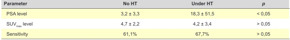
A 61.1% [33/(33 + 21)] 18F-FCH PET/CT sensitivity was found in the group of 55 patients not receiving HT, while a sensitivity of 67.7% [21/(21 + 10)] was found in the group of
patients receiving HT (p > 0.05) (Table 6).
Risk stratification
A 33.3% [3/(3 + 6)] increase in 18F-FCH PET/CT scan sensitivity was found in D’Amico (score of the risk of PCA recurrence) low-to-intermediate-risk patients, while a 55.2% [37/(37 + 30)] increase was found in high-risk patients (missing data from 30 patients made stratification
unavailable) (p > 0.05).
A 52.6% [20/(20 + 18)] sensitivity was found in high-risk
Figure 4 - Variation on 18F-FCH PET/CT sensitivity according to PSA levels.
PSA (ng/mL)
Sensitivity
(%)
0 90
80
70
60
50
40
30
20
10 100
0.2 - 2.0 2.1 - 10.0 10.1 - 30.0 > 30.1
Table 2 – Average PSA level and positive scan
Global Negative PET Positive PET p
Average PSA level 8.2 ± 29.1 2.0 ± 3.5 11.8 ± 36.2 < 0.05
Table 3 – Average PSA and disease location
Local recurrence Lymph node involvement Bone involvement p
Average PSA 5.6 ± 6.7 4.5 ± 3.3 7.3 ± 12.9 > 0.05
Table 4 – Average PSA level and local-regional versus distant metastases
Local-regional disease Distant metastases p
ARTIGO ORIGINAL
patients not receiving HT, while a 75% [12/(12 + 4)] sensitivity
was found in those receiving it (p > 0.05). This analysis was
not possible in low-to-intermediate risk patients due to the small number of patients in this group.
The average SUVmax of lesions was slightly higher in
high-risk patients, when compared to low-to-intermediate risk patients, even though statistically significant differences were
not found (4.4 ± 3.4 vs. 5.0 ± 3.3) (p > 0.05).
Predictive factors and sensitivity of the 18F-FCH PET/ CT scan
A logistic regression analysis was carried out in order to assess the impact of different factors in positive 18F-FCH PET/CT scans. Patient’s age, PSA recurrence level, level of PSA at diagnosis, Gleason score, tumour staging, HT and time between initial treatment and the 18F-FCH PET/CT scan were the variables included in the model.
Gleason score (B = 1.138 and p = 0.019) and the level of
PSA at recurrence (B = 1.42 and p = 0.037) were the only
statistically significant variables found in this analysis.
DISCUSSION
Recurrence is a common event in PCa upon initial therapy with a curative intent, with radical prostatectomy or
RT.32
PSA level is very relevant in follow-up, allowing for an early detection of biochemical recurrence. Nevertheless, it does not allow for the identification of the disease locations, namely the differentiation between a local and regional recurrence and a disease with a distant spread or between
a single lesion and a spreading disease,33 which are crucial
for a tailored therapy.34
Prostate biopsy has limitations due to its invasive nature as well as to potential sampling errors. Conventional imaging techniques, although well established in clinical practice, have some limitations regarding the detection of
disease locations, especially as regards bone involvement.35
Approximately 70% of lymphadenopathies have small
dimensions (long axis < 8 mm).36 This is a limitation as
regards the sensitivity of any imaging technique based on morphological criteria, such as CT or MRI.
An accurate identification of any lymph node involvement is particularly important, due to the influence on 5-year disease-free survival, ranging from 20-30% with multiple lymph node metastases compared to 75-80% with a single
metastasis.37
Bone scintigraphy has been used in detection of bone involvement. It has however a limited sensitivity and mostly a low specificity and usually there is the need for using other imaging techniques in order to clarify the results of scintigraphy.
The 18F-FCH PET/CT scan is a non-invasive whole-body multi-organ study combining anatomical and functional information, now increasingly being used in imaging PCa
assessment.38 Although it seems useful in the initial clinical
staging, it has been shown as especially useful in re-staging
of patients with biochemical recurrence.39 It is mainly useful
in the early stage of biochemical recurrence, allowing for the selection of patients that may benefit from salvage therapy, as well as for the assessment of patients with biochemical recurrence receiving HT, due to the likely presence of
extra-pelvic involvement.39
Different studies have assessed the diagnostic accuracy of this technique in the presence of biochemical recurrence of PCa, showing widely-varying sensitivities
ranging between 43 and 95%.5,7,10,12,14,24,40-43 A recent study
has found a high sensitivity and high negative predictive value of 18F-FCH PET/CT scan. In this study, a group of patients with biochemical recurrence remained in complete remission over a 6.5-year follow-up, upon removal of a single lymphadenopathy identified by the 18F-FCH PET/CT
scan.44 These were promising results, although further
long-term studies are necessary to confirm these results. Other studies have assessed the influence of different factors such as the stage of the disease, Gleason score,
HT, PSA level and PSA doubling time.13,25-28,45 The accuracy
of 18F-FCH PET/CT scan in biochemical recurrence has
been well recognized46 even though some studies have
described that sensitivity is influenced by HT, as well as
by the PSA level.47 This relationship led to a debate on a
PSA threshold for this technique to be performed. Different PSA thresholds were described in literature and, as the
Table 5 – Comparison between SUVmax levels within different malignancies in patients receiving or not receiving HT
Parameter Global Under HT No HT p
Local recurrence 3.3 ± 2.1 4.0 ± 2.6 5.1 ± 2.7 > 0.05
Lymph node involvement 4.0 ± 3.8 3.7 ± 3.2 4.4 ± 3.5 > 0.05
Bone involvement 7.0 ± 4.7 9.6 ± 3.8 6.7 ± 3.3 > 0.05
Table 6 – Correlation between PSA, SUVmax levels and 18F-FCH PET/CT sensitivity in patients receiving or not receiving HT
Parameter No HT Under HT p
PSA level 3,2 ± 3,3 18,3 ± 51,5 < 0,05
SUVmax level 4,7 ± 2,2 4,2 ± 3,4 > 0,05
ARTIGO ORIGINAL technique has been increasingly used in clinical practice, these levels have been progressively lowered. In 2006, this
procedure was questionable for PSA levels under 5 ng/
mL.48 Even though 2 ng/mL is the most frequently accepted
threshold (and therefore also adopted in our study), it is currently recognized that the 18F-FCH PET/CT scan may be useful from 1 ng/mL and we may even consider it with
lower PSA levels and high PSA doubling times.22
Sound results were obtained in our study with the use of the 18F-FCH PET/CT scan in re-staging patients with PCa and biochemical recurrence, allowing for the identification of metastatic PCa, as well as for metastasis location (lymph node and bone involvement) in a significant percentage of cases (28% of the patients and 44% of positive scans). Globally, a 63.2% sensitivity was found and increased to 80% when considering only the patients with PSA > 2 ng/ mL. In total, 38 (35.5%) patients with a single lesion were detected, 26 (68.4%) with local recurrence, eight (21.1%) with lymph node involvement and four (10.5%) with bone involvement, with a clear impact in therapeutic approach. From the variables included in the logistic regression analysis, (patient’s age, PSA recurrence level, PSA at diagnosis, Gleason score, staging of the disease, HT and time between initial treatment and the time when scan was
performed), only Gleason score (B = 1.138 and p = 0,019)
and PSA recurrence level (B = 1.42 and p = 0.037) were
statistically significant.
Pre-clinical studies showed a lower choline accumulation in hormone-sensitive cells upon HT, an effect that was not
found in hormone-resistant cells.49 Different mechanisms
were proposed to explain for the reduction in SUVmax upon
HT, namely 1) reduced volume of the lesion (reducing the signal due to a partial volume effect), 2) reduced levels of cell proliferation and 3) under-regulation of the expression of genes regulating the lipid metabolism, including a lower active choline transporter or lower choline-kinase enzyme
activity.50-55 However, HT did not show the same effects in
castration-resistant patients.
In our study, the sensitivity of 18F-FCH PET/CT scan,
as well as levels of SUVmax were slightly higher in the group
of patients under HT, even though statistically significant
differences were not found in both parameters (p > 0.05). In
addition, HT was not found as a positive predictive variable of 18F-FCH PET/CT scan in logistic regression analysis. Nevertheless, it should be mentioned that this analysis was limited by the small number of patients in whom HT was documented.
Despite these limitations, these results suggest that HT does not significantly affect 18F-FCH uptake in castration-resistant patients. Therefore, pre-scan HT withdrawal in patients with biochemical recurrence seems unnecessary. However, further studies are needed to clarify the mechanisms by which 18F-FCH uptake is inhibited by HT. The major limitations of this study regard its retrospective nature as well as the fact that most of the lesions were not histologically confirmed. Histological confirmation of every lesion is not feasible due to practical and ethical issues.
Lesions were considered as malignant based on clinical and laboratory, as well as imaging follow-up. However, different lesions may respond differently to therapy, which may have biased the results.
New advances in PCa imaging are constantly emerging. The PET/CT scan with Prostate Specific Membrane
Antigen (PSMA) labelled with 68Ga is a very promising
technique, which seems to combine high sensitivity, even in patients with low PSA, with high specificity. In addition,
the introduction of beta-emitting (such as the 177Lu) labelled
PSMA molecules provides for oriented molecular diagnostic and therapeutic approach (theragnostics) that may change
treatment of metastatic PCa.56
CONCLUSION
Sound results were found in our study regarding the use of 18F-FCH PET/CT scan in re-staging of patients with biochemical recurrence of PCa. The use of this technique allowed for the distinction between local-regional and systemic disease, an information relevant to therapeutic outcomes.
A strong positive correlation was found between the sensitivity of this procedure and recurrent PSA level. The 18F-FCH PET/CT scan provided for important diagnostic data in patients with low PSA levels and a significant increase in sensitivity was found in patients with PSA > 2 ng/mL.
A slightly higher sensitivity of 18F-FCH PET/CT scan was found in high-risk patients, when compared to low-to-intermediate-risk patients (according to the D’Amico classification), even though a statistically significant difference was not achieved.
Slightly higher sensitivity and SUVmax levels were found
in the group of patients under HT, even though a statistically significant difference was not found. These results suggest that diagnostic information is not negatively influenced by HT in castration-resistant patients and therefore pre-scan HT withdrawal seems unnecessary in this group of patients.
HUMAN AND ANIMAL PROTECTION
The authors declare that the followed procedures were according to regulations established by the Ethics and Clinical Research Committee and according to the Helsinki Declaration of the World Medical Association.
DATA CONFIDENTIALITY
The authors declare that they have followed the protocols of their work centre on the publication of patient data.
CONFLICTS OF INTEREST
The authors declare that there were no conflicts of interest in writing this manuscript.
FINANCIAL SUPPORT
ARTIGO ORIGINAL
REFERENCES
1. Boccon-Gibod L, Djavan W, Hammerer P, Hoeltl W, Kattan M, Prayer-Galetti T. Management of prostate-specific antigen relapse in prostate cancer: a European Consensus. Int J Clin Pract. 2004;58:382-90. 2. Platz E, Giavannucci E. Prostate cancer. Cancer epidemiology and
prevention. 3rd ed. New York: Oxford University Press: Schottenfeld D, Fraumeni Jr JF, editors; 2006:1151-65.
3. Dillioglugil O, Leibman B, Kattan M, Seale-Hawkins C, Wheeler T, Scardino P. Hazard rates for progression after radical prostatectomy for clinically localized prostate cancer. Urology. 1997;50:93-9.
4. Chondrogiannis S, Marzola M, Ferreti A, Grassetto G, Maffione AM, Rampin L, et al. Is the detection rate of 18F-choline PET/CT influenced by androgen deprivation therapy? Eur J Nucl Mol Imaging. 2014;41:1293-300.
5. Picchio M, Messa C, Landoni C, Gianolli L, Sironi S, Brioschi M, et al. Value of 11C-choline-positron emission tomography for re-staging prostate cancer: a comparison with 18F-fluorodeoxyglucose-positron emission tomography. J Urol. 2003;169:1337-40.
6. Langsteger W, Heinisch M, Fogelman I. The role of fluorodeoxyglucose, 18F-dihydroxiphenylalanine, 18F-choline and 18F-fluoride in bone imaging with emphasis on prostate and breast. Semin Nucl Med. 2006;36:73-92.
7. Cimitan M, Bortolus R, Morassut S, Canzonieri V, Garbeglio A, Baresic T, et al. [18F]fluorocholine PET/CT imaging for the detection of recurrent cancer at PSA relapse: experience in 100 consecutive patients. Eur J Nucl Med Mol Imaging. 2006;33:1387-98.
8. Reske S, Blumstein N, Neumaier B, Gottfried HW, Finsterbusch F, Kocot D, et al. Imaging prostate cancer with [11C]choline PET/CT. J Nucl Med. 2006;47:1249-54.
9. Kwee S, Wei H, Sesterhenn I, Yun D, Coel M. Localization of primary prostate cancer with dual-phase [18F]fluorocholine PET. J Nucl Med. 2006;47:262-9.
10. Schmid D, John H, Zweifel R, Cservenyak T, Westera G, Goerres GW, et al. Fluorocholine PET/CT in patients with prostate cancer: initial experience. Radiology. 2005;235:623-8.
11. Schiavina R, Scattoni V, Castelluci P, Picchio M, Corti B, Briganti A, et al. [11C]Choline positron emission tomography/computerized tomography for preoperative lymph-node staging in intermediate-risk and high-risk prostate cancer: comparison with clinical staging normograms. Eur Urol. 2008;54:392-401.
12. de Jong I, Pruim J, Elsinga P, Vaalburg W, Mensink H. [11C]choline positron emission tomography for the evaluation after treatment of localized prostate cancer. Eur Urol. 2003;44:32-8.
13. Heinisch M, Dirisamer A, Loidl W, Stoiber F, Gruy B, Haim S, et al. Positron emission tomography/computed tomography with [18F] fluorocholine for restaging of prostate cancer patients: meaningfull PSA <5 ng/ml? Mol Imaging Biol. 2006;8:43-8.
14. Husarik D, Mirabell R, Dubs M, John H, Giger OT, Gelet A, et al. Evaluation of [18F]choline PET/CT for staging and restaging of prostate cancer. Eur J Nucl Med Mol Imaging. 2008;35:253-63.
15. Beheshti M, Imamovic L, Broinger G, Vali R, Waldenberger P, Stoiber F, et al. [18F]choline PET/CT in the preoperative staging of prostate cancer in patients with intermediate or high risk of extracapsular disease: a prospective study of 130 patients. Radiology. 2010;254:925-33. 16. Scattoni V, Picchio M, Suardi N, Messa C, Freschi M, Roscigno M, et al.
Detection of lymph-node metastases with integrated [11C]choline PET/ CT in patients with PSA failure after radical retro-pubic prostatectomy: results confirmed by open pelvic-retroperitoneal lymphadenectomy. Eur Urol. 2007;52:423-9.
17. Abuzallouf S, Dayes I, Lukka H. Baseline staging of newly diagnosed prostate cancer: a summary of the literature. J Urol. 2014;171:2122-7. 18. Hricak H, Choyke P, Eberhardt S, Leibel S, Scardino P. Imaging prostate
cancer: a multidisciplinary prespective. Radiology. 2007;243:28-53. 19. Kent C. Regulation of phosphatidylcholine biosynthesis. Prog Lipid Res.
1990;29:87-105.
20. Fox JJ, Schöder H, Larson SM. Molecular imaging of prostate cancer. Curr Opin Urol. 2012;22:320-7.
21. Kitajima K, Murphy RC, Nathan MA, Froemming AT, Hagen CE, Takahashi N, et al. Detection of recurrent prostate cancer after radical prostatectomy: comparison of 11C-choline PET/CT with pelvic multiparametric MR imaging with endorectal coil. J Nucl Med. 2014;55:223-32.
22. Brenot-Rossi I. Mise au point: TEP-choline et cancer de la prostate. Prog Urol. 2014;24:3-8.
23. Beheshti M, Haim S, Zakavi R, Steinmair M, Waldenberger P, Kunit T,
et al. Impact of 18F-choline PET/CT in prostate cancer patients with biochemical recurrence: influence of androgen deprivation therapy and correlation with PSA kinetics. J Nucl Med. 2013;54:833-40.
24. Vees H, Buchegger F, Albrecht S, Khan H, Husarik D, Zaidi H, et al. [18F]choline and/or [11C]acetate positron emission tomography: detection of residual or progression subclinical disease at very low prostate-specific antigen values (<1 ng/ml) after radical prostatectomy. BJU Int. 2007;99:1415-20.
25. Krause B, Souvatzoglou M, Tuncel M, Herrmann K, Buck AK, Praus C, et al. The detection rate of [11C]choline PET/CT depends on the serum PSA value in patients with biochemical recurrence of prostate cancer. Eur J Nucl Med Mol Imaging. 2008;35:18-23.
26. Giovacchini G, Picchio M, Coradeschi E, Garcia Parra R, Briganti A, Gianolli L, et al. Predictive factors of [11C]choline PET/CT in patients with biochemical failure after radical prostatectomy. Eur J Nucl Med Mol Imaging. 2010;37:301-309.
27. Giovacchini G, Picchio M, Scattoni V, al e. PSA doubling time for prediction of [11C]choline PET/CT findings in prostate cancer patients with biocheminal failure after radical prostatectomy. Eur J Nucl Med Mol Imaging. 2010;37:1106-16.
28. Giovacchini G, Picchio M, Coradeschi E, Scattoni V, Bettinardi V, Cozzarini C, et al. [11C]choline uptake with PET/CT for the initial diagnosis of prostate cancer: relation to PSA levels, tumour stage and anti-androgenic therapy. Eur J Nucl Med Mol Imaging. 2008;35:1065-73. 29. Fuccio C, Schiavina R, Castellucci P, Rubello D, Martorana G, Celli M, et
al. Androgen deprivation therapy influences the uptake of [11C]choline in patients with recurrent prostate cancer: the preliminary results of a sequential PET/CT study. Eur J Nucl Med Mol Imaging. 2011;38:1985-9. 30. Moul J. Prostate specific antigen only progression of prostate cancer. J
Urol. 2000;163:1632-42.
31. Roach III M, Hanks G, Thames Jr H, Schellhammer P, Shipley W, Sokol G. Defining biochemical failure following radiotherapy with or without hormonal therapy in men with clinically localized prostate cancer: recommendations of the RTOG-ASTRO Phoenix consensus conference. Int J Radiat Oncol Biol Phys. 2006;65:965-74.
32. Freedland SJ, Presti JC, Amling CL, Kane CJ, Aronson WJ, Dorey F, et al. Time trends in biochemical recurrence after radical prostatectomy: results of the SEARCH database. Urology. 2003;61:736-41.
33. Evangelista L, Zattoni F, Guttilla A, Saladini G, Colletti PM, Rubello D. Choline PET or PET/CT and biochemical relapse of prostate cancer: a systematic review and meta-analysis. Clin Nucl Med. 2013;38:305-14. 34. Apolo AB, Pandit-Taskar N, Morris MJ. Novel tracers and their
development for the imaging of metastatic prostate cancer. J Nucl Med. 2008;49:2031-41.
35. de Jong IJ, Pruim J, Elsinga PH, Vaalburg W, Mensink HJ. Preoperative staging of pelvic lymph nodes in prostate cancer by 11C-choline PET. J Nucl Med. 2003;44:331-5.
36. Murphy G, Haider M, Ghai S, Sreeharsha B. The expanding role of MRI in prostate cancer. AJR Am J Roentgenol. 2013;201:1229-38. 37. Paño B, Sebastià C, Buñesch L, Mestres J, Salvador R, Macías NG, et
al. Pathways of lymphatic spread in male urogenital pelvic malignancies. Radiographics. 2011;311:135-60.
38. Krause BJ, Souvatzoglou M, Treiber U. Imaging of prostate cancer with PET/CT and radioactively labeled choline derivates. Urol Oncol. 2013;31:427-35.
39. Castellucci P, Ceci F, Graziani T, Schiavina R, Brunocilla E, Mazzarotto R, et al. Early Biochemical Relapse After Radical Prostatectomy: Which Prostate Cancer Patient May Benefit from a Restaging 11C-Choline PET/CT Scan Before Salvage Radiation Therapy? J Nucl Med. 2014;55:1424-9.
40. Rinnab L, Blumstein N, Mottaghy F, Hautmann RE, Küfer R, Hohl K, et al. [11C]choline positron-emission tomography/computed tomography and transrectal ultrasonography for staging localized prostate cancer. BJU Int. 2007;99:1421-6.
41. Chiti A, Picchio M. The rising PET: the increasing use of choline PET/CT in prostate cancer. Eur J Nucl Med Mol Imaging. 2011;38:53-4. 42. Rinnab L, Mottaghy F, Blumstein NM, Reske SN, Hautmann RE,
Hohl K, et al. Evaluation of [11C]choline positron emission/computed tomography in patients with increasing prostate-specific antigen levels after primary treatment for prostate cancer. BJU Int. 2007;100:786-93. 43. Pelosi E, Arena V, Skanjeti A, Pirro V, Douroukas A, Pupi A, et al. Role
ARTIGO ORIGINAL
44. Winter A, Henke RP, Wawroschek F. Targeted salvage lymphadenectomy in patients treated with radical prostatectomy with biochemical recurrence: complete biochemical response without adjuvant therapy in patients with low volume lymph node recurrence over a long-term follow-up. BMC Urol. 2015;15:4.
45. Schillaci O, Calabria F, Tavolozza M, Caracciolo CR, Finazzi Agrò E, et al. Influence of PSA, PSA velocity and PSA doubling time on contrast-enhanced [18F]choline PET/CT detection rate in patients with rising PSA after radical prostatectomy. Eur J Nucl Med Mol Imaging. 2012;39:589-96.
46. Ceci F, Castellucci P, Graziani T, Schiavina R, Brunocilla E, Mazzarotto R, et al. 11C-choline PET/CT detects the site of relapse in the majority of prostate cancer patients showing biochemical recurrence after EBRT. Eur J Nucl Med Mol Imaging. 2014;41:878-86.
47. Giovacchini G, Picchio M, Scattoni V, Garcia Parra R, Briganti A, Gianolli L, et al. PSA doubling time for prediction of [(11)C]choline PET/CT findings in prostate cancer patients with biochemical failure after radical prostatectomy. Eur J Nucl Med Mol Imaging. 2010;37:1106-16. 48. Heinisch M, Dirisamer A, Loidl W, Stoiber F, Gruy B, Haim S, et al.
Positron emission tomography/computed tomography with F-18-fluorocholine for restaging of prostate cancer patients: meaningful at PSA < 5 ng/ml? Mol Imaging Biol. 2006;8:43-8.
49. Hara T, Bansal A, DeGrado T. Effect on hypoxia on the uptake of [methyl-3H]choline, [1-14C]acetate and [18F]FDG in cultured prostate cancer cells. Nucl Med Biol. 2006;99:977-84.
50. Giovacchini G, Picchio M, Coradeschi E, Scattoni V, Bettinardi V,
Cozzarini C, et al. [(11)C]choline uptake with PET/CT for the initial diagnosis of prostate cancer: relation to PSA levels, tumour stage and anti-androgenic therapy. Eur J Nucl Med Mol Imaging. 2008;35:1065-73. 51. Nakashima J, Imai Y, Tachibana M, Baba S, Hiramatsu K, Murai M.
Effects of endocrine therapy on the primary lesion in patients with prostate carcinoma as evaluated by endorectal magnetic resonance imaging. Cancer. 1997;80:237-41.
52. Swinnen JV, Verhoeven G. Androgens and the control of lipid metabolism in human prostate cancer cells. J Steroid Biochem Mol Biol. 1998;65:191-8.
53. Mueller-Lisse UG, Swanson MG, Vigneron DB, Hricak H, Bessette A, Males RG, et al. Time-dependent effects of hormone-deprivation therapy on prostate metabolism as detected by combined magnetic resonance imaging and 3D magnetic resonance spectroscopic imaging. Magn Reson Med. 2001;46:49-57.
54. Yoshimoto M, Waki A, Obata A, Furukawa T, Yonekura Y, Fujibayashi Y. Radiolabeled choline as a proliferation marker: comparison with radiolabeled acetate. Nucl Med Biol. 2004;31:859-65.
55. Breeuwsma AJ, Pruim J, Jongen MM, Suurmeijer AJ, Vaalburg W, Nijman RJ, et al. In vivo uptake of [11C]choline does not correlate with cell proliferation in human prostate cancer. Eur J Nucl Med Mol Imaging. 2005;32:668-73.