R E S E A R C H
Open Access
BRAF
V600E mutation in Juvenile
Xanthogranuloma family neoplasms of the
central nervous system (CNS-JXG): a revised
diagnostic algorithm to include pediatric
Erdheim-Chester disease
J. Picarsic
1*, T. Pysher
2, H. Zhou
2, M. Fluchel
3, T. Pettit
4, M. Whitehead
5, L. F. Surrey
6, B. Harding
6, G. Goldstein
7,
Y. Fellig
8, M. Weintraub
9, B. C. Mobley
10, P. M. Sharples
11, M. L. Sulis
12, E. L. Diamond
13, R. Jaffe
14, K. Shekdar
15and
M. Santi
6Abstract
The family of juvenile xanthogranuloma family neoplasms (JXG) with ERK-pathway mutations are now classified within the “L”(Langerhans) group, which includes Langerhans cell histiocytosis (LCH) and Erdheim Chester disease (ECD). Although the BRAF V600E mutation constitutes the majority of molecular alterations in ECD and LCH, only three reported JXG neoplasms, all in male pediatric patients with localized central nervous system (CNS) involvement, are known to harbor the BRAF mutation. This retrospective case series seeks to redefine the clinicopathologic spectrum of pediatric CNS-JXG family neoplasms in the post-BRAF era, with a revised diagnostic algorithm to include pediatric ECD. Twenty-two CNS-JXG family lesions were retrieved from consult files with 64% (n= 14) having informative BRAFV600E mutational testing (molecular and/or VE1 immunohistochemistry). Of these, 71% (n= 10) were pediatric cases (≤18 years) and half (n= 5) harbored the BRAFV600E mutation. As compared to theBRAF wild-type cohort (WT), theBRAF V600E cohort had a similar mean age at diagnosis [BRAFV600E: 7 years (3–12 y), vs. WT: 7.6 years (1–18 y)] but demonstrated a stronger male/female ratio (BRAFV600E: 4 vs WT: 0.67), and had both more multifocal CNS disease (BRAFV600E: 80% vs WT: 20%) and systemic disease (BRAFV600E: 40% vs WT: none). Radiographic features of CNS-JXG varied but typically included enhancing CNS mass lesion(s) with associated white matter changes in a subset of BRAF V600E neoplasms. After clinical-radiographic correlation, pediatric ECD was diagnosed in theBRAFV600E cohort. Treatment options varied, including surgical resection, chemotherapy, and targeted therapy with BRAF-inhibitor dabrafenib in one mutated case. BRAF V600E CNS-JXG neoplasms appear associated with male gender and aggressive disease presentation including pediatric ECD. We propose a revised diagnostic algorithm for CNS-JXG that includes an initial morphologic diagnosis with a final integrated diagnosis after clinical-radiographic and molecular correlation, in order to identify cases of pediatric ECD. Future studies with long-term follow-up are required to determine if pediatric BRAF V600E positive CNS-JXG neoplasms are a distinct entity in the L-group histiocytosis category or represent an expanded pediatric spectrum of ECD.
Keywords:BRAFV600E, Juvenile Xanthogranuloma, JXG, Central nervous system, CNS, Pediatric, Erdheim-Chester disease, ECD, LCH
© The Author(s). 2019Open AccessThis article is distributed under the terms of the Creative Commons Attribution 4.0 International License (http://creativecommons.org/licenses/by/4.0/), which permits unrestricted use, distribution, and reproduction in any medium, provided you give appropriate credit to the original author(s) and the source, provide a link to the Creative Commons license, and indicate if changes were made. The Creative Commons Public Domain Dedication waiver (http://creativecommons.org/publicdomain/zero/1.0/) applies to the data made available in this article, unless otherwise stated. * Correspondence:[email protected]
A portion of this work was presented at the annual Histiocyte Society Meeting in Lisbon, Portugal on October 2018.
1Department of Pathology, University of Pittsburgh School of Medicine,
Introduction
In the most recent revised classification of histiocytic disorders, [21], cutaneous juvenile xanthogranuloma (JXG) lesions and those JXG lesions with a systemic component, but not associated with a molecular alter-ation, are categorized separately into the cutaneous or “C”-group histiocytosis. However, extracutaneous JXG lesions with mitogen activated pathway kinase (MAPK) / extracellular-signal-regulated kinase (ERK) pathway activating mutations are now categorized into the Langerhans “L-group” histiocytosis, including three rare BRAF V600E JXG “L-group” neoplasm [56]. In this revised classification, Langerhans cell histiocytosis (LCH) and Erdheim Chester Disease (ECD), are also categorized in the “L group” of histiocytic neoplasms. At the far ends of their phenotypic spectra, LCH, ECD, and JXG all have distinct clinical and pathologic features; however, this shared categorization was proposed based on similar molecular alterations, mixed LCH/ECD histio-cytic presentations in adult cases, and accumulating data supporting a common hematopoietic precursor, at least between adult LCH and ECD [21]. However, pediatric extracutaneous JXG with MAPK molecular alterations as an L-group histiocytosis, has been less studied in relation to its possible shared origins with LCH and pediatric ECD [10, 16, 38, 40, 46, 51] Furthermore, while the BRAF V600E mutation constitutes the majority of molecular alterations in ECD and LCH [3, 5, 30, 53], only three reported JXG neoplasms, all in male pediatric patients with localized central nervous system (CNS) involve-ment, are known to harbor the BRAF mutation; how-ever, none showed evidence of systemic disease or a prior history of LCH [56].
In general, CNS-JXG neoplasm are rare, often requir-ing surgical resection or chemotherapy [13, 36, 55, 58] and do not have the propensity to regress spontaneously, unlike their cutaneous JXG counterpart [58]. CNS-JXG neoplasm range from isolated CNS lesions to multi-focal CNS lesions to those associated with systemic disease [6,13, 22,26,27,36,58]. In adults, CNS based neoplasms with a JXG or xanthogranuloma pathologic phenotype are often the first and most debilitating manifestation of ECD. They are often a challenge to diagnose and have a generally poor prognosis; how-ever, in adults these neoplasms often have an excellent response to inhibitor therapy [15,24,48]. In children, both systemic JXG with CNS involvement and CNS-limited JXG also appear to have poorer outcomes, as compared to pediatric JXG without CNS disease; however, none of these prior pediatric JXG studies have investigated the BRAF mutational status [13,58].
Furthermore, the current revised classification of histiocytes [21] has created a divide between the JXG family of neoplasms with molecular alterations
(L-group) and those without molecular alterations (C-group). Standing alone, this grouping does not have particular clinical significance, especially given that both C-group and even R-group histiocytic lesions now also harbor MAPK-pathway activated mutations [16,25,28,44,49, 52]. Furthermore, the World Health Organization (WHO) recommends that CNS neo-plasms have an initial morphologic report followed by an integrated final diagnosis after molecular studies are completed [42]. The aim of this study is to revisit the pathology and incidence ofBRAFV600E mutations in pediatric CNS-JXG neoplasms in order to propose a revised diagnostic algorithm that requires the integra-tion of pathology, molecular, clinical, and radiographic findings for a comprehensive final diagnosis, in the hope of advancing clinical management and treatment options.
Materials and methods
Cases: inclusion and exclusion criteria
Following institutional review board approval (University of Pittsburgh IRB number PRO12110055), we retrieved cases from our pathology consult files for CNS-based JXG family lesions, which includes previously published pediatric cases [14,55,57]. In our initial inclusion criteria, we included all cases that were diagnosed as a JXG family neoplasm by morphology and immunophenotype, as previously described [8,50,59]. In brief, JXG family neo-plasms range from 1) small to intermediate-sized mono-nuclear histiocytes, to 2) abundant foamy, xanthomatous (i.e. lipidized) histiocytes and Touton giant cells, to 3) those lesions resembling benign fibrous histiocytoma with a predominance of spindle-shaped cells and fibrosis with lesser quantity of foamy histiocytes and giant cells, while also including 4) oncocytic cells with abundant glassy pink cytoplasm (i.e reticulohistiocytoma” subtype). Under the microscope JXG and ECD share similar morphologic patterns and a shared immunophenotype (i.e., positive: CD163, CD68, CD14, Factor 13a, fascin, typically S100 negative, and CD1a and Langerin negative). Both JXG and ECD can be diagnosed as “JXG family” on pathologic grounds alone, with the distinction of ECD made by cor-relating appropriate clinical and radiographic features, as previously described [15].
Immunohistochemistry
Immunohistochemistry was performed on 3μm thick formalin fixed paraffin embed (FFPE) sections using commercially available antibodies: CD163, CD68 PGM1, CD14, Factor XIIIa, Fascin, Ki-67, S100, CD1a, Langerin, and Braf-VE1 (Table1).
BRAF V600E assessment
BRAF status was assessed either by DNA-based studies and/or immunohistochemistry with a clinically validated
BRAF V600E (VE1) immunohistochemical stain (Table1). Previous in-house validation and others have shown a very high correlation with molecular status when diffuse 2–3+ intensity granular staining is present in > 10% of while negative cases had either complete absence of staining, weak/faint granular staining (1+) or staining in only rare, single scattered cells, often not possessing the morphology of a histiocytic cell [41]. For those that had molecular testing around the time of diagnosis, a variety of methodologies were used, dependent on the
Fig. 1Flow Diagram of Cases. A total of 10 Pediatric CNS-JXG neoplasms with informative BRAF status are included in primary analysis
Table 1Immunohistochemistry of histiocytic neoplasms
Antibody (Source) Clone Dilution Antigen Retrieval
(Ventana proprietary reagents)
Detection
(Ventana proprietary reagents)
CD163 (Novocastra) 10D6 1:200 uCC1 mild iView DAB
CD68 (Dako) PG-M1 1:100 uCC1 mild iView DAB
CD14 (Cell Marque) EPR3653 1:100 uCC1 standard Optiview DAB
Factor XIIIα(GeneTex) Polyclonal 1:250 Protease iView DAB
Fascin (Dako) 55 K-2 1:500 uCC1 mild iView DAB
Ki-67 (Dako) MIB-1 1:25 uCC1 mild iView DAB
S100 (Dako) Polyclonal 1:3000 uCC1 mild iView DAB
CD1α(Immunotech) O1O 1:5 uCC1 mild uV DAB
Langerin (Leica) 12D6 1:100 uCC1 standard uV DAB
referring institution. A single sample (case 3) under-went PCR Sanger Sequence locally at our institution on available FFPE consult block material of the CNS-JXG lesion, along with Braf-VE1 immunostaining. Briefly, a manual microdissection was performed on this sample (> 50% tumor cells present). DNA was iso-lated using standard laboratory procedure with optical density readings. For the detection of the mutation, Light Cycler Platform (Roche Molecular Systems, Inc. Pleasanton, California) was used to amplify BRAF exon 15 codons 599–601 sequences. Post-PCR melt curve analysis was used to detect mutation and confirmed with Sanger Sequencing of the PCR product on ABI3130 (Applied Biosystems, Thermo Fisher Scien-tific, Waltham, Massachusetts). The limit of detection was approximately 10–20% of alleles with mutation present in background of normal DNA.
Results
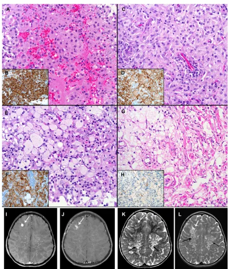
We identified 22 CNS lesions with JXG phenotype that met our initial inclusion criteria (Fig. 1), designated as CNS-JXG. Fourteen CNS-JXG cases (64%) had inform-ative molecular status for theBRAF V600E point muta-tion, which included 10 pediatric CNS-JXG neoplasms that were included in the primary analysis. The overall mean age was 7.3 years (range: 1–18 y) with a male/fe-male ratio of 1.5 (Table2). Within such a small cohort it is difficult to ascertain clinically relevant, statistical dif-ference between the pediatric BRAF V600E (n = 5) and BRAF wild-type (n = 5) CNS-JXG cohorts, but certain trends were noted. There was a similar mean age in both cohorts [BRAF V600E: 7 years (3–12 y) vs BRAF wild-type 7.6 years (1–18 years)]. While the overall male/fe-male ratio in the pediatric CNS-JXG cohort was male/fe-male predominant (1.5), the BRAF V600E cohort had more males (male/female ratio: 4.0), as compared to the wild type cohort (male/female ratio: 0.67). The pediatric BRAF V600E cohort also had more cases of multifocal CNS disease [BRAFV600E: 3/5 (60%) vs theBRAF wild-type: 1/5 (20%)] along with associated CNS white matter changes and enhancement of nodular lesions (Table 2). The two cases with systemic disease were BRAF V600E positive (Table2). One had multifocal CNS-JXG disease, including intracranial, sellar, dural, ventricular, and cav-ernous sinus involvement, along with bilateral long bone sclerosis and confirmatory bone biopsy also with the BRAFV600E mutation (case 3). Thus the integrated final diagnosis with pathology and radiographic correlation was that of pediatric ECD, as previously published [14]. The second case also had systemic disease, with an as-sociated cutaneous BRAF V600E positive JXG lesions. Symmetric CNS white matter changes were present on MRI (Fig. 2i-l), along with an enhancing parenchymal mass; however, there was no evidence of bone
involvement or other classic features of ECD. One of the BRAF V600E cases with multifocal CNS lesions had visual decline and panhypopituitarism from sellar/ optic chiasm-based masses, while the other had result-ant encephalomalacia and brain atrophy with progres-sive developmental delay and was started on hospice care six years after initial presentation (Table 2). In contrast, the BRAF wild-type cohort had more isolated CNS lesions without mention of associated symmetric white matter changes or reported systemic disease; however, one of these cases did not have long term follow-up after diagnosis (Table 2). The BRAF wild type group also did not have further molecular testing or phosphorylated-ERK staining.]
Pathologic features of the pediatric BRAF informative cohort
Table 2Pediatric CNS-JXG cases withBRAFV600E status with clinical, imaging, and treatment findings
Case BRAF V600E Status
Sex Age at diagnosis (y)
Radiology/ Location Systemic Disease Treatment and follow-up
PediatricBRAF-V600E CNS-JXG patients (n= 5).
1 Mutated M 3 Unifocal: Pituitary stalk thickening and loss of bright spot on MRI.
No; LCH staging did not reveal additional lesions, including PET and CT of chest and abdomen
Presumptive MRI diagnosis of LCH with DI, started on LCH III based Rx (Prednisone and vinblastine) with mild progression of the lesion and suprasellar extension at 12 weeks. Bx performed with Rx continued for 12 mo. Stable thickening of pituitary at 2.5 y following Rx completion. No other lesions noted with central diabetes insipidus.
2 Mutated F 4 Multifocal: Enhancing, dominant, right frontal lobe lesion, along with T2 hypointense cerebral, cerebellar, and brain stem nodules with background of symmetric T2 white matter
hyperintensity.
Yes-systemic with JXG BRAF-V600E positive cutaneous lesions; but: no imaging suggestive of classic ECD lesions
Clofarabine and dexamethasone (cycle 4, 4 months after initial biopsy), demonstrating clinical and radiographic improvement
3* Mutated M 7 Multifocal: Enhancing intracranial, dural plaque-like thickening, sellar/suprasellar, 4th ventricle, posterior fossa, cavernous sinus
Yes–ECD confirmed on bone scan and bx with bilateral tibial sclerosing lesions
Partial resection/debulking with progression after 6 mo. 2 cycles clofarabine with progression. After dx of ECD, started on Anakinra 2 mg/kg. At 2 y f/u MRI improvement of CNS and osseous lesions.
4 Mutated M 9 Multifocal: Enhancing Intraventricular and subependymal masses (bilateral), enlarged pituitary, Increased T2 signal pons, WM of cerebellum including dentate nuclei, post-enhanced T1 signal increase of basal ganglia (bilateral), expansion of cavernous sinuses (bilateral) (Ddx included LCH)
N–Multiple body imaging including MRI of spine, negative skeletal survey
Progressive ataxia since 4.5 yo. Imaging 4 mo after bx with extensive enhancing lesions suggestive of‘perivascular spread’abnormal WM signal (FLAIR): bihemispheric subcortical areas (subinsular, thalami, basal ganglia), WM of temporal/parietal/occipital lobe, and WM of cerebellar hemispheres, brain stem, mesial temporal (Comment concerning for malignant histiocytosis or demyelinating disease). 23 mo after bx: Encephalomalacia, atrophic changes of the brain, bright T2 both in WM of cerebellum, no enhancing lesions 4 y after bx: Developmental delay with ventriculomegaly and periventricular WM T2 prolongation; Persistent, stable cerebellar WM T1 and T2 prolongation without enhancing lesions 5 yr after dx: Lost to f/u in hospice care
5 Mutated M 12 Multifocal: Large sellar mass (pituitary and optic chiasm), (enhancing) two dural based temporal masses, two lateral intraventricular masses and cerebellar ND-white changes.
No; After tissue diagnosis: no imaging s/o of ECD lesions. Subsequent MRI showed ND changes in cerebellum
DI since age 7yo, then 4th cranial nerve palsy, decreased visual acuity. MRI and Bx (originally called RDD) with subsequent resection of temporal mass; following 9 mo later with surgical decompression of optic chiasm and panhypopituitarism with cognitive decline. Started on dabrafenib (100 mg ×2/day) with dramatic clinical response. MRI 2 years on I-Rx showed no new lesions and moderate reduction tumor size. No other new lesions noted; with occasional CSF drainage through Ommaya reservoir for intermittent headache.
Pediatric BRAF wild type CNS-JXG patients (n= 5).
6** Wild-type
M 1 Unifocal: Cerebellopontine Angle with encasement of cranial nerves V and VI; MRI heterogenous T1 iso to
hypointense and T2 hypointense with
No Initial radiographic concern for
Therapy and outcomes of the pediatric BRAF informative cohort
Treatment options in pediatric CNS-JXG cases were variable showing a combination of both surgical excision and systemic chemotherapy (Table 2). For most BRAF V600E CNS-JXG cases, theBRAF mutational status was not known at the time of initial diagnosis. Treatments included the following: LCH III based protocol with prednisone/vinblastine for 12 months in unifocal CNS disease of the hypothalamic-pituitary axis, clofarabine and dexamethasone for systemic JXG with multifocal CNS-JXG, anakinra for pediatric ECD that had previ-ously progressed on prednisone/vinblastine for 6 weeks, cladribine for 6 cycles, and clofarabine for 2 cycles [14], and BRAF-inhibitor therapy with dabrafenib for a multi-focal CNS disease, which was aggressive and refractory to first line therapy. In this last case, dabrafenib showed
an immediate and dramatic clinical response, including complete resolution of hyperventilation and weaning from dexamethasone with interval MRI at 2 months, 4 months and 15 month of therapy, along with a reduction in intracranial size and no new lesions. (Table2). Case 4 did not have a known BRAFV600E prospectively in his course with progressive CNS white matter disease in the years following excision. The wild-type cases also had surgical resection with initial prednisone/vinblastine and then cladribine in one case with multifocal lesions and prednisone/vinblastine in a unifocal lesion of the cere-bellopontine angle of a 1-year-old (Table2).
Discussion
This retrospective case series characterizes the largest series to date of BRAF V600E mutated pediatric JXG family neoplasms, all of which were first diagnosed with Table 2Pediatric CNS-JXG cases withBRAFV600E status with clinical, imaging, and treatment findings(Continued)
Case BRAF V600E Status
Sex Age at diagnosis (y)
Radiology/ Location Systemic Disease Treatment and follow-up
contrast enhancement year with no evidence of recurrent
disease. Unchanged postsurgical appearance of the brain including linear/nodular enhancement along the medial aspect of the left posterior fossa resection cavity that is favored to represent postsurgical change.
7 Wild-type
F 2 Unifocal Thalamus No Right hemiplegia at presentation that
persists. IV methylprednisolone, follow-ed by oral Prednisolone at presentation that was weaned. No subsequent treatment. Followed with q6 months and now annual MRI brain surveillance scanning with no significant change in the size of the lesion and no new lesions in brain or elsewhere at 4 y follow-up.
8^ Wild-type
F 3 Multifocal: Cranial nerve/trigeminal involvement with Meckel’s cave, cavernous sinus, orbit, dural, skull base extension
No Subtotal resection with progression and
leptomeningeal spread at 9 mo and 12 mo f/u, managed with prednisone/ vinblastine and then cladribine. 18 mo after chemotherapy and 3 yr after surgery, clinically stable with marked shrinkage of multifocal tumor without adjuvant therapy
9 Wild-type
M 14 Unifocal: Intraparenchymal right temporal lobe
No Presentation with new onset seizures,
hyponatremia, F/u: NA
10 Wild-type
F 18 Unifocal: Enhancing (homogenously) dural based lesions within the right frontal region (4.4 cm) with regional mass effect and vasogenic edema without restricted diffusion
NA No significant prior medical history
presents with 2 wk. history of progressive worsening bifrontal headaches, dizziness, and intermittent left periorbital and hand paresthesia. After resection, chronic, recurrent headache. 2 year post-resection MRI head surveillance imaging no recurrent tumor
Legend:MMale,FFemale,yyear,yoyears old,momonths,bidbis in die (twice a day),ECDErdheim Chester Disease ,LCHLangerhans cell histiocytosis,DI diabetes insipidus,bxbiopsy,dxdiagnosis,bxbiopsy,s/osuggestive of,f/ufollow-up,NAnot available,Rxtreatment,NDneurodegenerative,WMwhite matter *Consult cases with subsequent published results: *Ref 16 (Diamond et al.Blood2016). ^Ref 55 (Tamir et al.J Clin Neurosci2013). **Ref 57 (Tittman et al.
]
CNS disease and share a striking young male predomin-ance with aggressive disease. As compared to the three previous reported BRAF mutated cases [56] and our BRAF-wild type case, there was a similar age distribution throughout, but overall more boys are represented in the BRAF V600E cohort. Radiographically, the majority of BRAF V600E CNS-JXG neoplasms had multifocal CNS disease, often with contrast enhancement and a subset were noted to have background white matter changes, suggestive of neurodegeneration, which is also a feature shared in cases of CNS-ECD and CNS-LCH [15, 45]. Two of ourBRAFV600E CNS-JXG cases also presented with systemic disease, including one classic pediatric ECD with long bone involvement and one case with cutaneous JXG and associated CNS white matter disease. In both cases, the non-CNS systemic lesions also dem-onstrated theBRAFV600E mutation. Treatment options varied in this case series, but those with BRAF V600E may benefit from targeted inhibitor therapy, especially in aggressive or refractory disease and may halt progressive decline from histiocytosis-associated neurodegeneration, which is now recognized as aBRAF V600E driven pro-gress [32, 43, 45]. Together with previous published cases [56], our findings support the classification of CNS-JXG neoplasms withBRAFV600E into the current “L group” histiocytic neoplasm category [21], with all CNS-JXG neoplasms benefiting from upfront molecular testing including MAPK/ERK pathway mutations and possibly also ALK fusions/mutations [12]. Thus for clinical-pathologic relevance in CNS lesions, we propose that the neuropathologist first focus on an accurate diagnosis of CNS-JXG neoplasm. Recognizing the varied histologic subtypes and shared immunophenotype with ECD is foremost. Following this, integration with molecular testing and clinical/radiographic staging will allow for a more comprehensive, integrated final diagno-sis, similar to the current WHO process for other CNS neoplasms. Furthermore, recognizing malignant cytology [47] or a previous diagnosis of leukemia/lymphoma in the same patient [9], or an associated histiocytosis in-cluding LCH [38] (either concurrently with the CNS-JXG neoplasm or previously diagnosed in the same pa-tient) is also imperative, as all three of these instances
will have different and distinct outcomes. This study specifically excluded such cases, including mixed histio-cytosis, which needs further investigation to understand whether BRAF V600E mixed pediatric CNS LCH-JXG lesions also share a common hematopoietic precursor, similar to adult BRAF V600E LCH-ECD histiocytosis [4,34]. Thus, by encompassing a comprehensive diagnostic algorithm for CNS-JXG neoplasms with morphology, mo-lecular, clinical, and radiographic correlation, the neuro-pathologist will enable a heightened awareness amongst the clinical team for appropriate management and treat-ment, including prevention ofBRAFV600E driven neuro-degeneration, similar to LCH [45].
The pediatric BRAF V600E CNS-JXG neoplasms in this series share histologic and variable clinical/radio-graphic overlap with adult ECD cases, including one classic pediatric ECD. The otherBRAFmutant cases, in-cluding the systemic cutaneous case with CNS-white matter changes, are suggestion of pediatric ECD, despite no diagnostic long bone involvement or other classic radiographic ECD findings, as described in adults [15]. In fact, pediatric ECD may present differently than adults and often experience a delay in diagnosis from months to years, given the rare reporting in the litera-ture [37–39]. Since there are so few pediatric examples, it may be difficult to know the full clinical-radiographic spectrum of pediatric ECD, which in part may be due to underreporting in the pre-BRAF era. While in an adult, a nodular parenchymal BRAF V600E CNS-JXG diag-nosed neoplasm with background CNS white matter changes and a cutaneous BRAF mutated xanthogranu-loma lesion is highly suggestive of ECD [23], in children this presentation is not as well recognized as a form of pediatric ECD, especially in the pre-BRAF era [7]. In children, isolated skin JXG lesion are not known to harbor the BRAF V600E mutation (i.e., grouped as “C group” lesions) [49, 56]; however, in adults a cutaneous BRAFV600E xanthogranuloma is highly correlative with ECD, especially xanthelasmas, and should immediately prompt further clinico-radiographic investigation for ECD after biopsy diagnosis [15]. Thus, we propose that the same should be true for pediatric CNS-JXG lesions in which a morphologic diagnosis is only the first step (See figure on previous page.)
Fig. 2Morphologic, BRAF-VE1 expression, and CNS radiographic features ofBRAFV600E CNS-JXG neoplasm. Various histiologic patterns in one
lesion including:aEpithelioid histiocytes (h&e) withbstrong (3+) diffuse BRAF-VE1 staining of histiocytescPlump, pale histiocyes withd moderate (2+) diffuse BRAF-VE1 staining including some foamy histiocytes.eMore abundant foamy/xanthomatous histiocytes withfvariable moderate (2+) to weak (1+) to focally BRAF-VE1 negative xanthomatous histiocytes, andgfibrohistiocytic areas with only weak (1+) BRAF-VE1 staining in focal histioyctes with others negative. Original magnifications at 400x.i-l. MRI imaging showingiT1 axial with contrast pre-biopsy with dominant focal enhancing lesion in the right frontal lobe (white arrow) andjstatus post-excisional biopsy.kT2 axial with extensive confluent, nearly symmetric white matter T2 hyper-intensity throughout the cerebral hemispheres, with a posterior predominance, and a mottled
in diagnosis. While our CNS-JXG pediatric patient, with an associated BRAF V600E skin lesion did not have classic radiographic stigmata of ECD and has thus far responded to clofarabine and dexamethasone with clinical and radiographic improvement, the background radiographic features suggestive of ECD-related neurode-generation should be further followed in this setting. Fur-thermore, two otherBRAFV600E positive CNS-JXG cases in our series also had features suggestive of ECD with progressive multifocal CNS disease resulting in cognitive decline, including brain atrophy. Despite the lack of long bone sclerosis or other classic ‘adult-type’ ECD findings, our cases not only share similarities with the aggressive cognitive decline that are observed in adult ECD, but also share radiographic features including associated white matter changes and brain atrophy [15,18,23,29,45].
For these reasons, adult ECD cases with CNS involve-ment are generally associated with poor prognosis [2]. Similarly, in one of the largest studies of previously pub-lished CNS-JXG [58], there was a higher rate (18.6%) of mortality/morbidity in both the isolated CNS-JXG neo-plasms and those associated with systemic disease, as compared to the low mortality/morbidity (1–2%) of JXG in general [13, 36]. However, none of these previous JXG studies or registries included molecular testing, which would likely help further stratify patients, given our emerging data. In fact, one aggressive multifocal BRAF V600E CNS-JXG in this series, originally diag-nosed in the pre-BRAF era, had poor prognosis with a rapidly progressive CNS disease with transition to hos-pice care, while the other BRAF V600E case, diagnosed prospectively, benefited from initiation with upfront BRAF-inhibitor therapy and had a dramatic and quick clinical response.
This type of immediate and favorable response is simi-lar to the BRAF- and MAPK-inhibitor therapy in both adult ECD and LCH patients [17, 19, 24, 31]. However, this study was not designed to assess what constitutes best treatment protocols. It rather only highlights the lack of standard treatment protocols among the various cases. Treatment for CNS-JXG lesions should first take into account the final integrated diagnosis based on ac-curate morphologic diagnosis with molecular correlation and clinical/radiographic staging. However, in order to draw meaningful conclusions and develop consensus guidelines, long-term systematic study of these rare patients with follow-up is needed. To this end the His-tiocyte Society’s International Rare Histiocytic Disorders Registry (NCT02285582) and subsequent prospective studies are poised to help accomplish this endeavor.
In the post-BRAF era, we now turn our attention to the molecular classification of histiocytic neoplasms as an area of ongoing, active investigation, which now includes extracutaneous JXG with BRAF V600E and
MAPK pathway mutations, in addition to LCH and ECD with BRAF V600E mutations, and even rare re-ports of RDD with BRAF V600E [25, 44]. Thus, the question of whether the L-group histiocytic group should include only LCH/ECD or whether a more in-clusive category of “MAPK-pathway activated histiocy-toses”should now exist for all groups will need further discussion. Nonetheless, histology remains a seminal discriminator, as many other CNS tumors carry the BRAF V600E mutation, including both primary CNS (i.e., pleomorphic xanthoastrocytoma, ganglioglioma, pilocytic astrocytoma, papillary craniopharyngioma) and metastatic CNS tumors (i.e., melanoma, carcin-omas including colorectal cancer). Thus, it is of fore-most importance that the pathologist accurately diagnose these histiocytic neoplasms, with heightened awareness of their varied histopathologic patterns within the rubric of JXG family neoplasms, which may include ECD [8, 59, 60]. The radiologist must also be aware of their varied radiographic presentations as focal, multifocal, and possible association with white matter changes and brain atrophy, which can further progress years after surgical excision of the main en-hancing parenchymal lesion. We advocate applying a consistent JXG-immunostain panel, including molecu-lar based immunostains that will aid in the pathologic diagnosis of these neoplasms, given their variable mor-phologic features. It is also important to exclude other histiocytosis, including LCH both by morphology and CD1a/Langerin immunostains and RDD by morph-ology of large RDD histiocytes (with and without emperipolesis) with diffuse, dark S100/fascin immu-nostains [50]. At least one case in our series carried an erroneous diagnosis of RDD based on a subset of scat-tered S100 positive cells. Typically S100 immunostain has limited value in the CNS lesions with high back-ground staining; however, a subset of CNS-JXG cases in this series had variable light nuclear and cytoplasmic S100 staining in the lesional histiocytes, with and with-out emperipolesis. This light staining pattern with S100 in a subset of JXG cells should be distinguished from CNS-RDD, which has strong/diffuse S100 and fascin staining of lesional histiocytes and lacks Factor XIIIa staining. Scattered RDD-like cells with emperi-polesis and variable light S100 staining has been previ-ously noted in cutaneous JXG-family lesions [33, 54]. Furthermore BRAF V600E mutations have also been identified in rare cases of RDD [25, 44], including a variant BRAF mutation with CNS disease [52], which further emphasizes that morphology combined with molecular are useful for accurate diagnosis.
MAPK pathway mutations. An immunohistochemical stain for phosphorylated ERK (p-ERK) is commercially available which can provide additional evidence for up-regulation of the MAPK pathway as evidenced by diffuse expression in the majority of histiocytes [11,35]. Unfor-tunately, many cases had no additional material to per-form pERK staining. As advocated in other histiocytoses, especially those that fail standard therapy [1], the finding of MEK-ERK pathway mutations and/or upregulation by pERK may allow for more directed, targeted therapy with improved outcomes. While targeted therapy is not necessarily curative in most cases [20], it does provide a rapid and sustaining clinical response across the“L”group histiocytosis [16,19,32] in which there is immediate clin-ical response. In addition, it has value in CNS based dis-ease not amenable to complete resection and/or in those cases that do not respond to traditional therapy protocols, including histiocytosis- associated neurodegeneration.
Conclusion
BRAF V600E CNS-JXG neoplasms appear enriched in male children, associated with multifocal parenchymal CNS lesions, background CNS white matter changes, and associatedBRAFV600E positive systemic disease manifes-tations in a subset, which may in turn help expand the spectrum of pediatric ECD in the post-BRAF era. A coherent multidisciplinary approach is needed for best diagnosis, including an accurate and timely pathologic diagnosis, prospective molecular investigation, and subse-quent radiographic whole-body staging to evaluate disease extent, similar to adult CNS-ECD. We propose a refine-ment to diagnosis of CNS-JXG based on pathology, molecular, radiology, and clinical correlation with a com-prehensive diagnostic algorithm that has relevance to both clinical management and treatment protocols and is also in line with the current 2016 WHO model of reporting CNS tumors [42]. An initial morphologic diagnosis would first report histology, along with any associated results from a well-validated molecular based immunostain (i.e. BRAF VE1, pERK), if available. Only after DNA-based molecular testing with sensitive testing techniques and clinical/radiographic staging are complete should an inte-grated final diagnosis be rendered, with description of specific sites of involvement and molecular integration. For example, in case 3 the initial morphologic diagnosis would read as: CNS-JXG, BRAF VE1 immunostain posi-tive. Then the final integrated diagnosis may read as: Pediatric ECD (adult-type) with involvement of brain and long bones,BRAFV600E positive. Such an integrated final diagnosis in CNS-JXG neoplasms will allow for refinement of management with tailored treatment protocols and possible expansion of the spectrum of pediatric ECD, based on pathology, molecular and clinical/radiographic correlation in the post-BRAF era.
Abbreviations
CNS:Central nervous system; ECD: Erdheim Chester disease;
ERK: Extracellular-signal-regulated kinase; JXG: Juvenile xanthogranuloma family; LCH: Langerhans cell histiocytosis; MAPK: Mitogen activated pathway kinase; RDD: Rosai -Dorfman-Destombes disease dorfman disease
Acknowledgements
We would like to thank Ms. Lori Schmitt, ASAP for her technical excellence in the validation and quality of BRAF V600E immunostain (VE1) and all of the clinicians and pathologists who allowed us to review their rare and unique cases in consultation, including, but not limited to Dr. Justine Kahn at Columbia University and Dr. Eva LØbner Lund at Rigshospitalet.
Authors’contributions
JP and MS designed study and drafted manuscript, TP, HZ, MF, TP, MW, LS, BH, GG, YF, MW, BM, PS, MLS, ELD, RJ, and KS contributed cases and help revise the manuscript. All authors read and approved the final manuscript.
Funding
The project described was supported by the National Institutes of Health through Grant Number UL1TR000005 and CA88041 and was also supported by the Department of Pathology, University of Pittsburgh School of Medicine.
Availability of data and materials
All data generated or analyzed during this study are included in this published article and its supplementary information files.
Ethics approval and consent to participate
A waiver of HIPAA Authorization and waiver of consent were approved for this study by the University of Pittsburgh Institutional Review Board (PRO12110055).
Consent for publication Not required; see above.
Competing interests
The authors declare that they have no competing interests.
Author details
1Department of Pathology, University of Pittsburgh School of Medicine,
UPMC Children’s Hospital of Pittsburgh, Pittsburgh, PA, USA.2Department of
Pathology, University of Utah, Primary Children’s Hospital, Salt Lake City, UT, USA.3Department of Pediatric Hematology-Oncology, University of Utah,
Primary Children’s Hospital, Salt Lake City, UT, USA.4Children’s Hematology
Oncology Centre, Christchurch Hospital, Christchurch, New Zealand.
5Department of Pathology, Christchurch Hospital, Christchurch, New Zealand. 6Department of Pathology and Laboratory Medicine, Children’s Hospital of
Philadelphia, Philadelphia, PA, USA.7Department of Pediatric
Hematology-Oncology, Hadassah University Hospital, Jerusalem, Israel.
8Department of Pathology, Hadassah Hebrew University Medical Center,
Jerusalem, Israel.9Acquired Brain Injury Service, Alyn Pediatric and
Adolescent Rehabilitation Hospital, Jerusalem, Israel.10Department of
Pathology, Vanderbilt Hospital, Nashville, USA.11Department of Pediatric
Neurology, Bristol Royal Hospital for Children, Bristol, England.12Department
of Pediatrics, Memorial Sloan Kettering Cancer Center, New York City, USA.
13Department of Neurology, Memorial Sloan Kettering Cancer Center, New
York, NY, USA.14Department of Pathology, University of Pittsburgh School of
Medicine, UPMC Magee Women’s Hospital, Pittsburgh, PA, USA.
15Department of Radiology, Children’s Hospital of Philadelphia, Philadelphia,
PA, USA.
Received: 27 August 2019 Accepted: 14 September 2019
References
2. Arnaud L, Hervier B, Neel A, Hamidou MA, Kahn JE, Wechsler B, Perez-Pastor G, Blomberg B, Fuzibet JG, Dubourguet F et al (2011) CNS involvement and treatment with interferon-alpha are independent prognostic factors in Erdheim-Chester disease: a multicenter survival analysis of 53 patients. Blood 117:2778–2782.https://doi.org/10.1182/blood-2010-06-294108 3. Badalian-Very G, Vergilio JA, Degar BA, MacConaill LE, Brandner B, Calicchio
ML, Kuo FC, Ligon AH, Stevenson KE, Kehoe SM et al (2010) Recurrent BRAF mutations in Langerhans cell histiocytosis. Blood 116:1919–1923.https://doi. org/10.1182/blood-2010-04-279083
4. Bellinato F, Maurelli M, Colato C, Balter R, Girolomoni G, Schena D (2019) BRAF V600E expression in juvenile xanthogranuloma occurring after Langerhans cell histiocytosis. Br J Dermatol 180:933–934.https://doi.org/10.1111/bjd.17420 5. Berres ML, Lim KP, Peters T, Price J, Takizawa H, Salmon H, Idoyaga J, Ruzo
A, Lupo PJ, Hicks MJ et al (2014) BRAF V600E expression in precursor versus differentiated dendritic cells defines clinically distinct LCH risk groups. J Exp Med 211:669–683.https://doi.org/10.1084/jem.20130977
6. Bostrom J, Janssen G, Messing-Junger M, Felsberg JU, Neuen-Jacob E, Engelbrecht V, Lenard HG, Bock WJ, Reifenberger G (2000) Multiple intracranial juvenile xanthogranulomas. Case report J Neurosurg 93:335–341. https://doi.org/10.3171/jns.2000.93.2.0335
7. Botella-Estrada R, Sanmartin O, Grau M, Alegre V, Mas C, Aliaga A (1993) Juvenile xanthogranuloma with central nervous system involvement. Pediatr Dermatol 10:64–68
8. Burgdorf WH, Zelger B (1996) The non-Langerhans' cell histiocytoses in childhood. Cutis 58:201–207
9. Castro EC, Blazquez C, Boyd J, Correa H, de Chadarevian JP, Felgar RE, Graf N, Levy N, Lowe EJ, Manning JT Jr et al (2010) Clinicopathologic features of histiocytic lesions following ALL, with a review of the literature. Pediatr Dev Pathol 13:225–237.https://doi.org/10.2350/09-03-0622-oa.1
10. Chakraborty R, Hampton OA, Abhyankar H, Zinn DJ, Grimes A, Skull B, Eckstein O, Mahmood N, Wheeler DA, Lopez-Terrada D et al (2017) Activating MAPK1 (ERK2) mutation in an aggressive case of disseminated juvenile
xanthogranuloma. Oncotarget.https://doi.org/10.18632/oncotarget.17521 11. Chakraborty R, Hampton OA, Abhyankar H, Zinn DJ, Grimes A, Skull B,
Eckstein O, Mahmood N, Wheeler DA, Lopez-Terrada D et al (2017) Activating MAPK1 (ERK2) mutation in an aggressive case of disseminated juvenile xanthogranuloma. Oncotarget 8:46065–46070.https://doi.org/10. 18632/oncotarget.17521
12. Chang KTE, Tay AZE, Kuick CH, Chen H, Algar E, Taubenheim N, Campbell J, Mechinaud F, Campbell M, Super L et al (2019) ALK-positive histiocytosis: an expanded clinicopathologic spectrum and frequent presence of KIF5B-ALK fusion. Mod Pathol 32:598–608.https://doi.org/10.1038/s41379-018-0168-6 13. Dehner LP (2003) Juvenile xanthogranulomas in the first two decades of
life: a clinicopathologic study of 174 cases with cutaneous and extracutaneous manifestations. Am J Surg Pathol 27:579–593 14. Diamond EL, Abdel-Wahab O, Durham BH, Dogan A, Ozkaya N, Brody L,
Arcila M, Bowers C, Fluchel M (2016) Anakinra as efficacious therapy for 2 cases of intracranial Erdheim-Chester disease. Blood 128:1896–1898.https:// doi.org/10.1182/blood-2016-06-725143
15. Diamond EL, Dagna L, Hyman DM, Cavalli G, Janku F, Estrada-Veras J, Ferrarini M, Abdel-Wahab O, Heaney ML, Scheel PJ et al (2014) Consensus guidelines for the diagnosis and clinical management of Erdheim-Chester disease. Blood 124:483–492.https://doi.org/10.1182/blood-2014-03-561381 16. Diamond EL, Durham BH, Haroche J, Yao Z, Ma J, Parikh SA, Wang Z, Choi J,
Kim E, Cohen-Aubart F et al (2016) Diverse and targetable kinase alterations drive Histiocytic neoplasms. Cancer discovery 6:154–165.https://doi.org/10. 1158/2159-8290.cd-15-0913
17. Diamond EL, Durham BH, Ulaner GA, Drill E, Buthorn J, Ki M, Bitner L, Cho H, Young RJ, Francis JH et al (2019) Efficacy of MEK inhibition in patients with histiocytic neoplasms. Nature 567:521–524. https://doi.org/10.1038/s41586-019-1012-y
18. Diamond EL, Hatzoglou V, Patel S, Abdel-Wahab O, Rampal R, Hyman DM, Holodny AI, Raj A (2016) Diffuse reduction of cerebral grey matter volumes in Erdheim-Chester disease. Orphanet J Rare Dis 11:109–109.https://doi.org/ 10.1186/s13023-016-0490-3
19. Diamond EL, Subbiah V, Lockhart AC, Blay JY, Puzanov I, Chau I, Raje NS, Wolf J, Erinjeri JP, Torrisi J et al (2018) Vemurafenib for BRAF V600-mutant Erdheim-Chester disease and Langerhans cell Histiocytosis: analysis of data from the histology-independent, phase 2, open-label VE-BASKET study. JAMA Oncol 4:384–388.https://doi.org/10.1001/ jamaoncol.2017.5029
20. Eckstein OS, Visser J, Rodriguez-Galindo C, Allen CE (2019) Clinical responses and persistent BRAFV600E + blood cells in children with LCH treated with MAPK pathway inhibition. Blood. https://doi.org/10.1182/blood-2018-10-878363
21. Emile JF, Abla O, Fraitag S, Horne A, Haroche J, Donadieu J, Requena-Caballero L, Jordan MB, Abdel-Wahab O, Allen CE et al (2016) Revised classification of histiocytoses and neoplasms of the macrophage-dendritic cell lineages. Blood 127:2672–2681.https://doi.org/10.1182/blood-2016-01-690636
22. Ernemann U, Skalej M, Hermisson M, Platten M, Jaffe R, Voigt K (2002) Primary cerebral non-Langerhans cell histiocytosis: MRI and differential diagnosis. Neuroradiology 44:759–763.https://doi.org/10.1007/s00234-002-0819-6 23. Estrada-Veras JI, O'Brien KJ, Boyd LC, Dave RH, Durham B, Xi L, Malayeri AA,
Chen MY, Gardner PJ, Alvarado-Enriquez JR et al (2017) The clinical spectrum of Erdheim-Chester disease: an observational cohort study. Blood Adv 1:357–366.https://doi.org/10.1182/bloodadvances.2016001784 24. Euskirchen P, Haroche J, Emile JF, Buchert R, Vandersee S, Meisel A (2015)
Complete remission of critical neurohistiocytosis by vemurafenib. Neurol Neuroimmunol Neuroinflamm 2:e78.https://doi.org/10.1212/nxi. 0000000000000078
25. Fatobene G, Haroche J, Helias-Rodzwicz Z, Charlotte F, Taly V, Ferreira AM, Abdo ANR, Rocha V, Emile JF (2018) BRAF V600E mutation detected in a case of Rosai-Dorfman disease. Haematologica 103:e377–e379.https://doi. org/10.3324/haematol.2018.190934
26. Flach DB, Winkelmann RK (1986) Juvenile xanthogranuloma with central nervous system lesions. J Am Acad Dermatol 14:405–411
27. Fulkerson DH, Luerssen TG, Hattab EM, Kim DL, Smith JL (2008) Long-term follow-up of solitary intracerebral juvenile xanthogranuloma. Case report and review of the literature. Pediatr Neurosurg 44:480–485.https://doi.org/ 10.1159/000180303
28. Garces S, Medeiros LJ, Patel KP, Li S, Pina-Oviedo S, Li J, Garces JC, Khoury JD, Yin CC (2017) Mutually exclusive recurrent KRAS and MAP2K1 mutations in Rosai-Dorfman disease. Mod Pathol 30:1367–1377.https://doi.org/10. 1038/modpathol.2017.55
29. Grois N, Prayer D, Prosch H, Lassmann H (2005) Neuropathology of CNS disease in Langerhans cell histiocytosis. Brain 128:829–838.https://doi.org/ 10.1093/brain/awh403
30. Haroche J, Charlotte F, Arnaud L, von Deimling A, Helias-Rodzewicz Z, Hervier B, Cohen-Aubart F, Launay D, Lesot A, Mokhtari K et al (2012) High prevalence of BRAF V600E mutations in Erdheim-Chester disease but not in other non-Langerhans cell histiocytoses. Blood 120:2700–2703.https://doi. org/10.1182/blood-2012-05-430140
31. Haroche J, Cohen-Aubart F, Emile J-F, Arnaud L, Maksud P, Charlotte F, Cluzel P, Drier A, Hervier B, Benameur N et al (2013) Dramatic efficacy of vemurafenib in both multisystemic and refractory Erdheim-Chester disease and Langerhans cell histiocytosis harboring the BRAF V600E mutation. Blood 121:1495–1500. https://doi.org/10.1182/blood-2012-07-446286
32. Haroche J, Cohen-Aubart F, Emile JF, Maksud P, Drier A, Toledano D, Barete S, Charlotte F, Cluzel P, Donadieu J et al (2015) Reproducible and sustained efficacy of targeted therapy with vemurafenib in patients with BRAF(V600E)-mutated Erdheim-Chester disease. J Clin Oncol 33:411–418.https://doi.org/ 10.1200/jco.2014.57.1950
33. Haynes ESE, Guo H, Jaffe R, and Picarsic J. (2017) S100
Immunohistochemistry In 65 Localized Juvenile Xanthogranuloma Family Lesions: Understanding Staining Patterns With Clinicopathologic Correlation. Spring 2017 Meeting of the Society for Pediatric Pathology. Pediatric and Developmental Pathology, City, pp 526–562, A543
34. Hervier B, Haroche J, Arnaud L, Charlotte F, Donadieu J, Neel A, Lifermann F, Villabona C, Graffin B, Hermine O et al (2014) Association of both Langerhans cell histiocytosis and Erdheim-Chester disease linked to the BRAF V600E mutation: a multicenter study of 23 cases. Blood.https://doi. org/10.1182/blood-2013-12-543793
35. Holck S, Nielsen HJ, Pedersen N, Larsson LI (2015) Phospho-ERK1/2 levels in cancer cell nuclei predict responsiveness to radiochemotherapy of rectal adenocarcinoma. Oncotarget 6:34321-34328
36. Janssen D, Harms D (2005) Juvenile xanthogranuloma in childhood and adolescence: a clinicopathologic study of 129 patients from the Kiel pediatric tumor registry. Am J Surg Pathol 29:21–28
38. Kim S, Lee M, Shin HJ, Lee J, Suh YL (2016) Coexistence of intracranial Langerhans cell histiocytosis and Erdheim-Chester disease in a pediatric patient: a case report. Childs Nerv Syst 32:893–896.https://doi.org/10. 1007/s00381-015-2929-6
39. Kumandas S, Kurtsoy A, Canoz O, Patiroglu T, Yikilmaz A, Per H (2007) Erdheim Chester disease: cerebral involvement in childhood. Brain Dev 29: 227–230.https://doi.org/10.1016/j.braindev.2006.08.013
40. Lee LH, Gasilina A, Roychoudhury J, Clark J, McCormack FX, Pressey J, Grimley MS, Lorsbach R, Ali S, Bailey M et al (2017) Real-time genomic profiling of histiocytoses identifies early-kinase domain BRAF alterations while improving treatment outcomes. JCI Insight 2:e89473.https://doi.org/ 10.1172/jci.insight.89473
41. Lopez Nunez O, Schmitt L, Picarsic J (2018 ) Diagnostic Value of BRAF VE1 Immunohistochemistry as a Reliable Marker of Neoplasms Harboring BRAF V600E Mutation in Pediatric and Histiocytic Neoplasms. A17. 2018 SPP fall abstracts. Pediatr Dev Pathol 0: 1093526618806423.https://doi.org/10.1177/ 1093526618806423
42. Louis DN, Perry A, Reifenberger G, von Deimling A, Figarella-Branger D, Cavenee WK, Ohgaki H, Wiestler OD, Kleihues P, Ellison DW (2016) The 2016 World Health Organization classification of tumors of the central nervous system: a summary. Acta Neuropathol 131:803–820.https://doi.org/10.1007/ s00401-016-1545-1
43. Mass E, Jacome-Galarza CE, Blank T, Lazarov T, Durham BH, Ozkaya N, Pastore A, Schwabenland M, Chung YR, Rosenblum MK et al (2017) A somatic mutation in erythro-myeloid progenitors causes neurodegenerative disease. Nature 549:389–393.https://doi.org/10.1038/nature23672 44. Mastropolo R, Close A, Allen SW, McClain KL, Maurer S, Picarsic J (2019)
BRAF V600E mutated Rosai-Dorfman-Destombes disease and Langerhans cell histiocytosis with response to BRAF inhibitor. Blood Adv 3:1848–1853. https://doi.org/10.1182/bloodadvances.2019000093
45. McClain KL, Picarsic J, Chakraborty R, Zinn D, Lin H, Abhyankar H, Scull B, Shih A, Lim KPH, Eckstein O et al (2018) CNS Langerhans cell histiocytosis: common hematopoietic origin for LCH-associated neurodegeneration and mass lesions. Cancer 124:2607–2620.https://doi. org/10.1002/cncr.31348
46. Meyer P, Graeff E, Kohler C, Munier F, Bruder E (2018) Juvenile xanthogranuloma involving concurrent iris and skin: clinical, pathological and molecular pathological evaluations. Am J Ophthalmol Case Rep 9:10– 13.https://doi.org/10.1016/j.ajoc.2017.09.004
47. Orsey A, Paessler M, Lange BJ, Nichols KE (2008) Central nervous system juvenile xanthogranuloma with malignant transformation. Pediatr Blood Cancer 50:927–930.https://doi.org/10.1002/pbc.21252
48. Pan Z, Kleinschmidt-DeMasters BK (2017) CNS Erdheim-Chester disease: a challenge to diagnose. J Neuropathol Exp Neurol 76:986–996.https://doi. org/10.1093/jnen/nlx095
49. Picarsic J, Alaggio R, Jaffe R, Diamond EL, Durham B, Abdel-Wahab O (2019) Abstracts from the 34th annual meeting of the Histiocyte society Lisbon, Portugal, October 22–23, 2018. 66:e27548.https://doi.org/10.1002/pbc.27548 50. Picarsic J, Jaffe R (2017) Pathology of Histiocytic disorders and neoplasms and
related disorders. In: Abla O. JGe (ed) Histiocytic disorders. Springer, City, pp 3–50 51. Pinney SS, Jahan-Tigh RR, Chon S (2016) Generalized eruptive histiocytosis
associated with a novel fusion in LMNA-NTRK1. Dermatol Online J 22. 52. Richardson TE, Wachsmann M, Oliver D, Abedin Z, Ye D, Burns DK, Raisanen
JM, Greenberg BM, Hatanpaa KJ (2018) BRAF mutation leading to central nervous system rosai-dorfman disease. Ann Neurol 84:147–152.https://doi. org/10.1002/ana.25281
53. Rollins BJ (2015) Genomic alterations in Langerhans cell Histiocytosis. Hematol Oncol Clin North Am 29:839–851.https://doi.org/10.1016/j.hoc.2015.06.004 54. Ruby KN, Deng AC, Zhang J, LeBlanc RE, Linos KD, Yan S (2018)
Emperipolesis and S100 expression may be seen in cutaneous
xanthogranulomas: a multi-institutional observation. J Cutan Pathol.https:// doi.org/10.1111/cup.13285
55. Tamir I, Davir R, Fellig Y, Weintraub M, Constantini S, Spektor S (2013) Solitary juvenile xanthogranuloma mimicking intracranial tumor in children. J Clin Neurosci 20:183–188.https://doi.org/10.1016/j.jocn.2012.05.019 56. Techavichit P, Sosothikul D, Chaichana T, Teerapakpinyo C, Thorner PS,
Shuangshoti S (2017) BRAF V600E mutation in pediatric intracranial and cranial juvenile xanthogranuloma. Hum Pathol 69:118–122.https://doi.org/ 10.1016/j.humpath.2017.04.026
57. Tittman SM, Nassiri AM, Manzoor NF, Yawn RJ, Mobley BC, Wellons JC, Rivas A (2019) Juvenile xanthogranuloma of the cerebellopontine angle: a case
report and review of the literature. Otolaryngol Case Rep 12:100124.https:// doi.org/10.1016/j.xocr.2019.100124
58. Wang B, Jin H, Zhao Y, Ma J (2016) The clinical diagnosis and management options for intracranial juvenile xanthogranuloma in children: based on four cases and another 39 patients in the literature. Acta Neurochir 158:1289– 1297.https://doi.org/10.1007/s00701-016-2811-7
59. Weitzman S, Jaffe R (2005) Uncommon histiocytic disorders: the non-Langerhans cell histiocytoses. Pediatr Blood Cancer 45:256–264.https://doi. org/10.1002/pbc.20246
60. Zelger BW, Sidoroff A, Orchard G, Cerio R (1996) Non-Langerhans cell histiocytoses. A new unifying concept. Am J Dermatopathol 18:490–504
Publisher’s Note