Structure
of
the
Nanobody-Stabilized
Active
State
of
the
Kappa
Opioid
Receptor
TaoChe,1SusrutaMajumdar,2SaheemA.Zaidi,3PaulineOndachi,4JohnD.McCorvy,1ShengWang,1PhilipD.Mosier,5 RajendraUprety,2 EyalVardy,1 BrianE.Krumm,1 GyeWonHan,6 Ming-YueLee,6,7,8 ElsPardon,9,10 JanSteyaert,9,10 Xi-PingHuang,11 RyanT.Strachan,1 AlexandraR.Tribo,1 GavrilW.Pasternak,2 F.IvyCarroll,4 RaymondC.Stevens,3,6 VadimCherezov,6 VsevolodKatritch,3,6 DanielWacker,1,*andBryanL.Roth1,11,12,13,*
1DepartmentofPharmacology,UniversityofNorthCarolinaatChapelHillSchoolofMedicine,ChapelHill,NC27599,USA
2MolecularPharmacologyProgramandDepartmentofNeurology,MemorialSloanKetteringCancerCenter,NewYork,NY10065,USA 3DepartmentofBiologicalSciences,BridgeInstitute,UniversityofSouthernCalifornia,LosAngeles,CA90089,USA
4CenterforOrganicandMedicinalChemistry,ResearchTriangleInstitute,ResearchTrianglePark,NC27709,USA
5DepartmentofMedicinalChemistryandInstituteforStructuralBiology,DrugDiscoveryandDevelopment,VirginiaCommonweath University,Richmond,VA23298,USA
6DepartmentofChemistry,BridgeInstitute,UniversityofSouthernCalifornia,LosAngeles,CA90089,USA
7SchoolofMolecularSciences,BiodesignCenterforAppliedStructuralDiscovery,BiodesignInstitute,ArizonaStateUniversity,Tempe, AZ85287,USA
8InstituteofNaturalResourcesandEnvironmentalAudits,NanjingAuditUniversity,Nanjing,China 9StructuralBiologyBrussels,VrijeUniversiteitBrussel,1050Brussels,Belgium
10VIB-VUBCenterforStructuralBiology,VIB,1050Brussels,Belgium
11NationalInstituteofMentalHealthPsychoactiveDrugScreeningProgram(NIMHPDSP),SchoolofMedicine,UniversityofNorthCarolinaat ChapelHillSchoolofMedicine,ChapelHill,NC27599,USA
12DivisionofChemicalBiologyandMedicinalChemistry,EshelmanSchoolofPharmacy,UniversityofNorthCarolinaatChapelHill, ChapelHill,NC27599,USA
13LeadContact
*Correspondence:[email protected](D.W.),[email protected](B.L.R.)
https://doi.org/10.1016/j.cell.2017.12.011
SUMMARY
The
k
-opioid receptor (KOP) mediates the actions of
opioids with hallucinogenic, dysphoric, and
anal-gesic activities. The design of KOP analanal-gesics devoid
of hallucinatory and dysphoric effects has been
hin-dered by an incomplete structural and mechanistic
understanding of KOP agonist actions. Here, we
pro-vide a crystal structure of human KOP in complex
with the potent epoxymorphinan opioid agonist
MP1104 and an active-state-stabilizing nanobody.
Comparisons between inactive- and active-state
opioid receptor structures reveal substantial
confor-mational changes in the binding pocket and
intracel-lular and extracelintracel-lular regions. Extensive structural
analysis and experimental validation illuminate key
residues that propagate larger-scale structural
rear-rangements and transducer binding that,
collec-tively, elucidate the structural determinants of KOP
pharmacology, function, and biased signaling. These
molecular insights promise to accelerate the
struc-ture-guided design of safer and more effective
k
-opioid receptor therapeutics.
INTRODUCTION
Thek-opioid receptor (KOP) and closely related mu-opioid re-ceptor (MOP) and delta-opioid rere-ceptor (DOP) are
G-protein-coupled receptors (GPCRs) for endogenous opioid peptides (dynorphins, endorphins, and enkephalins, respectively). KOP was originally identified as the receptor for hallucinogenic syn-thetic (Martin et al., 1976; Pfeiffer et al., 1986) and naturally occurring (Roth et al., 2002) opioids. KOP also functions as the principal opioid receptor subtype responsible for mediating the myriad actions of dynorphin and dynorphin-related peptides on stress, addiction, emotion, and perception (Bruchas et al., 2010; Chavkin et al., 1982). In fact, KOP has emerged as an alter-native molecular target for the creation of safer analgesics ( Bru-chas and Roth, 2016) given the side effects associated with MOP agonists including respiratory depression, tolerance, depen-dence, and constipation. Recent studies have uncovered further potential therapeutic areas for KOP ligands, such as affective disorders and addiction-related behaviors (Bruchas et al., 2010; Bruchas and Roth, 2016). Consistent with the therapeutic promise of biased agonism (i.e., the selective activation of bene-ficial pathways over deleterious signaling pathways) (Kenakin and Christopoulos, 2013; Urban et al., 2007), activating Gi/o pro-tein-mediated pathways downstream of KOP, while avoiding arrestin-mediated signaling, hold promise in designing safer KOP therapeutics devoid of the dysphoric and hallucinatory ac-tions of conventional KOP agonists (Brust et al., 2016; Spetea et al., 2017; White et al., 2015).
attenuated the agonist dissociation rate by approximately 6-fold, whereas Nb6 accelerated agonist dissociation >2-fold (Figure S1D). Importantly, BRET studies revealed that Gai1 dose-dependently inhibits KOP/Nb39 interactions (Figure S1E; Table S1), whileb-arrestin2 modestly promotes Nb39 binding to KOP (Figure S1E;Table S1), confirming previous suggestions thatb-arrestins and G proteins recognize different receptor con-formations (Wacker et al., 2017a).
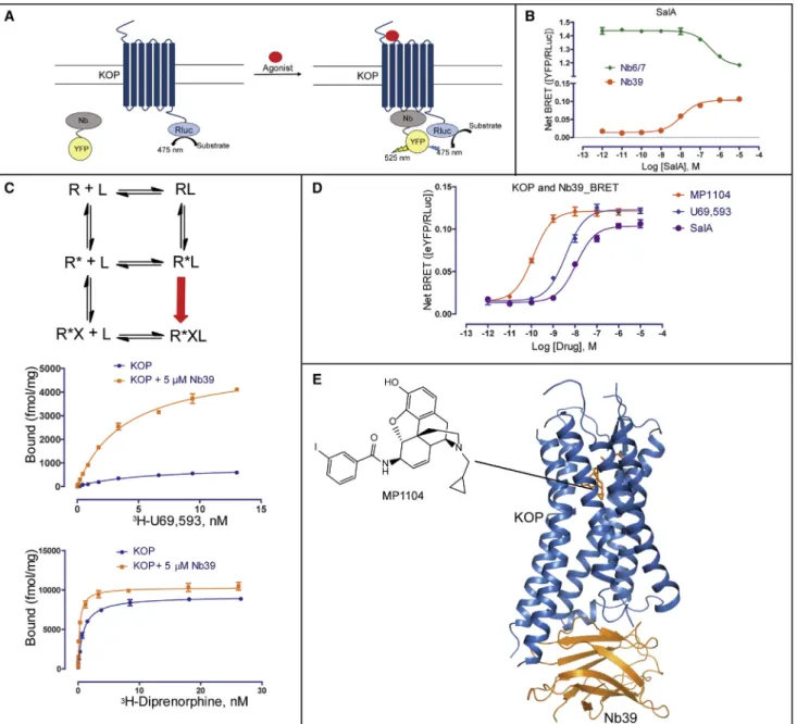
To identify a ligand suitable for crystallization of a Nb39-stabi-lized KOP active state, we tested several ligands in the BRET assay and found that the epoxymorphinan MP1104 (Figure 1D) (Va´radi et al., 2015) displayed the highest potency and efficacy for Nb39 recruitment to KOP. MP1104 has picomolar KOP bind-ing affinity (Figure S1F) and is a potent KOP, MOP, and DOP agonist (Figure S1G).
As Nb39 and MP1104 cooperatively promote a stable active-state KOP, we were able to determine the X-ray crystal structure of a KOP-MP1104-Nb39 complex (Figure 1E). Crystals were ob-tained using a newly engineered human KOP construct with an N-terminal thermostabilized apocytochrome b562RIL (BRIL) (Chun et al., 2012) to increase receptor expression, and to facil-itate crystallization (seeSTAR Methodsfor details). Binding and functional assays showed that this KOP crystallization construct retains high affinity for MP1104 and elicits MP1104-mediated Gi activation similar to wild-type (WT) KOP (Figures S2A and S2B). The structure of the KOP-MP1104-Nb39 complex was deter-mined to 3.1 A˚ resolution in the space group P21with two mono-mers per asymmetric unit (Figure S2C and S2D;Table 1). While we observed strong density for KOP, Nb39, and MP1104 ( Fig-ure S2E), no electron density was observed for the likely disor-dered N-terminal BRIL fusion protein as reported for the NOP receptor (Thompson et al., 2012) and the nanobody-stabilized
b2AR structures (Rasmussen et al., 2011a).
Large-Scale Structural Changes in Active-State KOP Next, we analyzed overall helical movements by comparing the inactive-state KOP-JDTic structure (PDB ID: 4DJH) (Wu et al., 2012) and the active-state KOP-MP1104-Nb39 complex ( Fig-ure 2A) and observed substantial rearrangements in the relative positions of the helices. These include an overall contraction of the extracellular portion in the active-state KOP, with extracel-lular loop (ECL) 2, and transmembrane helices (TMs) 4–6 moving closer to the receptor core (Figure 2B). The orthosteric pocket of the active-state structure shows a10% reduced volume when compared with that of the inactive-state KOP-JDTic complex (945 A˚3in active KOP vs.1,049 A˚3in inactive KOP) (Figure 2D). Similar inward movements of helices and a contraction of the pocket were also observed in the active-state MOP structure (Figure 2E), and other active-state GPCR structures (Figure S2F) (Huang et al., 2015; Kruse et al., 2013; Rasmussen et al., 2011b). These conserved structural rearrangements may indicate a general activation mechanism among many Class A GPCRs whereby contraction of the helical bundle on the extracellular side facilitates an opening of the helical bundle on the intracel-lular side which accommodates the binding of transducers (and vice versa). The larger contraction in the KOP orthosteric site (10%,104 A˚3) compared to MOP (6%, 58 A˚3) is likely due to the more compact positions of TM2, TM3, TM6 and inactive-state structures of all four opioid receptors (MOP, KOP,
DOP,andnociceptin[NOP])(Fenaltietal.,2014,2015;Granier etal.,2012;Manglik etal.,2012;Thompsonetal.,2012;Wu etal.,2012) have provided unprecedented molecular insights into opioid receptor structure, a molecular understanding of KOPactivationremainselusive.Thisislargelyduetoour insuffi-cient molecular understanding of active-state GPCRs, because obtaining crystal structures of such GPCR states remains challenging.
To facilitate a deeper understanding of KOP function and, moregenerally,ofGPCRactivation,wereporttheactive-state crystal structure of KOP in complex with a high-affinity agonist andanactive-state-stabilizingnanobody(Nb)at3.1A˚resolution. A comparison with other opioid receptor structures identifies residuescriticalforKOPactivationandilluminateskeymolecular determinants of subtype selectivity and signaling bias.
RESULTS
ANanobody-StabilizedActive-StateKOPStructure Weadoptednanobodytechnologytoobtainanactive-state-like crystal structure of KOP, since structure determination of signaling complexes remains incredibly challenging. Nb—small single-chainantibodies—havefacilitatedthestructural charac-terization of high-affinity agonist states of the b2-adrenergic
(b2AR) (Rasmussenetal.,2011a), M2 muscarinic (M2R) (Kruse
et al., 2013), and MOP (Huang et al., 2015) receptors, by mimickingthebindingofsignaltransducers.Weraised nano-bodies by injecting KOP liposomes (Vardyetal.,2015) bound to the agonist salvinorin A (SalA) (Rothetal.,2002) into a llama andusedphagedisplaytoidentifynineclonesfromthese im-mune libraries (Pardonetal.,2014). To verify KOP-nanobody binding, we measured nanobody recruitment to unliganded, agonist-bound, and antagonist-bound KOP using biolumines-cenceresonanceenergytransfer(BRET)(Figure 1A).Onlythe identical clones Nb6 and Nb7 (Nb6/7) from the KOP-SalA library as well as the MOP active-state-stabilizing nanobody Nb39 (Huangetal.,2015) bound KOP in our assay (Figures1B and S1A). Consistent with a preference for inactive KOP, we observedstrong basal BRETbetweenNb6/7 and unliganded KOP (Figure1B); we also found that the selective KOP agonist SalAreducedtheBRETratio,whiletheselectiveKOPantagonist JDTic (Thomasetal.,1998) reversed SalA’s effect (FigureS1B). Remarkably,Nb39was recruitedinan agonist-and efficacy-dependent manner to KOP by the full agonist SalA and partial agonistdiprenorphine,whiletheantagonistJDTichadnoeffect (Figure S1C). These findingsimplied that Nb39 stabilizes an active-state conformation of KOP as reported for MOP (Huang etal.,2015).
Figure 1. Identification of Inactive- and Active-State-Stabilizing Nanobodies and Overall Structure of the KOP-MP1104-Nb39 Complex (A) Cartoon of receptor-nanobody (Nb) interaction monitored by bioluminescence resonance energy transfer (BRET). Agonist-stimulated GPCR is bound by active-state-stabilizing nanobody causing BRET signal. Nanobodies are tagged with C-terminal YFP, and GPCRs are tagged with C-terminal Rluc (Renilla luciferase).
(B) BRET measurement of SalA-mediated nanobody recruitment at KOP. The clones Nb6 and Nb7 (half maximal effective concentration [EC50] = 341±15 nM), which have the same protein sequence, show dissociation from the receptor upon SalA stimulation, indicating that Nb6/7 is pre-bound to KOP. The BRET signal from the KOP-Nb39 complex (EC50= 11.3±1.1 nM) increases with increasing SalA concentrations, indicating that Nb39 recognizes active-state KOP (n = 3). (C) Top: Scheme of extended ternary complex model of GPCR activation. R, receptor; L, ligand; R*, active-state receptor; X, transducer or transducer mimetics. Middle and Bottom: Measurement of saturation binding at KOP with or without Nb39. High-affinity binding sites increase about 6-fold for the full agonist 3
H-U69,593 in the presence of Nb39 (Kd = 2.80±0.06 nM; Bmax= 5,262±138 fmol/mg) compared to KOP alone (Kd = 5.41±0.05 nM; Bmax= 860±35 fmol/mg) (n = 3). High-affinity binding sites increase by10% for the partial agonist3
H-diprenorphine in the presence of Nb39 (Kd = 0.26±0.02 nM; Bmax= 10,315± 195 fmol/mg) compared to KOP alone (Kd = 0.84±0.05 nM; Bmax= 9,191±120 fmol/mg) (n = 3).
(D) Ligand-mediated Nb39 binding to KOP measured BRET. EC50for Nb39 recruitment: MP1104, 0.12±0.02 nM; U69,593, 3.78±0.04 nM; SalA, 11.2±0.8 nM (n = 3).
These rearrangements are stabilized by several key KOP-Nb39 interactions (Figure S2G) whereby Nb39 is inserted into an intra-cellular receptor crevice that presumably serves as a binding pocket for key signal transducer residues. This interface is conserved among opioid receptors (Figure S2H), explaining why the MOP-originated Nb39 also stabilizes KOP. We also observed a conformation of KOP that is consistent with a general expansion of the intracellular binding site to accommodate transducers, reminiscent of those described for G protein, ar-restin, or nanobody-stabilized active-state structures of b2AR (Rasmussen et al., 2011a, 2011b), M2R (Kruse et al., 2013), rhodopsin (Kang et al., 2015), and MOP (Huang et al., 2015).
Molecular Determinants for KOP Ligand Binding and Agonist Efficacy
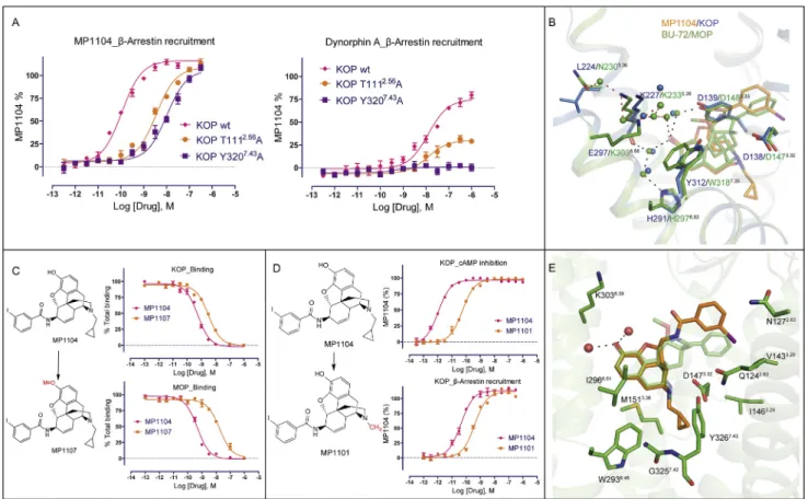
Next, we characterized ligand-receptor contacts of the agonist MP1104 and the antagonist JDTic bound to the KOP orthosteric pocket. We found that the JDTic and MP1104 core scaffolds as-sume distinctive poses (Figure 3A), albeit with common features typical for opioid ligands: (1) anchored in the receptor binding pocket through a salt bridge to D1383.32in TM3, (2) interacting with TM5 through a phenolic group, and (3) forming interactions with TM2/3 via chemically diverse moieties.
The antagonist JDTic and the agonist MP1104 both form a salt bridge between their respective amine moieties and D1383.32of the receptor as observed in many GPCR-ligand complexes ( Fig-ure 3A). The larger distance of this salt bridge (3.0 A˚) compared to similar interactions in the KOP-JDTic (2.6 A˚), and MOP-BU-72-Nb39 (2.7 A˚) structures implies a weaker ionic interaction be-tween MP1104 and KOP. Our mutagenesis study confirmed that, similar to SalA which does not contain a basic amine, MP1104 maintains high binding affinity (Figure 3B) but attenu-ated functional activity at the D1383.32A mutant, whereas Dynorphin A 1–17 binding and functional activity is abolished (Vardy et al., 2015) (Table S2). D1383.32also forms a hydrogen bond network with T1112.56 and Y3207.43 in KOP-MP1104-Nb39 that is likely critical for full KOP activation as mutation of these residues strongly attenuates or abolishes b-arrestin2 recruitment mediated by MP1104 or Dynorphin A 1–17, respec-tively (Figure S3A).
The MP1104 and JDTic phenolic groups extend toward TM5 forming water-mediated hydrogen bonds with the backbone carbonyl oxygen of K2275.39–as seen in other opioid receptor structures (Fenalti et al., 2014; Huang et al., 2015; Manglik et al., 2012; Wu et al., 2012). This interaction has been proposed to mimic that of the N-terminal tyrosine found in endogenous opioid peptides (Fenalti et al., 2015; O’Connor et al., 2015). Because of the lower resolution of the KOP-MP1104-Nb39 structure, we do not observe the waters involved in this interac-tion, but could predict their position by energy-based water modeling algorithms. (Figure S3B). Support for this prediction comes from studies where we replaced the hydroxyl groups ( OH) on the MP1104 scaffold with a methoxy group ( OCH3), resulting in decreased KOP affinity (Figure S3C) as observed for similar JDTic modifications (Urbano et al., 2014). The me-thoxy substitution, however, affects MOP binding affinity more severely than KOP, and these findings suggest that interactions with TM5 can be exploited for KOP selective ligand design. Table 1. Data Collection and Refinement Statistics
Structure BRIL-KOP-MP1104-Nb39
Data Collection
APS, GMCA/CAT 23ID-B/D, 1.033 A˚, 10-mm microfocus beam
Crystals 21
Resolution (A˚) 46.94–3.10 (3.27–3.10)
Space group P21
Complexes/ASU 2
Unit cell dimensionsa, b, c(A˚) 62.3, 150.8, 100.2
a,b,g() 90, 105.7, 90
No. total reflections 89,325 (10,237)
No. unique reflections 30,278 (4,159)
Multiplicity 3.0 (2.5)
Completeness (%) 93.6 (88.2)
Mean I/s(I) 4.0 (0.9)
Rmerge(%) 18.7 (96.1)
CC1/2(%) 98.8 (53.7)
Refinement Statistics
Resolution used in refinement (A˚) 46.94–3.10 (3.21–3.10)
No. reflections used in refinement 30,255 (2,708)
No. reflections used for R-free 1,505 (138)
R-work (%) 25.4 (24.5)
R-free (%) 27.5 (28.3)
Number of Atoms Complex A Complex B
KOP 2117 2013
Nb39 959 917
MP1104 33 33
Lipids 42 28
Overall B-Factors (A˚2) Complex A Complex B
KOP 82.6 82.9
Nb39 92.0 90.7
MP1104 80.0 82.5
Lipids 113.4 121.0
Model Statistics
RMSD Bond (A˚) 0.010
RMSD Bond () 0.95
Ramachandran favored (%)a 94.1
Ramachandran allowed (%)a 5.9
Ramachandran outliers (%)a 0.0
Rotamer outliers (%)a 1.1
Molprobity scorea 1.8
The highest-resolution shell is shown in parentheses. aAs defined in MolProbity.
TM7aroundtheagonistintheactive-stateKOPstructure,and the deeper pocket in the KOP inactive state, stabilized by the unique chemotype of JDTic.
MP1104’s cyclopropylmethyl group extends into a hydropho-bic pocket at the bottom of the orthosteric site, similar to the iso-propyl moiety in JDTic (Figure 3C). This hydrophobic pocket has been proposed to play an important role in determining agonist or antagonist activity at MOP (Huang et al., 2015). Our analysis indicates that the connection between ligand-receptor interac-tions within this hydrophobic pocket of opioid receptors and
corresponding ligand efficacy may not be as straightforward as previously proposed. Compared to MP1104’s cyclopropyl-methyl group, BU-72 in the MOP/BU-72 complex has a cyclopropyl-methyl substituent at its tertiary amine, and thus does not fully extend into this hydrophobic pocket (Figure 3C) (Huang et al., 2015). We observed several contacts between MP1104 and residues of this hydrophobic pocket including hydrophobic interactions Figure 2. Large-Scale Structural Changes between Inactive and Active KOP
(A) Structural alignment of active (blue) and inactive (PDB: 4DJH, gray) KOP shows TM6 outward displacement of10 A˚.
(B) Extracellular view highlights contraction of the TMs and extracellular loops upon binding to MP1104 (orange) versus JDTic (purple). Distances were measured between Caatoms of I581.31
, Q1152.60 , Q213ECL2
, D2235.35 , L2996.60
, and S3107.33 .
(C) Intracellular view shows expansion of the 7TM bundle upon binding of MP1104 and Nb39 versus JDTic, with particularly pronounced movements in TM5-7 and ICL2. Distances were measured between the Caatoms of K2545.66
, D2666.27 , Y3307.53
, and D168ICL2
. Nb39 and T4L fusion proteins have been omitted for clarity.
(D and E) Reduction of orthosteric site volumes in KOP and MOP upon activation. Superimposed pockets for inactive (PDB: 4DJH, gray) (1,049 A˚3
) and active (blue) (945 A˚3
) KOP (D). Superimposed pockets for MOP, inactive (PDB: 4DKL, gray) 1,112 A˚3
and active (PDB: 5C1M, green) 1,053 A˚3
(E). Volumes were calculated for the pockets of 4 superimposed receptors and uniformly delimited between the level of extracellular lipid layer boundary (as predicted by OMP database) and the Caatom of conserved residue W6.48
MP1104’sb-arrestin2 recruitment potency (Figures 3D and 3E; Tables S3andS4). Similar effects were also observed for other tested KOP agonists (Tables S3and S4), suggesting that this pocket is a general node in relating structural changes in the binding pocket to the engagement of transducers. Importantly, substituting MP1104’s cyclopropylmethyl with a methyl group Figure 3. MP1104 Interactions in the Active-State KOP Binding Pocket
(A) Binding pose comparison of MP1104 (orange) in the active-state KOP (blue) compared with JDTic (purple) in the inactive state (gray, PDB: 4DJH). Main interactions involved MP1104 and binding pocket residues are shown, with hydrogen bonds depicted as dashed lines (black).
(B) Comparison of MP1104 or SalA binding affinity at KOP WT and KOP D1383.32A mutant using3H-diprenorphine (n = 3). SeeTable S2for values.
(C) Top: Major interactions between the cyclopropylmethyl group of MP1104 (orange) and the hydrophobic pocket of active KOP (blue). Bottom: Comparison of binding pose between MP1104 (orange) and BU-72 (green) in KOP and MOP shows that MP1104 extends into the hydrophobic pocket but BU-72 does not. (D and E) Mutations of hydrophobic pocket residues (W2876.48
L, G3197.42
L, and Y3207.43
L) strongly affect MP1104’s G protein activation (D) andb-arrestin2 recruitment (E), as measured by cyclic AMP (cAMP) inhibition and Tango assay, respectively (n = 3). SeeTables S3andS4for values.
See alsoFigure S3.
between the cyclopropylmethyl group and the aromatic ring of the Y3207.43side chain, the side chain of W2876.48, and the back-boneofG3197.42(Figure3C).
resulted in a >15-fold reduction of KOP agonist potency ( Fig-ure S3D). Since MP1104 potently activates all three canonical opioid receptors (Figure S1E), we docked MP1104 into the MOP active state and observed a similar orientation as BU-72, with MP1104’s cyclopropylmethyl group extending into MOP’s hydrophobic pocket (Figure S3E).
Collectively our results indicate that the precise orientation of the rigid and bulky morphinan scaffold within the binding pocket is critical for determining ligand efficacy/potency via minor changes in contact forces or tensions generated by substituents. The orientation within the pocket likely depends on (1) the hybrid-ization of intramolecular bonds that determines the angles be-tween the functional modules of the compound and (2) receptor subtype specific interactions. Accordingly, even small modifica-tions to identical scaffolds can subtly affect a compound’s bind-ing mode and, thereby, its potency and/or efficacy, as observed for other GPCR ligands (Wacker et al., 2017b).
Potential Mechanisms of KOP Activation
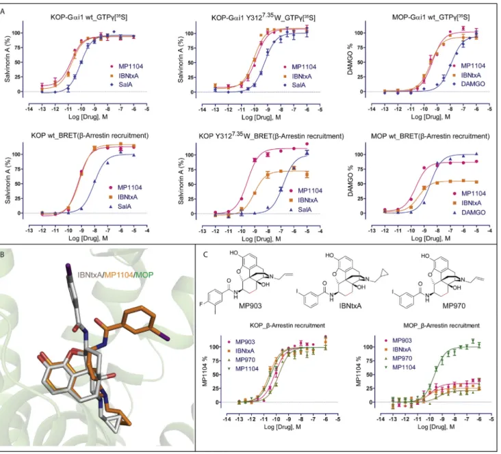
To clarify molecular mechanisms of KOP activation, we investi-gated in detail how conformational changes are likely propa-gated between ligand and transducer binding sites (Figure 4A).
We observed several rearrangements in the MP1104-bound or-thosteric site, specifically in the anchoring D3.32YYNM3.36motif (Figure 4B), which is conserved in opioid receptors and formed by TM3 residues that have previously been proposed as a hub of GPCR structural rearrangements (Venkatakrishnan et al., 2013). MP1104’s cyclopropylmethyl group interacts with the conserved W2876.48residue of the CWxP motif within hydropho-bic pocket (Figure 3C), and with M1423.36of the D3.32YYNM3.36 motif. W2876.48, which is located at the center of an aromatic clus-ter involved in GPCR activation (Holst et al., 2010; Shi et al., 2002), forms hydrophobic contacts with F2836.44, thus coupling the ligand binding site to the P5.50-I3.40-F6.44motif, a central ‘‘micro-switch’’ for the activation of many GPCRs (Katritch et al., 2013). Facilitated by KOP’s ligand binding site contraction in the active state, P2385.50moves inward and I1463.40changes its side chain rotameric state, which likely promotes the rotation of F2836.44 (Figure 4C). This rotation has been linked to the TM6 swivel motion causing receptor opening on the intracellular side for transducer binding (Figure 2A) (Katritch et al., 2013; Rasmussen et al., 2011a; Valentin-Hansen et al., 2012; Wacker et al., 2013).
Changes in TM3 residues also appear to connect the orthos-teric and sodium pocket, the NPxxY motif, and larger scale acti-vation-related changes in TM7. The sodium pocket is located between TM2, TM3 and TM7 and accommodates a single so-dium ion, which acts as a negative allosteric modulator at opioid receptors (Fenalti et al., 2014; Pasternak et al., 1975; Pert et al., 1973) and most other GPCRs (Katritch et al., 2014). In KOP, D1052.50, N1413.35and S1453.39form the sodium pocket, which collapses upon activation and instead D1052.50 forms a hydrogen bond with S1453.39 (Figure 4D). The N1413.35 side chain changes its rotameric state in the active state of KOP ( Fig-ure 4D) similar to MOP (Huang et al., 2015), and directly connects changes in the orthosteric site to the sodium pocket. As in DOP (Fenalti et al., 2014), N3.35 forms a water-mediated hydrogen bond with D3.32 and directly coordinates sodium. Through a hydrogen bond between D1052.50 and N3267.49, the sodium
pocket is further directly coupled to the NPxxY motif at the intra-cellular tip of TM7, which has been implicated in propagating structural changes in the orthosteric site through a sequence of conformational changes leading to larger scale helical move-ments (Katritch et al., 2014; Valentin-Hansen et al., 2012). Furthermore, upon KOP activation, the NPxxY motif residues N3227.45and Y3307.53move toward the receptor core by 1.7 A˚ and 3.9 A˚, respectively, with the hydroxyl group of Y3307.53 mov-ing as much as 7.8 A˚ (Figure 4E), which is consistent with similar activation-related changes in other class A GPCRs (Katritch et al., 2013).
At the intracellular end of TM3, we observed additional struc-tural rearrangements in the conserved DRY motif, which has been shown to directly interact with signal transducers through R1563.50. Specifically, we observed that the salt bridge between R1563.50and D1553.49, which is characteristic for many inactive-state structures, is broken in the KOP active inactive-state, and instead R1563.50points toward the receptor core, forming a hydrogen-bond with Y2465.58(Figure 4F). A similar configuration of R3.50 has previously been observed in the structures ofb2AR bound to a heterotrimeric G protein (Rasmussen et al., 2011b), adeno-sine A2Areceptor bound to a thermostabilized ‘‘mini-Gs’’ ( Car-penter et al., 2016), opsin bound to a C-terminal peptide of transducin (Scheerer et al., 2008), and rhodopsin bound to visual arrestin (Kang et al., 2015).
Together, our observations confirm and extend previous sug-gestions that structural changes in TM3 residue interfaces are critical for coupling ligand-mediated changes in the orthosteric site and the transducer interface (Venkatakrishnan et al., 2013). This hypothesis is further strengthened by the finding that an N1413.35A mutation converts several antagonists into full ago-nists (Figure 4G).
Structural Determinants for Biased Signaling and Subtype Selectivity
Pathway selective KOP ligands are not only important tools for elucidating receptor mechanisms and uncovering novel re-ceptor physiology, they are also therapeutically desirable. It is becoming clear that KOP’s analgesic and anti-pruritic effects appear to be G protein mediated, while many of the undesirable actions of KOP agonists may be mediated by arrestin-ergic and other non-canonical pathways (Bruchas and Roth, 2016; Brust et al., 2016; White et al., 2015). We thus performed structure-activity relationship (SAR) and docking experiments to identify and further characterize the molecular determinants for biased signaling at KOP.
this position (Figure 5A). Since the IBNtxA docking pose in KOP suggests a similar receptor interaction through a stronger direct or water-mediated bond to Y3127.35(3.4 A˚ distance) ( Fig-ure 5A), we hypothesized that differences at position 7.35 could explain IBNtxA’s functional dissimilarities between MOP and KOP. Indeed, when we mutated KOP’s Y3127.35to a tryptophan to mimic MOP’s configuration, we observed a pathway-selective attenuation of IBNtxA’s arrestin recruitment potency, while Figure 4. Activation Signal Propagation within KOP Motifs
(A) Close-ups highlight activation-related conformational changes in key receptor motifs and show connection of structural changes from orthosteric site to the cytoplasmic transducer binding site.
(B–F) Conformational changes between active KOP (blue) and inactive KOP (gray) are highlighted for MP1104 binding pocket (B), P-I-F motif (C), sodium binding pocket (D), NPxxY motif (E), and DRY motif (F).
(G) KOP N1413.35
A mutation switches classic opioid receptor antagonists (left) into full agonists (right) in Tango-arrestin recruitment (n = 3). KOP WT: MP1104 (red), EC50= 0.071±0.008 nM, Emax= 100±2; KOP N1413.35
A: MP1104 (red), EC50= 0.027±0.008 nM, Emax= 100±2, naltrexone (orange), EC50= 6.53± 0.90 nM, Emax= 107±3, naloxone (light green), EC50= 12.75±1.50 nM, Emax= 93±2, 6b-naltrexol (green), EC50= 22.47±1.8 nM, Emax= 120±2.
and IBNtxA’s chair configuration, caused by their respective unsaturatedandsaturatedC-rings(Figure 5A).IBNtxA’s iodo-benzamidemoietyispointedtowardtheextracellularregionin both receptors, instead of toward TM2 and TM3 as for MP1104. InKOP,MP1104’siodobenzamidemoietyappearssta-bilizedby a weak, water-mediated hydrogen bond between the ligand’s carbonylandY3127.35(3.6and4.1A˚distance inmole-culesA and B respectively), while MOP contains W3207.35
MP1104 was only slightly affected (Figure 5D). Moreover, we observe similar functional profiles for several analogs that all contain saturated C-rings (Figure S4C).
In addition to the desired signaling properties, subtype selec-tivity is critical for developing safer therapeutics with reduced side effects. To identify features responsible for selective KOP Figure 5. Structural Insights for the Design of Biased and Selective KOP Ligands
(A) Chemical structures of MP1104 and IBNtxA. Chemical differences are highlighted by color. Comparison of iodobenzamide binding pose between MP1104 (orange, top) and docked IBNtxA (white, bottom) in KOP (blue). Y3127.35
in KOP forms a hydrogen bond with amide oxygen of both compounds.
(B) MP1104 (red) and IBNtxA (orange) are balanced full agonists in KOP measuring in cAMP inhibition and Tango-arrestin recruitment. Gi: MP1104, EC50= 0.003± 0.001 nM, Emax= 97±1; IBNtxA, EC50= 0.002±0.001 nM, Emax= 96±1. Arrestin: MP1104, EC50= 0.035±0.010 nM, Emax= 115±8; IBNtxA, EC50= 0.032± 0.010 nM, Emax= 113±12. Bias factor toward G protein: 0.6 and 1.5 for MP1104 and IBNtxA, respectively (n = 3).
(C) IBNtxA (orange) displays G-protein-biased activity in MOP, whereas MP1104 (red) appears balanced in cAMP inhibition and Tango-arrestin recruitment. Gi: MP1104, EC50= 0.04±0.010 nM, Emax= 103±6; IBNtxA, EC50= 0.056±0.012 nM, Emax= 99±6. Arrestin: MP1104, EC50= 0.55±0.03 nM, Emax= 126±7; IBNtxA, EC50= 0.10±0.03 nM, Emax= 23±4. Bias factor toward G protein: 1.6 and 12 for MP1104 and IBNtxA, respectively (n = 3).
(D) The KOP Y3127.35
W mutant (orange) shows slightly reduced MP1104-mediated arrestin recruitment and strongly reduced IBNtxA-mediated arrestin recruitment compared to KOP WT (red) (n = 3). MP1104/KOP WT: EC50= 0.035±0.010 nM, Emax= 119±9; MP1104/KOP Y3127.35W: EC50= 0.055±0.02 nM, Emax= 111±4. IBNtxA/KOP WT: EC50= 0.03±0.01 nM, Emax= 112±10; IBNtxA/KOP Y3127.35
W: EC50= 0.20±0.06 nM, Emax= 52±4.
(E) Binding affinity of nalfurafine (Ki = 0.32±0.02 nM in KOP and 4.20±0.21 nM in MOP) and compound 18 (Ki = 1.50±0.05 nM in KOP and 533±65 nM in MOP) (n = 3).
DISCUSSION
Here, we present the nanobody-stabilized active-state structure of KOP and provide detailed molecular insights into KOP activa-tion, opioid receptor selectivity, and biased signaling.
While we observed large-scale conformational changes in the KOP active state reminiscent of those seen in other GPCRs, we also find distinct rearrangements in the KOP’s ligand binding pocket. A detailed analysis of these differences highlights limita-tions of prior ‘‘message-address’’-based hypotheses (Larson et al., 2000), which postulated that opioid receptor selectivity and efficacy are determined by distinctive compound moieties. Previous studies suggested that larger hydrophobic amine sub-stituents on prototype morphinans conferred opioid antagonism and thereby provided a roadmap for the design of selective agonists and antagonists. Instead, our findings indicate that combinatorial interactions with conserved and non-conserved residues specify ligand selectivity, pharmacology, efficacy and signaling bias.
Using a combination of structural analysis, molecular docking, binding, and functional studies, we identified important opioid receptor residues involved in conferring different patterns of biased signaling between opioid receptor subtypes. For instance, we showed that replacing Y7.35in the binding pocket of KOP with W7.35 found in MOP, transforms the balanced KOP agonist IBNtxA into a G-protein-biased ligand thereby mimicking its activity at MOP. The finding that residues at the same position confer differential signaling patterns is consistent with observations that a ligand may elicit very different patterns of activity between two receptors. Molecular insights into these mechanisms ultimately may be exploited for the design of poly-pharmacological ligands with ‘‘customized’’ activities at each target.
Taken together, these findings not only expand our general mechanistic framework of GPCR activation but also provide mo-lecular insights into KOP structure and function. Given the urgent need to develop safer opioid medications in an effort to battle the growing opioid epidemic, these molecular insights could greatly accelerate the design of novel KOP ligands through structure-enabled technologies and large-scale virtual ligand screening.
STAR+METHODS
Detailed methods are provided in the online version of this paper and include the following:
d KEY RESOURCES TABLE
d CONTACT FOR REAGENT AND RESOURCE SHARING
d EXPERIMENTAL MODEL AND SUBJECT DETAILS d METHOD DETAILS
B Generation of human KOP receptor crystallization construct
B Discovery and purification of nanobodies
B Expression and purification of KOP
B Lipidic cubic phase crystallization
B Data collection, structure solution and refinement
B cAMP inhibition assay.
B Tango arrestin recruitment assay actions, we next docked representative agonists U69,593,
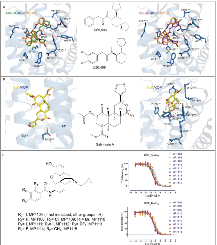
U50,488, and SalA into the current active-state structure. U69,593 and U50,488 (FigureS5A) show similar binding poses, forming ionic interactions between the pyrrolidinyl/amide nitrogen and D1383.32, while exhibiting steric overlap with the MP1104 crystallographic pose. One major difference of U69,593or U50,488frompeptides and morphinanopioids is the lack of phenol moiety, which interacts with H2916.52 and Y1393.33 residuesintheclassical opioids. Accordingly, muta-tions in these residues to Ala had no significant impact on ligand bindingto U69,593, while Dynorphin A binding wasreduced by>10-fold and 5-fold, respectively(Vardy etal., 2013).At the opposite side of the pocket, the phenyl ring of both U69,593andU50,488occupiesthesamepocketasthe iodo-phenyl moiety in the MP1104-KOP structure and the linker amide ofbothalsoformsahydrogenbondwithQ1152.60.SalA’s dock-ing mode (FigureS5B) is consistent with previous studies ( Cun-ninghametal.,2011;Vardyetal.,2013;Wuetal.,2012;Yan etal.,2009). In our current model, Y3127.35forms a hydrogen bondwiththecarbonyloxygenatomofSalAatthe1-position (seeFigureS5Bforatomnumbering).TheSalA2-positionacetyl group forms hydrophobic interactions with the aromatic ring of Y3127.35 and may hydrogen bond with the neighboring Y3137.36hydroxyl group. The 4-position methyl ester of SalA is situatedinasmall,largelyenclosedpocketwhereitisstabilized by an H-bond with the C210ECL2backbone NH group. The furan ring ofSalA engagesinedge-face aromaticinteractions with Y1393.33. It is noteworthy that these docking poses based on the active-state KOP structure are somewhat different from those derived from the inactive-state KOP-JDTic structure (Vardyetal.,2013), highlighting the unique conformation of the active-statebindingpocket.
Efforts to improve KOP selectivity by modifying MP1104’s or IBNtxA’siodobenzamidegroupwereunsuccessful(FigureS5C). Instead, we adopted a novel strategy which emerged from our earlierstructure-guided observation that modifications of the morphinanphenolicgroupaffectsbindingaffinityforKOPless than for MOP (FigureS3C).
B GTPg[35S] assay
B Bioluminescence Resonance Energy Transfer (BRET) assay
B Radioligand binding and ligand dissociation assays
B Molecular modeling
B MP1104 analog synthesis
d QUANTIFICATION AND STATISTICAL ANALYSES
B Dose response, log(t/KA) calculation and ligand bias quantification
d DATA AND SOFTWARE AVAILABILITY
B Data Resources
SUPPLEMENTAL INFORMATION
Supplemental Information includes five figures and five tables and can be found with this article online athttps://doi.org/10.1016/j.cell.2017.12.011.
ACKNOWLEDGMENTS
This work was supported by NIH grants (RO1MH61887 and U19MH82441), the NIMH Psychoactive Drug Screening Program Contract, and the Michael Hooker Distinguished Chair of Pharmacology (to B.L.R.), as well as grants from the National Institute on Drug Abuse to B.L.R., R.C.S., V.C., and V.K. (DA035764), to V.K. (DA038858), to G.W.P. (DA0624), and to S.M. (DA034106). This research was also funded by the Mayday Foundation and the Peter F. McManus Trust (to G.W.P.), (DA034106) (S.M.), and an NIH/NCI Cancer Center support grant (P30 CA008748) (to Memorial Sloan Kettering Cancer Center). We thank INSTRUCT, part of the European Strategy Forum on Research Infrastructures (ESFRI), and the Research Foundation - Flanders (FWO) for their support to the nanobody discovery and thank Nele Buys and Katleen Willibal for the technical assistance. We also thank R.H.J. Olsen for providing the KOP-Gai1 and MOP-Gai1 constructs. We gratefully acknowl-edge M.J. Miley and the UNC Macromolecular Crystallization Core for advice and use of their equipment for crystal harvesting and transport, which is sup-ported by the National Cancer Institute (award number P30CA016086). We thank J. Smith and R. Fischetti and the staff of GM/CA@APS, which has been funded with Federal funds from the National Cancer Institute (ACB-12002) and the National Institute of General Medical Sciences (AGM-12006). This research used resources of the Advanced Photon Source, a U.S. Depart-ment of Energy (DOE) Office of Science User Facility, operated for the DOE Office of Science by Argonne National Laboratory (contract no. DE-AC02-06CH11357).
AUTHOR CONTRIBUTIONS
T.C. designed the experiments; expressed and screened the nanobodies, re-ceptor constructs, and rere-ceptor ligands; crystallized the nanobody-rere-ceptor complex; collected the diffraction data; performed ligand binding and func-tional assays; analyzed all data, and prepared the manuscript. S.M. designed the MP1104 analogs and edited the manuscript. S.A.Z. analyzed the structure and performed the docking experiments. P.O. synthesized MP1104. J.D.M. helped with the binding assay, analyzed the data, and prepared the manu-script. S.W. helped with protein expression and crystal optimization. P.D.M. conducted the SalA modeling. R.U. synthesized MP1104 analogs under S.M.’s supervision. E.V. generated the KOP-SalA liposomes for immunization. B.E.K. helped with the diffraction data collection and processing. G.W.H. refined the structure. M.-Y.L. helped with crystallization. J.S. and E.P. immu-nized llamas, generated the nanobody library, and selected and identified all nanobodies. X.-P.H. helped with the functional assays. R.T.S. developed the GTPg[35
S] assay. A.R.T. helped with the kinetic measurement of GPCR signaling. G.W.P. edited the manuscript. F.I.C. provided the MP1104 com-pound. R.C.S. edited the manuscript. V.C. analyzed the data and prepared the manuscript. V.K. analyzed the data, supervised the docking experiments, and prepared the manuscript. D.W. designed experiments, supervised the structure determination strategy, helped with diffraction data collection,
pro-cessed the diffraction data, solved and refined the structure, analyzed the data, and prepared the manuscript. B.L.R. designed the experiments, was responsible for the overall project strategy and management, and prepared the manuscript.
DECLARATION OF INTERESTS
All authors declare no competing interests.
Received: July 10, 2017 Revised: October 11, 2017 Accepted: December 6, 2017 Published: January 4, 2018
REFERENCES
Abagyan, R., Totrov, M., and Kuznetsov, D. (1994). Icm - a new method for pro-tein modeling and design - applications to docking and structure prediction
from the distorted native conformation. J. Comput. Chem.15, 488–506.
Adams, P.D., Afonine, P.V., Bunko´czi, G., Chen, V.B., Davis, I.W., Echols, N., Headd, J.J., Hung, L.W., Kapral, G.J., Grosse-Kunstleve, R.W., et al. (2010). PHENIX: a comprehensive Python-based system for macromolecular struc-ture solution. Acta Crystallogr. D Biol. Crystallogr.66, 213–221.
Best, R.B., Zhu, X., Shim, J., Lopes, P.E.M., Mittal, J., Feig, M., and Mackerell, A.D., Jr. (2012). Optimization of the additive CHARMM all-atom protein force
field targeting improved sampling of the backbone4,cand side-chainc(1)
andc(2) dihedral angles. J. Chem. Theory Comput.8, 3257–3273.
Black, J.W., and Leff, P. (1983). Operational models of pharmacological ago-nism. Proc. R. Soc., Lond., Ser. B220, 141–162.
Bruchas, M.R., and Roth, B.L. (2016). New technologies for elucidating opioid receptor function. Trends Pharmacol. Sci.37, 279–289.
Bruchas, M.R., Land, B.B., and Chavkin, C. (2010). The dynorphin/kappa opioid system as a modulator of stress-induced and pro-addictive behaviors. Brain Res.1314, 44–55.
Brust, T.F., Morgenweck, J., Kim, S.A., Rose, J.H., Locke, J.L., Schmid, C.L., Zhou, L., Stahl, E.L., Cameron, M.D., Scarry, S.M., et al. (2016). Biased ago-nists of the kappa opioid receptor suppress pain and itch without causing sedation or dysphoria. Sci. Signal.9, ra117.
Caffrey, M., and Cherezov, V. (2009). Crystallizing membrane proteins using lipidic mesophases. Nat. Protoc.4, 706–731.
Carpenter, B., Nehme´, R., Warne, T., Leslie, A.G., and Tate, C.G. (2016). Struc-ture of the adenosine A(2A) receptor bound to an engineered G protein. NaStruc-ture 536, 104–107.
Chavkin, C., James, I.F., and Goldstein, A. (1982). Dynorphin is a specific
endogenous ligand of the kappa opioid receptor. Science215, 413–415.
Chu, R., Takei, J., Knowlton, J.R., Andrykovitch, M., Pei, W., Kajava, A.V., Steinbach, P.J., Ji, X., and Bai, Y. (2002). Redesign of a four-helix bundle pro-tein by phage display coupled with proteolysis and structural characterization
by NMR and X-ray crystallography. J. Mol. Biol.323, 253–262.
Chun, E., Thompson, A.A., Liu, W., Roth, C.B., Griffith, M.T., Katritch, V., Kunken, J., Xu, F., Cherezov, V., Hanson, M.A., and Stevens, R.C. (2012). Fusion partner toolchest for the stabilization and crystallization of G protein-coupled receptors. Structure20, 967–976.
Cunningham, C.W., Rothman, R.B., and Prisinzano, T.E. (2011). Neurophar-macology of the naturally occurring kappa-opioid hallucinogen salvinorin A.
Pharmacol. Rev.63, 316–347.
Emsley, P., Lohkamp, B., Scott, W.G., and Cowtan, K. (2010). Features and development of Coot. Acta Crystallogr. D Biol. Crystallogr.66, 486–501.
Endoh, T., Tajima, A., Izumimoto, N., Suzuki, T., Saitoh, A., Suzuki, T., Narita, M., Kamei, J., Tseng, L.F., Mizoguchi, H., and Nagase, H. (2001). TRK-820, a selective kappa-opioid agonist, produces potent antinociception in
cynomol-gus monkeys. Jpn. J. Pharmacol.85, 282–290.
Eugene Kellogg, G., and Abraham, D.J. (2000). Hydrophobicity: is LogP(o/w)
Lomize, M.A., Lomize, A.L., Pogozheva, I.D., and Mosberg, H.I. (2006). OPM:
orientations of proteins in membranes database. Bioinformatics22, 623–625.
Majumdar, S., Grinnell, S., Le Rouzic, V., Burgman, M., Polikar, L., Ansonoff, M., Pintar, J., Pan, Y.X., and Pasternak, G.W. (2011). Truncated G protein-coupled mu opioid receptor MOR-1 splice variants are targets for highly potent
opioid analgesics lacking side effects. Proc. Natl. Acad. Sci. USA 108,
19778–19783.
Majumdar, S., Subrath, J., Le Rouzic, V., Polikar, L., Burgman, M., Nagakura, K., Ocampo, J., Haselton, N., Pasternak, A.R., Grinnell, S., et al. (2012). Syn-thesis and evaluation of aryl-naloxamide opiate analgesics targeting truncated
exon 11-associatedmopioid receptor (MOR-1) splice variants. J. Med. Chem.
55, 6352–6362.
Manglik, A., Kruse, A.C., Kobilka, T.S., Thian, F.S., Mathiesen, J.M., Sunahara, R.K., Pardo, L., Weis, W.I., Kobilka, B.K., and Granier, S. (2012). Crystal
struc-ture of them-opioid receptor bound to a morphinan antagonist. Nature485,
321–326.
Manglik, A., Lin, H., Aryal, D.K., McCorvy, J.D., Dengler, D., Corder, G., Levit, A., Kling, R.C., Bernat, V., Hu¨bner, H., et al. (2016). Structure-based discovery of opioid analgesics with reduced side effects. Nature537, 185–190.
Martin, W.R., Eades, C.G., Thompson, J.A., Huppler, R.E., and Gilbert, P.E. (1976). The effects of morphine- and nalorphine- like drugs in the nondepen-dent and morphine-depennondepen-dent chronic spinal dog. J. Pharmacol. Exp. Ther. 197, 517–532.
McCoy, A.J., Grosse-Kunstleve, R.W., Adams, P.D., Winn, M.D., Storoni, L.C.,
and Read, R.J. (2007). Phaser crystallographic software. J. Appl. Cryst.40,
658–674.
Minor, W., Cymborowski, M., Otwinowski, Z., and Chruszcz, M. (2006). HKL-3000: the integration of data reduction and structure solution–from diffraction images to an initial model in minutes. Acta Crystallogr. D Biol. Crystallogr.62,
859–866.
Murshudov, G.N., Vagin, A.A., and Dodson, E.J. (1997). Refinement of macro-molecular structures by the maximum-likelihood method. Acta Crystallogr. D Biol. Crystallogr.53, 240–255.
Nagase, H., Imaide, S., Yamada, T., Hirayama, S., Nemoto, T., Yamaotsu, N., Hirono, S., and Fujii, H. (2012). Essential structure of opioidkreceptor agonist nalfurafine for binding tokreceptor 1: synthesis of decahydroisoquinoline
de-rivatives and their pharmacologies. Chem. Pharm. Bull. (Tokyo)60, 945–948.
O’Connor, C., White, K.L., Doncescu, N., Didenko, T., Roth, B.L., Czaplicki, G., Stevens, R.C., Wu¨thrich, K., and Milon, A. (2015). NMR structure and dynamics of the agonist dynorphin peptide bound to the human kappa opioid receptor.
Proc. Natl. Acad. Sci. USA112, 11852–11857.
Pardon, E., Laeremans, T., Triest, S., Rasmussen, S.G., Wohlko¨nig, A., Ruf, A., Muyldermans, S., Hol, W.G., Kobilka, B.K., and Steyaert, J. (2014). A general protocol for the generation of nanobodies for structural biology. Nat. Protoc. 9, 674–693.
Pasternak, G.W., Snowman, A.M., and Snyder, S.H. (1975). Selective enhancement of [3H]opiate agonist binding by divalent cations. Mol. Pharma-col.11, 735–744.
Pert, C.B., Pasternak, G., and Snyder, S.H. (1973). Opiate agonists and antag-onists discriminated by receptor binding in brain. Science182, 1359–1361.
Pfeiffer, A., Brantl, V., Herz, A., and Emrich, H.M. (1986). Psychotomimesis
mediated by kappa opiate receptors. Science233, 774–776.
Rasmussen, S.G., Choi, H.J., Fung, J.J., Pardon, E., Casarosa, P., Chae, P.S., Devree, B.T., Rosenbaum, D.M., Thian, F.S., Kobilka, T.S., et al. (2011a). Structure of a nanobody-stabilized active state of theb(2) adrenoceptor. Na-ture469, 175–180.
Rasmussen, S.G., DeVree, B.T., Zou, Y., Kruse, A.C., Chung, K.Y., Kobilka, T.S., Thian, F.S., Chae, P.S., Pardon, E., Calinski, D., et al. (2011b). Crystal
structure of theb2 adrenergic receptor-Gs protein complex. Nature 477,
549–555.
Roth, B.L., Baner, K., Westkaemper, R., Siebert, D., Rice, K.C., Steinberg, S., Ernsberger, P., and Rothman, R.B. (2002). Salvinorin A: a potent naturally
Fenalti,G.,Giguere,P.M.,Katritch,V.,Huang,X.P.,Thompson,A.A.,
Chere-zov,V.,Roth,B.L.,andStevens,R.C.(2014).Molecularcontrolofd-opioid re-ceptorsignalling.Nature506,191–196.
Fenalti,G.,Zatsepin,N.A.,Betti,C.,Giguere,P.,Han,G.W.,Ishchenko,A.,Liu,
W.,Guillemyn,K.,Zhang,H.,James,D.,etal.(2015).Structuralbasisfor
bifunctionalpeptide recognition at humand-opioidreceptor. Nat. Struct.
Mol.Biol.22,265–268.
Granier,S.,Manglik,A.,Kruse,A.C.,Kobilka,T.S.,Thian,F.S.,Weis,W.I.,and Kobilka,B.K.(2012).Structureofthed-opioidreceptorboundtonaltrindole. Nature485,400–404.
Holst,B.,Nygaard,R.,Valentin-Hansen,L.,Bach,A.,Engelstoft,M.S.,
Pe-tersen,P.S.,Frimurer,T.M.,andSchwartz,T.W.(2010).Aconservedaromatic
lockforthetryptophanrotamericswitchinTM-VIofseven-transmembrane
re-ceptors.J.Biol.Chem.285,3973–3985.
Huang,W.,Manglik,A.,Venkatakrishnan,A.J.,Laeremans,T.,Feinberg,E.N.,
Sanborn,A.L.,Kato,H.E.,Livingston,K.E.,Thorsen,T.S.,Kling,R.C.,etal.
(2015).Structural insights into m-opioid receptor activation. Nature 524,
315–321.
Kang,Y.,Zhou,X.E.,Gao,X.,He,Y.,Liu,W.,Ishchenko,A.,Barty,A.,White,
T.A.,Yefanov,O.,Han,G.W.,etal.(2015). Crystalstructureofrhodopsin
boundtoarrestinbyfemtosecondX-raylaser.Nature523,561–567.
Katritch,V.,Cherezov,V.,andStevens,R.C.(2013).Structure-functionofthe
Gprotein-coupledreceptorsuperfamily.Annu.Rev.Pharmacol.Toxicol.53,
531–556.
Katritch,V.,Fenalti,G.,Abola,E.E.,Roth,B.L.,Cherezov,V.,andStevens,
R.C.(2014).AllostericsodiuminclassAGPCRsignaling.TrendsBiochem.
Sci.39,233–244.
Kenakin,T.,andChristopoulos,A.(2013).Signallingbiasinnewdrug
discov-ery:detection,quantificationandtherapeuticimpact.Nat.Rev.DrugDiscov.
12,205–216.
Kenakin,T.,Watson,C.,Muniz-Medina,V.,Christopoulos,A.,andNovick,S.
(2012).Asimplemethodforquantifyingfunctionalselectivityandagonistbias.
ACSChem.Neurosci.3,193–203.
Kim,S.,Lee, J.,Jo,S.,Brooks,C.L.,3rd, Lee,H.S.,andIm,W.(2017).
CHARMM-GUIligandreaderandmodelerforCHARMMforcefieldgeneration
ofsmallmolecules.J.Comput.Chem.38,1879–1886.
Krivov,G.G.,Shapovalov,M.V.,andDunbrack,R.L.,Jr.(2009).Improved
pre-diction of protein side-chain conformations with SCWRL4. Proteins 77,
778–795.
Kroeze,W.K.,Sassano,M.F.,Huang,X.P.,Lansu,K.,McCorvy,J.D.,Gigue`re,
P.M.,Sciaky,N.,andRoth,B.L.(2015).PRESTO-Tangoasanopen-source
resourceforinterrogationofthedruggablehumanGPCRome.Nat.Struct.
Mol.Biol.22,362–369.
Kruse,A.C.,Ring,A.M.,Manglik,A.,Hu,J.,Hu,K.,Eitel,K.,Hu¨bner,H., Pardon,E.,Valant,C.,Sexton,P.M.,etal.(2013).Activationandallosteric
modulationofamuscarinicacetylcholinereceptor.Nature504,101–106.
Larson,D.L.,Jones,R.M.,Hjorth,S.A.,Schwartz,T.W.,andPortoghese,P.S.
(2000).Bindingofnorbinaltorphimine(norBNI)congenerstowild-typeand
mutantmuandkappaopioidreceptors:molecularrecognitionlociforthe
pharmacophoreandaddresscomponentsof kappaantagonists. J.Med.
Chem.43,1573–1576.
Laskowski,R.A.,MacArthur,M.W.,Moss,D.S.,andThornton,J.M.(1993).
PROCHECK:aprogramtocheckthestereochemicalqualityofprotein
struc-tures.J.Appl.Cryst.26,283–191.
Lee,J.,Cheng,X.,Swails,J.M.,Yeom,M.S.,Eastman,P.K.,Lemkul,J.A.,Wei, S.,Buckner,J.,Jeong,J.C.,Qi,Y.,etal.(2016).CHARMM-GUIinputgenerator
forNAMD,GROMACS,AMBER,OpenMM,andCHARMM/OpenMM
simula-tionsusingtheCHARMM36additiveforcefield.J.Chem.TheoryComput.
12,405–413.
Liu,W.,Wacker,D.,Gati,C.,Han,G.W.,James,D.,Wang,D.,Nelson,G.,
Weierstall,U.,Katritch,V.,Barty,A.,etal.(2013).Serialfemtosecond
occurring nonnitrogenous kappa opioid selective agonist. Proc. Natl. Acad.
Sci. USA99, 11934–11939.
Samama, P., Cotecchia, S., Costa, T., and Lefkowitz, R.J. (1993). A mutation-induced activated state of the beta 2-adrenergic receptor. Extending the
ternary complex model. J. Biol. Chem.268, 4625–4636.
Scheerer, P., Park, J.H., Hildebrand, P.W., Kim, Y.J., Krauss, N., Choe, H.W., Hofmann, K.P., and Ernst, O.P. (2008). Crystal structure of opsin in its G-pro-tein-interacting conformation. Nature455, 497–502.
Shi, L., Liapakis, G., Xu, R., Guarnieri, F., Ballesteros, J.A., and Javitch, J.A. (2002). Beta2 adrenergic receptor activation. Modulation of the proline kink
in transmembrane 6 by a rotamer toggle switch. J. Biol. Chem. 277,
40989–40996.
Smart, O.S., Womack, T.O., Flensburg, C., Keller, P., Paciorek, W., Sharff, A., Vonrhein, C., and Bricogne, G. (2012). Exploiting structure similarity in refine-ment: automated NCS and target-structure restraints in BUSTER. Acta Crys-tallogr. D Biol. CrysCrys-tallogr.68, 368–380.
Spahn, V., Del Vecchio, G., Labuz, D., Rodriguez-Gaztelumendi, A., Massaly, N., Temp, J., Durmaz, V., Sabri, P., Reidelbach, M., Machelska, H., et al. (2017). A nontoxic pain killer designed by modeling of pathological receptor
conformations. Science355, 966–969.
Spetea, M., Eans, S.O., Ganno, M.L., Lantero, A., Mairegger, M., Toll, L.,
Schmidhammer, H., and McLaughlin, J.P. (2017). Selectivekreceptor partial
agonist HS666 produces potent antinociception without inducing aversion af-ter i.c.v. administration in mice. Br. J. Pharmacol.174, 2444–2456.
Thomas, J.B., Mascarella, S.W., Rothman, R.B., Partilla, J.S., Xu, H., McCul-lough, K.B., Dersch, C.M., Cantrell, B.E., Zimmerman, D.M., and Carroll, F.I. (1998). Investigation of the N-substituent conformation governing potency and mu receptor subtype-selectivity in (+)-(3R,
4R)-dimethyl-4-(3-hydroxy-phenyl)piperidine opioid antagonists. J. Med. Chem.41, 1980–1990.
Thompson, A.A., Liu, W., Chun, E., Katritch, V., Wu, H., Vardy, E., Huang, X.P., Trapella, C., Guerrini, R., Calo, G., et al. (2012). Structure of the
nociceptin/or-phanin FQ receptor in complex with a peptide mimetic. Nature485, 395–399.
Urban, J.D., Clarke, W.P., von Zastrow, M., Nichols, D.E., Kobilka, B., Wein-stein, H., Javitch, J.A., Roth, B.L., Christopoulos, A., Sexton, P.M., et al. (2007). Functional selectivity and classical concepts of quantitative pharma-cology. J. Pharmacol. Exp. Ther.320, 1–13.
Urbano, M., Guerrero, M., Rosen, H., and Roberts, E. (2014). Antagonists of
the kappa opioid receptor. Bioorg. Med. Chem. Lett.24, 2021–2032.
Valentin-Hansen, L., Holst, B., Frimurer, T.M., and Schwartz, T.W. (2012). PheVI:09 (Phe6.44) as a sliding microswitch in seven-transmembrane (7TM)
G protein-coupled receptor activation. J. Biol. Chem.287, 43516–43526.
Van Der Spoel, D., Lindahl, E., Hess, B., Groenhof, G., Mark, A.E., and
Berend-sen, H.J.C. (2005). GROMACS: fast, flexible, and free. J. Comput. Chem.26,
1701–1718.
Va´radi, A., Marrone, G.F., Eans, S.O., Ganno, M.L., Subrath, J.J., Le Rouzic, V., Hunkele, A., Pasternak, G.W., McLaughlin, J.P., and Majumdar, S. (2015). Synthesis and characterization of a dual kappa-delta opioid receptor agonist analgesic blocking cocaine reward behavior. ACS Chem. Neurosci. 6, 1813–1824.
Vardy, E., Mosier, P.D., Frankowski, K.J., Wu, H., Katritch, V., Westkaemper, R.B., Aube´, J., Stevens, R.C., and Roth, B.L. (2013). Chemotype-selective
modes of action of k-opioid receptor agonists. J. Biol. Chem. 288,
34470–34483.
Vardy, E., Robinson, J.E., Li, C., Olsen, R.H.J., DiBerto, J.F., Giguere, P.M., Sassano, F.M., Huang, X.P., Zhu, H., Urban, D.J., et al. (2015). A new DREADD facilitates the multiplexed chemogenetic interrogation of behavior. Neuron86,
936–946.
Venkatakrishnan, A.J., Deupi, X., Lebon, G., Tate, C.G., Schertler, G.F., and Babu, M.M. (2013). Molecular signatures of G-protein-coupled receptors. Na-ture494, 185–194.
Wacker, D., Wang, C., Katritch, V., Han, G.W., Huang, X.P., Vardy, E., McCorvy, J.D., Jiang, Y., Chu, M., Siu, F.Y., et al. (2013). Structural features for functional selectivity at serotonin receptors. Science340, 615–619.
Wacker, D., Stevens, R.C., and Roth, B.L. (2017a). How ligands illuminate
GPCR molecular pharmacology. Cell170, 414–427.
Wacker, D., Wang, S., McCorvy, J.D., Betz, R.M., Venkatakrishnan, A.J., Levit, A., Lansu, K., Schools, Z.L., Che, T., Nichols, D.E., et al. (2017b). Crystal
struc-ture of an LSD-bound human serotonin receptor. Cell168, 377–389.e12.
Wang, S., Wacker, D., Levit, A., Che, T., Betz, R.M., McCorvy, J.D., Venkatak-rishnan, A.J., Huang, X.P., Dror, R.O., Shoichet, B.K., and Roth, B.L. (2017). D4 dopamine receptor high-resolution structures enable the discovery of selec-tive agonists. Science358, 381–386.
White, K.L., Robinson, J.E., Zhu, H., DiBerto, J.F., Polepally, P.R., Zjawiony, J.K., Nichols, D.E., Malanga, C.J., and Roth, B.L. (2015). The G protein-biased
k-opioid receptor agonist RB-64 is analgesic with a unique spectrum of activ-ities in vivo. J. Pharmacol. Exp. Ther.352, 98–109.
Wu, H., Wacker, D., Mileni, M., Katritch, V., Han, G.W., Vardy, E., Liu, W., Thompson, A.A., Huang, X.P., Carroll, F.I., et al. (2012). Structure of the human
k-opioid receptor in complex with JDTic. Nature485, 327–332.
Yan, F., Bikbulatov, R.V., Mocanu, V., Dicheva, N., Parker, C.E., Wetsel, W.C., Mosier, P.D., Westkaemper, R.B., Allen, J.A., Zjawiony, J.K., and Roth, B.L. (2009). Structure-based design, synthesis, and biochemical and pharmaco-logical characterization of novel salvinorin A analogues as active state probes
of the kappa-opioid receptor. Biochemistry48, 6898–6908.
Zheng, Z., Huang, X.P., Mangano, T.J., Zou, R., Chen, X., Zaidi, S.A., Roth, B.L., Stevens, R.C., and Katritch, V. (2017). Structure-based discovery of new antagonist and biased agonist chemotypes for the kappa opioid receptor.
STAR
+
METHODS
KEY RESOURCES TABLE
REAGENT or RESOURCE SOURCE IDENTIFIER
Antibodies
gp64-PE antibody Expression Systems Cat#97-201
Chemicals, Peptides, and Recombinant Proteins
AEBSF GoldBio Cat#A-540-5
Leupeptin Sigma Cat#L2884
E-64 AG Scientific Cat#E-2030
Aprotinin GoldBio Cat#A-655-100
Iodoacetamide Sigma Cat#I1149
n-dodecyl-beta-D-maltopyranoside (DDM) Anatrace Cat#D310
Cholesterol hemisucinate (CHS) Sigma Cat#C6512
TALON IMAC resin Clontech Cat#635507
1-Oleoyl-rac-glycerol (monoolein) Sigma Cat#M7765
Cholesterol Sigma Cat#C8667
Sodium luciferin Goldbio Cat#LUNCA-1g
BrightGlo Promega Cat#E2620
Coelenterazine-h Promega Cat#S2011
Poly-L-lysine Sigma Cat#P2636
Tetracycline Sigma Cat#T7660
Polyethyleneimine (PEI) solution Sigma Cat#P3143
Penicillin/Streptomycin Invitrogen Cat#15140-122
Hygromycin B KSE Scientific Cat#98-923
Zeocin Invitrogen Cat#R25005
3
H-U69,593 Perkin Elmer Cat#NET952
3H-Diprenorphine Perkin Elmer Cat#NET1121
Sf-900 II SFM Invitrogen Cat#10902096
ESF921 Expression Systems Cat#96-001-01
Production Boost Additive Expression Systems Cat#95-006-100
DMEM VWR Cat#45000-306
FBS VWR Cat#97068-085
Dialyzed FBS Omega Scientific Cat#FB-03
10xHBSS Invitrogen Cat#14065-056
Cellfectin II Reagent Invitrogen Cat#10362-100
TransIT-2020 Mirus Cat#MIR5400
Critical Commercial Assays
Bac-to-Bac Baculovirus Expression system Invitrogen Cat#A11100
QuikChange site-directed mutagenesis Agilent Technologies Cat#200519
Deposited Data
KOP-MP1104-Nb39 complex structure This paper PDB: 6B73
Experimental Models: Cell Lines
HEK293T ATCC Cat#CRL-3216
Sf9 cells Expression Systems Cat#94-001S
HTLA Dr. Richard Axel, Columbia Univ N/A
Software and Algorithms
COOT Emsley et al., 2010 www2.mrc-lmb.cam.ac.uk/personal/pemsley/coot
XDS N/A http://xds.mpimf-heidelberg.mpg.de/
CONTACT FOR REAGENT AND RESOURCE SHARING
Further information and requests for reagents should be directed to and will be fulfilled by the Lead Contact, Bryan L. Roth ([email protected]).
EXPERIMENTAL MODEL AND SUBJECT DETAILS
For KOP expression, we used the Sf9 insect cells derived from the parentalSpodoptera frugiperdacell line IPLB-Sf-21-AE (Expres-sion systems). Cells were grown in ESF 921 medium (Expres(Expres-sion systems) at 27C and 125 rpm. Nanobodies were expressed at 27C inE. coliWK6 (su-) cells in TB medium (Terrific Broth, Sigma). Nanobodies were induced with 1 mM IPTG (final concentration, Isopropylb-D-1-thiogalactopyranoside) when the bacteria density reached an OD600of 0.6-0.8 and bacteria cells were grown over-night at 170 rpm. For KOP functional assays, Human embryonic kidney (HEK) 293T (ATCC CRL-11268) cells were cultured in DMEM (Dulbecco’s Modified Eagle Medium). Wild-type or mutant KOP plasmids were transfected into HEK293T cells using the calcium pre-cipitation method.
METHOD DETAILS
Generation of human KOP receptor crystallization construct
Crystallization of the human KOP complex was done using an engineered receptor construct that was modified based on the KOP-T4L sequence (Wu et al., 2012). The final construct a) lacks N-terminal residues 1-53, b) lacks C-terminal residues 359-380, c) contains M1-L106 of the thermostabilized apocytochrome b562RIL (BRIL) fromE. coli(M7W, H102I, R106L) in place of receptor N terminus residues M1-H53, a glycine-serine linker was inserted between BRIL and receptor to facilitate crystallization. Further modifications are I135L mutation was introduced to increase expression; a haemagglutinin (HA) signal sequence followed by a FLAG tag at the N terminus, then a 10X His tag followed by a TEV protease site to enable purification by immobilized metal affinity chromatography.
Discovery and purification of nanobodies
KOP specific nanobodies were generated as described before (Pardon et al., 2014). In brief, one llama (Lama glama) was immunized six times with in total 0.5 mg purified BRIL-KOP DREADD (KOP D1383.32N) bound to SalA [KOP D1383.32N was used here because it has higher affinity with SalA than wild-type (Vardy et al., 2015)]. Four days after the final boost, blood was taken to isolate peripheral Continued
REAGENT or RESOURCE SOURCE IDENTIFIER
ICM-Pro 3.8-6 Abagyan et al., 1994 http://molsoft.com
Marvin v.15.11.23.0 ChemAxon https://www.chemaxon.com
Phaser McCoy et al., 2007 http://www.ccp4.ac.uk
Phenix Adams et al., 2010 https://www.phenix-online.org
Prime Schro¨dinger https://www.schrodinger.com/
PyMOL Schro¨dinger https://www.pymol.org/2/
Prism v.5.0 GraphPad Software N/A
REFMAC Murshudov et al., 1997 http://www.ccp4.ac.uk
Other
100 kDa molecular weight cut-off Vivaspin 20 concentrator
Sartorius Stedim Cat#VS2042
PD MiniTrap G-25 columns GE Healthcare Cat#28-9180-07
100 kDa molecular weight cut-off Vivaspin 500 centrifuge concentrator
Sartorius Stedim Cat#VS0142
96-well LCP glass sandwich set Marienfeld GmbH Cat#0890003
384-well black plates Greiner Bio-one GmbH Cat#781091
384-well white plates Greiner Bio-one GmbH Cat#781098
96-well black plates Greiner Bio-one GmbH Cat#655090
Meltilex Perkin Elmer Cat#1450-441
blood lymphocytes. RNA was purified from these lymphocytes and reverse transcribed by PCR to obtain cDNA. The resulting library was cloned into the phage display vector pMESy4 bearing a C-terminal hexa-His tag and a Glu-Pro-Glu-Ala-tag (EPEA-tag, also calledorCaptureSelectC-tag).SelectionswereperformedeitheronBRIL-KOPinliposomessolidphasecoateddirectlyonplates. Sixdifferentfamilieswereselectedbybiopanning.Aftertworoundsofselection,periplasmicextractsweremadeandsubjectedto ELISAscreens.ClonesgivingapositivesignalinELISAweresequencedandanalyzed.PlasmidsweretransformedtoE.coliWK6 cells, KOP specific nanobodies i.e., Nb6 and Nb7 were expressed and purified following steps 70-73 described in the previous pro-tocol (Pardonetal.,2014). Nb39 DNA sequence was synthesized (Integrated DNA Technologies, IDT) based on the protein sequence in the active-state MOP structure (PDB: 5C1M) (Huangetal.,2015), and was expressed and purified using the same protocols as Nb6/7.Nanobodieswereconcentratedanddesaltedtothebuffer:10mMHEPES,100mMNaCland10%Glycerolandstored at 80Cforfutureuse.
ExpressionandpurificationofKOP
High-titer recombinant baculovirus (> 109viral particles per ml) was generated using the Bac-to-Bac Baculovirus Expression System (Invitrogen). 5 mg of recombinant bacmid in 50 ml Sf-900 II SFM media (Invitrogen) and 3 ml Cellfectin II Reagent (Invitrogen) in another 50 ml Sf-900 II SFM media (Invitrogen) were incubated for 30 min. Recombinant baculovirus was obtained by transfecting theabovemixedsolutioninto400mlSf-900IISFMmediaincluding5x105settledSpodopterafrugiperda(Sf9)cells(Expression Sys-tems)ina12-wellplate(Corning).After5h,mediawasexchangedfor1mLSf-900IISFMmedia(Invitrogen)andincubatedfor5days at 27C. P0 viral stock with 109virus particles per ml was harvested as the supernatant and used to generate high-titer baculovirus stock by infection of 40 mL of Sf9 cells (cell density: 2-3 3 106cells/ml) and incubation for 3 days. Viral titers were determined by flow-cytometricanalysisofcellsstainedwithgp64-PEantibody(ExpressionSystems).ExpressionofKOPwascarriedoutbyinfectionof Sf9 cells at a cell density of 2.5 3 106cells/ml in ESF921 media (Expression Systems) with P1 or P2 virus at a MOI (multiplicity of infection) of 3. 5% production boost additive (PBA, Expression Systems) was added to maintain cell alive. Final concentration of 10 mM naltrexone was added to help the receptor trafficking. Cells were harvested by centrifugation at 48 h post infection, washed in1xPBS,andstoredat 80Cuntiluse.Cellswerefirstwashedbyresuspendingfrozencellpelletsinalow-saltbuffercontaining 10mMHEPES,pH7.5,10mMMgCl2,20mMKClandproteaseinhibitors(500mMAEBSF,1mME-64,1mMLeupeptin,150nM Apro-tinin).Membranespurificationwasfollowedby4repeatedcentrifugationinahighosmolaritybuffercontaining1.0MNaCl,10mM HEPES,pH7.5,10mMMgCl2,20mMKCl,toremovesolubleandmembraneassociatedproteins.Purifiedmembranesweredirectly flash-frozeninliquidnitrogenandstoredat 80Cforfutureuse.
Purifiedmembraneswereresuspendedinbuffercontaining10mMHEPES,pH7.5,10mMMgCl2,20mMKCl,150mMNaCl, 50mMMP1104(synthesizedinhouse),and1xproteaseinhibitors(500mMAEBSF,1mME-64,1mMLeupeptin,150nMAprotinin), and incubated at room temperature for 1 h. The sample was then transferred to 4C for 30 min. After another 30 min incubation in the presenceof2mg/mliodoacetamide(Sigma),membranes weresolubilizedin10mMHEPES,pH7.5,150mMNaCl,1%(w/v) n-dodecyl-b-D-maltopyranoside (DDM, Anatrace), 0.2% (w/v) cholesteryl hemisuccinate (CHS, Sigma), and protease inhibitors for 2 h at 4C. The supernatant was obtained by centrifugation at 150,000 3 g for 30 min and was incubated with 20 mM imidazole andTALONIMACresin(Clontech)overnightat4Cusingapproximately500mlresinforproteinpurifiedfrom1Lofcells.Theresin wasthenwashedwith10columnvolumes(cv)ofWashBufferI(50mMHEPES,pH7.5,800mMNaCl,0.1%(w/v)DDM,0.02%(w/v) CHS,20mMimidazole,10%(v/v)glycerol,and25mMMP1104,followedby10cvofWashBufferII(25mMHEPES,pH7.5,150mM NaCl, 0.05% (w/v) DDM, 0.01% (w/v) CHS, 10% (v/v) glycerol, and 25 mM MP1104). Proteins were eluted in 2.5 cv of Wash Buffer II + 250mMimidazole,concentratedina100kDamolecularweightcut-offVivaspin20concentrator(SartoriusStedim)to500ml,and imidazole was removed by desalting the protein over PD MiniTrap G-25 columns (GE Healthcare). The N-terminal 10 3His-tag wasremovedbyadditionofHis-taggedTEVprotease(Homemade)andincubationovernightat4C.Protease,cleavedHis-tag and uncleaved protein were removed by passing the suspension through equilibrated TALON IMAC resin (Clontech) and collecting theflow-through.ExcessiveNb39(KOP/Nb39m/m:1:2)wasthenaddedtotheproteinsampleandincubatedfor3h. KOP-MP1104-Nb39complexeswerethenconcentratedto30mg/mlwitha100kDamolecularweightcut-offVivaspin500centrifugeconcentrator (SartoriusStedim).Proteinpurityandmonodispersityweretestedbyanalyticalsize-exclusionchromatography.
Lipidiccubicphasecrystallization