CELL CYCLE TRANSCRIPTION CONTROL BY UBIQUITIN SIGNALING
Anthony P. Arceci
A dissertation submitted to the faculty at the University of North Carolina at Chapel Hill in partial fulfillment of the requirements for the degree of Doctor of Philosophy in the Curriculum
in Genetics and Molecular Biology in the School of Medicine
Chapel Hill 2019
ABSTRACT
Anthony P. Arceci: Cell Cycle Transcription Control by Ubiquitin Signaling (Under the direction of Michael J. Emanuele)
The cell cycle is a tightly regulated series of molecular events which dictates proliferation. Both the timely activation of genes through transcription and destruction of proteins through the ubiquitin-proteasome system are integral to normal cell cycles.
Dysregulation of these networks often underlie a variety of malignant diseases such as cancer. Forkhead box protein M1 (FOXM1) is an essential cell cycle transcription factor.
FOXM1 regulates a transcriptional network that controls the G2/M transition and G1/S transition. Additionally, aberrant upregulation of the FOXM1 transcriptional network is linked to a variety of cancers. The kinases which activate FOXM1 are well explored, but the influence of the ubiquitin-proteasome system on FOXM1 remains unclear. Here, I described the role that two such enzymes, the E3 ubiquitin ligase CUL4-VPRBP and the deubiquitinating enzyme (DUB) USP21, have on the stability and activity of FOXM1 in both normal and dysregulated cell cycles.
First, I demonstrate that FOXM1 degradation is enhanced by association with CUL4-VPRBP. Depletion of VPRBP enhances FOXM1 stability and causes mitotic entry defects. Interestingly, overexpression of VPRBP enhances both FOXM1 ubiquitination and
Second, I demonstrate that FOXM1 is protected from degradation through association with the DUB USP21. Knockdown or overexpression of USP21 is able to destabilize or stabilize FOXM1, respectively, through deubiquitination of FOXM1. USP21 is able to influence mitotic entry and proliferation through regulating the FOXM1 transcriptional network. Furthermore, USP21 and FOXM1 are both significantly amplified in basal-like breast cancer with the knockdown of both sensitizing cells to the chemotherapy paclitaxel thus describing a novel combination treatment for this disease.
ACKNOWLEDGEMENTS
Looking back on my journey to this point, it is clear to me that I did not arrive here without help and support along the way. Starting from the beginning of my academic journey, I would like to acknowledge my mother, Darlene, and friend, Ron, for believing in me from the start and helping me establish myself as I relocated from Winchendon, Massachusetts to Tucson, Arizona to take my first step in what would become a 12 year journey of discovery and learning. I would like to acknowledge my network of friends, Anthony, Armando, Brandon, Craig, Greg, Patrick and Wayne who became my band of brothers, helping me navigate through the journey.
I would also like to acknowledge Dr. Kathleen Dixon, who fostered my passion for scientific inquiry and set me on the path to becoming a PhD.
Dr. Angela Wandinger-Ness, Dr. Richard Cripps and Antonio Banuelos deserve recognition for providing me with the launching pad and experience to transition from the undergraduate level to the post-graduate level during my year at the University of New Mexico.
I would like to acknowledge my colleagues in the lab Allie, Jen, Raj, Taylor, Thomas and Xianxi who were not only important participants in the research to follow, but also a great group of people to work with over the years.
Finally, I would be remiss without recognizing my advisor, Dr. Michael Emanuele and the role that he played in guiding me to this point. Without your insight and knowledge to help me guide my research and humor and grace to make working in the lab fun, I surely would not have succeeded.
PREFACE
In each of the following chapters portions of the work described were completed by other talented scientists and exceptional colleagues. Additionally, most chapters describe work that has been published and that I have permission to use as part of this dissertation.
Chapter 2 describes work from a project which I made significant contributions to during my rotation and first year in the Emanuele lab and was ultimately completed by my colleague in the lab, Xianxi Wang. I contributed all experiments utilizing cell cycle analysis, RT-qPCR, and chromatin immunoprecipitation (ChIP). Additionally, I cloned some of the plasmids used in many of the cell biology assays described and provided critical revisions and suggestions to the manuscript throughout the peer-review process. Permission to include this article in its entirety is granted by the journal Molecular and Cellular Biology as described in their editorial policy (https://mcb.asm.org/content/editorial-policy).
Chapter 3 describes work from a project in which I designed, conducted and analyzed the vast majority of experiments, wrote the manuscript and made most critical revisions throughout the peer-review process. The work described in Chapter 3 was published as an open-access article in Cell Reports. As such, I retain the copyright to this article as an author to include it in this dissertation in its entirety per editorial guidelines
TABLE OF CONTENTS
LIST OF TABLES ... xii
LIST OF FIGURES ... xiii
LIST OF ABBREVIATIONS ... xiv
CHAPTER 1: INTRODUCTION ... 1
The Ubiquitin-Proteasome System ... 1
Background ... 1
Enzymes of the Ubiquitin-Proteasome System ... 2
E3 Ubiquitin Ligases ... 3
CRL4A and CRL4B ... 5
DUBs ... 6
USP21 ... 8
The Cell Cycle ... 9
Historical Background ... 9
Mechanisms of Control ... 10
Cell Cycle Transcription by FOXM1 ... 14
Background ... 14
Overview of FOXM1 Biology ... 15
Post-Translational Regulation of FOXM1 Through the Cell Cycle ... 16
Ubiquitin-dependent Regulation of FOXM1 ... 18
CHAPTER 2: VPRBP/DCAF1 REGULATES THE DEGRADATION AND
NONPROTEOLYTIC ACTIVATION OF THE CELL CYCLE TRANSCRIPTION
FACTOR FOXM1 ... 25
Introduction ... 25
FOXM1 Stability is Regulated by CRL4VPRBP ... 28
FOXM1 Interaction and Ubiquitylation by VPRBP ... 31
VPRBP is a FOXM1 Activator ... 33
VPRBP Protein is Upregulated in High-Grade Serous Ovarian Tumors ... 37
Discussion ... 37
Materials and Methods ... 41
Immunoblot Analysis ... 41
Immunoprecipitation ... 42
RT-qPCR ... 42
Luciferase Reporter Assays ... 42
Flow Cytometry ... 43
In Vivo Ubiquitination Assay ... 43
Expression and Binding Assay for VPRBP and FOXM1c ... 44
CHAPTER 3: FOXM1 DEUBIQUITINATION BY USP21 REGULATES CELL CYCLE PROGRESSION AND PACLITAXEL SENSITIVITY IN BASAL-LIKE BREAST CANCER ... 56
Introduction ... 56
Results ... 58
USP21 Binds and Alters FOXM1 Abundance ... 58
FOXM1 is a USP21 Substrate ... 60
USP21 Affects the FOXM1 Transcriptional Network and Proliferation ... 62
USP21 Affects Paclitaxel Response in Basal-like Breast Cancer ... 66
Discussion ... 68
Materials and Methods ... 70
Cell Culture ... 70
Transfections and Treatments ... 71
Molecular Cloning ... 71
Lentivirus Production ... 72
Immunoblotting ... 72
Immunoprecipitations ... 73
FOXM1 and USP21 In Vitro Binding Assay ... 73
FOXM1 Transcriptional Reporter Assay ... 74
Cell Cycle Progression into Mitosis ... 74
In Vitro Paclitaxel Sensitivity Assays ... 75
Tumor Xenograft ... 75
Proliferation Assays ... 76
In Vivo Deubiquitination Assay ... 76
In Vitro Deubiquitination Assay ... 77
RT-qPCR ... 77
TCGA Dataset Analysis ... 78
Other Statistical Analysis ... 79
CHAPTER 4: CONCLUSIONS AND FUTURE DIRECTIONS ... 97
LIST OF TABLES
Table 2.1 Antibodies used in experiments described in Chapter 2. ... 54
Table 2.2 Sequences of primers used in experiments described in Chapter 2. ... 55
Table 3.1 FOXM1 target gene enrichment correlated to USP21 amplification. ... 93
Table 3.2 siRNA and shRNA used in Chapter 3 experiments. ... 94
Table 3.3 Primers used in Chapter 3 experiments. ... 95
LIST OF FIGURES
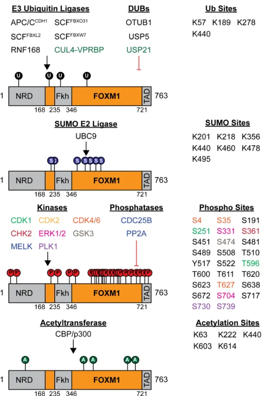
Figure 1.1 Overview of the ubiquitin-proteasome system. ... 23
Figure 1.2 FOXM1 post-translational modification landscape. ... 24
Figure 2.1 FOXM1 is regulated by CUL4, DDB1 and VPRBP. ... 45
Figure 2.2 FOXM1 is regulated by VPRBP. ... 46
Figure 2.3 VPRBP binds and ubiquitylates FOXM1 in vivo. ... 47
Figure 2.4 Examining the FOXM1-VPRBP interaction. ... 48
Figure 2.5 VPRBP activates the FOXM1 6xDB transcriptional reporter. ... 49
Figure 2.6 VPRBP regulates FOXM1 independent of binding to the CRL4 complex. ... 50
Figure 2.7 VPRBP depletion reduces FOXM1 target gene expression and the number of mitotic cells. ... 51
Figure 2.8 VPRBP associates with FOXM1 promoters and activates FOXM1 independent of CUL4... 52
Figure 2.9 Implications and cell cycle control and malignancy. ... 53
Figure 3.1 USP21 binds and regulates FOXM1 abundance. ... 80
Figure 3.2 Analysis of USP21-FOXM1 binding. ... 81
Figure 3.3 USP21 protects FOXM1 from proteasomal degradation. ... 82
Figure 3.4 USP21 controls FOXM1 ubiquitination and half-life. ... 83
Figure 3.5 USP21 affects growth through modulation of the FOXM1 transcriptional network. . 84
Figure 3.6 USP21 regulation of cell cycle and proliferation. ... 85
Figure 3.7 Amplification of FOXM1 and USP21 is linked to proliferation in BLBC. ... 86
Figure 3.8 USP21 knockdown sensitizes BLBC cells to paclitaxel. ... 87
Figure 3.9 Analysis of USP21 and proliferation in breast cancer. ... 88
LIST OF ABBREVIATIONS
AEBSF 4-(2-aminoethyl)-benzenesulfonly fluoride APC/C Anaphase promoting complex/cyclosome ATP Adenosine triphosphate
ATRA All-trans retinoic acid BLBC Basal-like breast cancer
BSA Bovine serum albumin
CAK CDK-activating kinase
CDI Cyclin-dependent kinase inhibitor CDK Cyclin-dependent kinase
ChIP Chromatin immunoprecipitation
CHX Cycloheximide
CNA Copy number alteration Co-IP Co-immunoprecipitation
CRL Cullin-RING ligase
CRL4 Cullin4-based ligase
CSN COP9 signalsome
DCAF DDB1 and CUL4-associated factor
DiGly Diglycine
DMEM Dulbecco’s modified Eagle’s medium
DMSO Dimethyl sulfoxide
dsRed Discosoma red fluorescent protein
DTT Dithithreitol
DUB Deubiquitinating enzyme
DWD DDB1 binding WD40 protein
EGFP Enhanced green fluorescent protein
ER Estrogen receptor
FACS Fluorescence activated cell sorting gDNA Genomic deoxyribonucleic acid
GISTIC Genome identification of significant targets in cancer GSEA Gene set enrichment analysis
HECT Homologous to the E6AP carboxyl terminus HGSOC High-grade serous ovarian cancer
HIV Human immunodeficiency virus HRP Horseradish peroxidase
IB Immunoblot
IP-MS/MS Immunoprecipitation-tandem mass spectrometry IPTG Isopropyl-b-D-thiogalactopyranoside
JAMM/MPN+ Jab1/Mov34/Mpr1 Pad1 N-terminal+ MCC Mitotic checkpoint complex
MINDY Motif interacting with Ub-containing novel DUB family MJD Machado-Josephin domain protease
mRNA Messenger ribonucleic acid
ORF Open reading frame OTU Ovarian tumor protease
P-H3 Phosphorylation on histone H3 serine 10 PBS Phosphate buffered saline
PCR Polymerase chain reaction PIP PCNA-interacting peptide motif PMSF Phenylmethylsulfonyl fluoride
PR Progesterone receptor
PTM Post-translational modification RING Really Interesting New Gene
RNA Ribonucleic acid
RPPA Reverse phase protein array
RT-qPCR Quantitative reverse transcription polymerase chain reaction SAC Spindle assembly checkpoint
SCF Skp, Cullin, F-box containing complex siRNA Small interfering ribonucleic acid SUMO Small ubiquitin-like modification TAD Transcriptional activation domain TCEP Tris(2-carboxyethyl)phosphine
TCGA The Cancer Genome Atlas
Ub Ubiquitin
CHAPTER 1: INTRODUCTION
The Ubiquitin-Proteasome System
Background
The ubiquitin-proteasome system (UPS) will play an important role in the work described in the subsequent chapters. As such, I would like to give a cursory overview of the history, mechanisms and general functions of the UPS in this section (Figure 1.1). A more thorough explanation of E3 ubiquitin ligases and cullin-RING ligase 4 will be described as they are importantly involved in the studies described in Chapter 2. Similarly, deubiquitinating enzymes and ubiquitin specific protease 21 (USP21) will be described in more detail as they are central to the studies described in Chapter 3.
The UPS describes a network of proteases which influence the abundance, stability and function of the vast majority of the proteins within the cell. It was long believed that most cellular proteins were stable and long-lived. It wasn’t until work done in the late 1970’s and early 1980’s that this paradigm began to shift. Hershko, Ciechanover and Rose elegantly
processes, how their dysregulation can lead to disease and how we may be able to leverage the UPS for therapeutic effect (6–9).
Enzymes of the Ubiquitin-Proteasome System
The signal by which this system communicates occurs through the conjugation of ubiquitin, a small 8.6 kDa regulatory protein, to the lysine’s (or methionine in N-terminal ubiquitin-ubiquitin linkages) of other proteins. This process is orchestrated by a cascade of enzymes described as E1 (ubiquitin-activating enzymes), E2 (ubiquitin-conjugating enzymes) and E3 (ubiquitin ligases) (10). The process of conjugating ubiquitin onto the lysine of a protein begins when the E1 enzyme generates a high-energy thiol ester intermediate with ubiquitin in a reaction requiring ATP (E1-S-ubiquitin) (11). From here, one of several E2 enzymes participates in a reaction with the E1-S-ubiquitin intermediate in which the ubiquitin is transferred in a thiol ester reaction to the E2 enzyme (E2-S-ubiquitin) (12). At this point, E2 enzymes bind into a complex with E3 ubiquitin ligase enzymes which also engage the substrate that is to be
ubiquitinated (10, 13). By keeping the substrate and the E2 enzyme conjugated to ubiquitin close to one another, the E3 ubiquitin ligases facilitate the transfer of ubiquitin onto the substrate. In this reaction, the C-terminal glycine of ubiquitin forms an amide linkage with the side chain of a lysine residue on the protein being ubiquitinated (14). This reaction can occur once, where only one ubiquitin is placed onto a substrate (monoubiquitination) (15), multiple times where a
Additionally, there is a class on enzymes known as deubiquitinating enzymes (DUBs) which antagonize this reaction and are able to bind substrates and remove ubiquitin directly from the protein itself or remove or edit polyubiquitin linkages (16). The specific ubiquitin code remaining on substrate proteins following interaction with the enzymes of the ubiquitin-proteasome system can have a variety of functional or signaling outcomes (10, 15).
One outcome of ubiquitin signaling, which is closely linked with polyubiquitin chain formation, is degradation. This occurs when ubiquitinated proteins interact with the 26S proteasome where they are digested into their component amino acids and the ubiquitin is recycled (10). Finally, there are a number of ubiquitin binding proteins (UBPs) within the cell which are able to bind and recognize the ubiquitin modifications on target proteins and determine downstream signaling outcomes for these ubiquitinated proteins (17).
E3 Ubiquitin Ligases
With around six hundred E3s encoded in the human genome (compared to two E1s and around fifty E2s), the E3 ubiquitin ligases represent the largest and most diverse set of enzymes within the broader family of enzymes comprising the ubiquitin-proteasome system (18). Due to this large number and the fact that E3 ubiquitin ligases are the enzymes which directly recognize unique substrates, most of the specificity in ubiquitin signaling is conferred through E3 ubiquitin ligase-substrate interactions. As such, the linking of specific substrates to their cognate E3 ubiquitin ligases remains of great interest for understanding how and which cell signaling pathways are influenced by the ubiquitin-proteasome system.
sequence and function. These are the RING (really interesting new gene)-domain E3s and the HECT (homologous to the E6AP carboxyl terminus)-domain E3s. The RING-domain E3 ubiquitin ligases are the largest of the two groups with around 300 RING-containing genes encoded by the human genome (18). In some RING-domain E3 ligases the substrate recognition site and the catalytic site is found within the same polypeptide, but for the vast majority of RING-domain E3 ligases, the substrate recognition module and the catalytic subunit are
separated into distinct polypeptides which come together to form a multi-subunit complex. More specifically, there are over two hundred modular cullin-RING ligases (CRLs) which can be formed from the combination of RING-domain containing proteins with a rigid cullin protein scaffold (19). The human genome encodes eight different cullins (CUL1, 2, 3, 4A, 4B, 5, 7 and 9), each of which engage a distinct set of subunits which are often referred to by various names (19). When fully assembled, E2-E3 CRLs are able to engage with substrates and participate in reactions that both initiate the addition of ubiquitin on a target substrate or elongate ubiquitin chains on a target substrate which result in substrates which are competent for subsequent degradation by the proteasome (20).
In addition to assembly dynamics, CRLs are regulated by the addition of a small,
able to be reset to a deNEDDylated state to engage new substrates but their activity is not compromised when they are indeed bound to substrate.
Additionally, there is another regulator of CRL complex formation known as CAND1. When CRLs are deNEDDylated, CAND1 can bind to the cullin scaffold and compete for binding directly with the E3 (23). When CAND1 predominates, this can block E3 binding and inhibits the cullin scaffold from engaging with other E3 partners. However, the addition of NEDD8 blocks the binding of CAND1 to the cullin scaffold and shifts the balance towards productive, fully assembled CRL complexes. This interplay between CAND1, E3s and NEDD8 is essential for the dynamic switching of E3s (24).
CRL4A and CRL4B
Of all the cullins, CUL4 is the only one which exists as a subfamily including 2 members, CUL4A and CUL4B which share over 80% sequence identity and functional redundancy (25). CUL4A and CUL4B engage with DNA damage binding protein 1 (DDB1) and CUL4-associated factors (DCAFs) to form active complexes (26, 27). CUL4A and CUL4B display differential expression based on tissue type with high expression observed in the thymus, testis, ovaries, T cells, multiple blood types as well as skeletal muscle (28). CRL4 promotes the ubiquitination of a number of substrates, both known (29) and predicted (27) involved in a range of physiological processes including DNA damage repair, chromatin remodeling, cell cycle progression,
Simian Virus 5 (35) and human immunodeficiency virus (HIV) (36) encode accessory proteins which are able to bind and engage CRL4 to degrade host proteins such as STAT1 (37), UNG2 (38, 39) and SAMHD1 (40–42). Intriguingly, HIV Vpr is able to engage CRL4 through
interaction with DCAF1/VPRBP which is sufficient to promote a G2 arrest and apoptosis (43–47) though the exact mechanism which promotes this arrest phenotype remains elusive.
Understanding the exact targets which exert this G2 arrest remains of great interesting in the field. Additionally, it has been discovered through genetic analysis of families who suffer from X-linked intellectual disabilities that mutations in the CUL4B gene are highly prevalent (48). More work remains to thoroughly validate and categorize specific CUL4 substrates and will significantly contribute to our understanding of cellular process which depend on CUL4 and how CUL4 dysregulation contributes to diseases.
DUBs
family, the Jab1/Mov34/Mpr1 Pad1 N-terminal+ (JAMM/MPN+) domain proteases (49–51), fall under this functional category.
DUBs can cleave the isopeptide bonds between ubiquitin molecules and modified proteins or the isopeptide bond which links ubiquitin molecules together in chains. There are a wide variety of other preferences which influence the specificity of DUB activity (52). Some examples of these specific preferences include: chain linkage type (i.e. K11 versus K48 versus K63 linkage) (53), end processing (distal-end versus inter-chain) (16, 53), chain length (54–56), number of ubiquitin molecules removed (monoubiquitin versus en bloc chain removal) (49, 57) and substrate recognition (58). Some DUBs can also remove small ubiquitin-like modifications such as SUMO (59), ISG15 (60) and NEDD8 (61). Abundance and localization of DUBs varies greatly and heavily influences the cellular processes which they regulate (62–64).
USP21
Ubiquitin specific peptidase 21 (USP21) is a 565 amino acid member of the USP family of DUBs. The USP21 gene is mapped to chromosome 1q23 (66). USP21 was first cloned from a human placenta cDNA library and was demonstrated to have the ability to remove both ubiquitin and NEDD8 conjugates (67), though reports on NEDD8 deconjugation remain mixed (68, 69). Like many USP family DUBs, USP21 utilizes a catalytically active triad of cysteine, histidine and asparagine to cleave ubiquitin isopeptide bonds. USP21 has been crystalized in complex with a linear di-ubiquitin probe where it adopts a “finger, palm, thumb” conformation around ubiquitin chains (68). The C-terminal domain extends out and interacts with the distal end of the ubiquitin molecule and the catalytically active site wraps around the ubiquitin isopeptide bond in a conformation similar to the region between the palm and thumb of a hand when gripping a handle (68).
stabilizing FOXP3 (83). Recently, USP21 has also been implicated in DNA damage repair through the stabilization of BRCA2 (84). USP21 expression is linked to cell proliferation with knockdown of USP21 slowing cell growth and tumor formation with USP21 overexpression enhancing growth and tumor formation (67, 74).
Intriguingly, the genomic loci of USP21 (1q23) is often amplified in a variety of cancers, though the exact oncogenes or reasons why this region seems to be preferentially selected for in cancers remains unclear (85). The exact mechanisms which regulate the activity of USP21 remain elusive. Future work describing how and when USP21 is activated and additional substrates it engages will greatly add to our knowledge of how USP21 broadly influences cell signaling pathways and how USP21 may contribute to the progression of disease.
The Cell Cycle
Historical Background
rediscovery of Mendel’s Laws on Inheritance, tightly linked hereditary transmission of genetic material to the cell growth and division (86). Following Watson and Crick’s discovery of the DNA double-helix structure and further work describing the time at which DNA replication occurs during the cell cycle, the cell cycle was broadly divided into the four phases which we use to describe the cell cycle today, S-phase, when DNA replication occurs, M phase or mitosis, when cells and their genetic material are split and divided, and the gap phases (G1 before S-phase and G2 following S-phase, but before mitosis) (86–88). Cell fusion experiments performed by Rao and Johnson in 1970 determined that there is a one-way directionality in the cell cycle where cells proceed from G1 to S to G2 to M (89). During the late 1970’s through the 1980’s the
essential molecular determinants of cell cycle progression were described (90–99). For their contributions in describing the molecular determinants of cell cycle control, Leland Hartwell, Timothy Hunt and Paul Nurse were awarded the 2001 Nobel Prize in Physiology or Medicine. Our understanding of the molecular determinants of cell cycle control continues to expand to this day as we determine how the integration of myriad factors and signaling pathways combine with the essential cell cycle control machinery to promote cell growth and division and how these processes, when aberrant, contribute to disease.
Mechanisms of Control
are, or leave the cell cycle and enter a state of quiescence (G0) (100, 101). At a molecular level, it is conventionally believed that the liberation of the E2F family of transcription factors from the retinoblastoma protein (Rb) is essential for the transcription of a variety of genes necessary to both accelerate and maintain the liberation of E2F from Rb and trigger the processes needed for DNA synthesis to occur. In order for E2Fs to become liberated from Rb, Rb must be
phosphorylated by cyclin D1 bound to cyclin-dependent kinases 4 and 6 (CDK4-6) (102). This phosphorylation event causes a conformational change in the Rb protein which frees E2F. This process is further maintained and accelerated by the increase in transcription of cyclin E1 (a transcriptional target of E2F), which when bound to CDK2 can promote the
hyperphosphorylation of Rb. In order for this process of Rb phosphorylation to begin, cyclin D1 levels must rise above the levels of the cyclin-dependent kinase inhibitors (CDIs) p16INK4A and p21WAF1 (103–107). Cyclin D1 levels typically peak during mid-G1, thus satisfying this
requirement. Similarly, p27KIP1, functions to restrain cyclin E1-CDK2 activity (107, 108). Enzymes of the UPS also modulate the G1/S transition in a variety of ways. APC/CCDH1 restrains the G1/S transition by ubiquitinating a number of substrates involved in the G1/S transition (109). Loss of CDH1 accelerates the progression from G1/S. Similarly, SCFCyclinF promotes the
transition from G1/S through targeting CDH1 for degradation, thus committing the cells to the S-phase transition (110, 111). Additionally, SCFSKP2 promotes the G1/S transition through the degradation of the previously mention CDIs, p27KIP1 and p21WAF1 (112–114).
until the DNA damage is recognized, repaired and fixed. There are a variety of DNA damage sensing and repair mechanisms activated under a variety of conditions at this point and also in G2 before the transition to mitosis occurs (116).
Following S-phase, the cells enter G2 where similar to G1, many transcriptional, translational, and post-translational process connecting a variety of signaling networks come together to prepare the cell to divide into two daughter cells. The critical cyclin regulating the G2/M transition is cyclin B1. Cyclin B1 binds to its cognate kinase partner CDK1 in the cytoplasm (117). At this point, a number of molecular events must occur as the cell proceeds towards mitosis to fully activate the cyclin B1-CDK1 complex. First, CDK1 must be
phosphorylated on T161 by CDK-activating kinase (CAK) and inhibitory phosphorylation on sites T14 and T15, orchestrated by the kinases WEE1 and Myt1 (118–121), must be removed by CDC25 (122–125). Second, active cyclin B1-CDK1 must be translocated from the cytoplasm to the nucleus in order to activate the processes necessary to complete mitosis (117). This largely occurs through phosphorylation events on cyclin B1 by kinases which include PLK1 (126, 127). These phosphorylation events block nuclear export factors, such as CRM1, from binding and exporting cyclin B1 and promotes the association with cyclin B1 with nuclear import factors to promote its import into the nucleus (117, 128–130). While in the nucleus, cyclin B1-CDK1 activate further activate transcription factors such as B-MYB and FOXM1 (131–133) to facilitate the creation of many of the necessary proteins to begin mitosis and also induce changes in the microtubule network, actin microfilaments and the nuclear lamin (131, 134–136).
is largely orchestrated by the localization and activity of a protein called condensin (137). Cohesin is mostly removed from the arms of the chromosomes and the mitotic spindle beings to form as centrioles polarize within the cells and microtubules begin to polymerize from the centrosomes.
Prometaphase is marked by nuclear envelope breakdown. This allows chromosomes to be accessed by the microtubules which connect to chromosomes at the kinetochores and ensure that sister chromatids have microtubule connections that will allow them to segregate to opposite poles. The activity of CDK1 and PLK1 are crucial for regulating centrosome and microtubule dynamics needed during this stage as well as in prophase (102, 138).
During metaphase, the chromosomes align on the metaphase plate, the equator between spindle attachments. The spindle assembly checkpoint (SAC) is active during this period and ensures that chromosome microtubule and spindle attachments have been made and that the chromosomes are properly aligned. Mechanistically, unattached kinetochore promotes the
creation of the mitotic checkpoint complex (MCC). This complex composed of the SAC proteins MAD2, BUBR1 and BUB3 bind the anaphase-promoting complex/cyclosome (APC/C) substrate recognition protein CDC20. APC/CCDC20 triggers the proteolytic degradation of cyclin B1 and securin, which are essential for the transition into anaphase (139, 140). The degradation of cyclin B1 inactivates CDK1, thus turning off the signal promoting the earlier stages of mitosis (141). The degradation of securin releases a protease called separase which degrades the cohesin ring holding sister chromatids together, thus allowing for their separation during anaphase (139, 142,
During anaphase, the sister chromatids are pulled apart due to the degradation of cohesin, which previously kept sister chromatids attached, the shortening of kinetochore microtubules, which causes the chromatids to move towards the spindle poles and the movement of non-kinetochore microtubules which further accelerate movement apart. Proteolytic destruction of many of the proteins essential for the G2/M transition continues during this time as the
APC/CCDC20 gives way to APC/CCDH1 thus leading to the destruction of additional mitotic proteins such as FOXM1, PLK1 and CDC20 itself (145).
During telophase, the chromosomes have reached their respective poles and are fully separated. The nuclear membrane then reforms around the chromosomes and the chromosomes begin to expand. Mitosis ends with cytokinesis which sees the cytoplasm separated from one cell to two cells.
Cell Cycle Transcription by FOXM1
Background
Overview of FOXM1 Biology
As previously mentioned, changes in transcriptional activity are central to establishing and ordering the distinct phases of the cell cycle as cells proceed from G1 to mitosis. One of these critical transcription factors is forkhead box protein M1 (FOXM1). FOXM1 was first cloned and characterized 25 years ago and is also known as MPP2, HFH-11, FKHL16, WIN and Trident as it was uniquely named following independent cloning of various groups in the mid 1990’s (146–149). Further analysis of FOXM1 placed it within the forkhead box (FOX) family of evolutionarily conserved transcription factors (150). FOX family transcription factors are characterized by a common DNA binding motif with a preference for recognizing the “TAAACA” nucleic acid sequence, though this is not as predictive for FOXM1 promoter binding than it is with other FOX family proteins (151). Expression of FOXM1 oscillates throughout the cell cycle where levels are very low through most of G1, begin to rise at the G1/S transition, continue to rise through G2 and peak during the G2/M transition where levels
precipitously drop due to rapid proteolytic degradation caused by the APC/CCDH1 (152, 153). As such, FOXM1 is only expressed in proliferating cells where it functions as a
proliferation-promoting transcription factor regulating the expression of a variety of genes associated with the G2/M transition and to a lesser extent, the G1/S transition (133, 154–156). Due to its central role in orchestrating the expression of a transcriptional network of genes needed for cell cycle
Post-Translational Regulation of FOXM1 Through the Cell Cycle
FOXM1 activity is tightly regulated throughout the cell cycle by a variety of post-translational modifications including phosphorylation, ubiquitination, SUMOylation and
acetylation (Figure 1.2). During G1, FOXM1 levels are low due to proteolytic degradation caused by the APC/CCDH1 (152, 153). As cells proceed closer to the G1/S transition, FOXM1 levels begin to rise, likely due to the inactivation of APC/CCDH1 at this point (110). Before becoming activated, the inhibitory N-terminal domain of FOXM1 must be phosphorylated, which releases it from association with its C-terminal transactivation domain (160). Though not entirely clear which kinases specifically phosphorylate the N-terminal domain to promote FOXM1 activation, it has been reported that FOXM1 is phosphorylated by cyclin D1-CDK4/6 and cyclin E1-CDK2 during the G1/S transition (161, 162). This phosphorylation accomplishes a number of goals. First, it liberates FOXM1 from association with Rb (161, 163). Second, it allows for the recruitment of the p300/CBP transcriptional coactivator complex to FOXM1 which promotes FOXM1 acetylation (161, 164). This enhances the ability of FOXM1 to transcribe a number of genes essential for the G1/S transition and DNA damage repair (when further phosphorylated by CHK2) (162, 165, 166). Third, the phosphorylation of FOXM1 seems to promote its
During S-phase, FOXM1 levels continue to rise and nuclear translocation is enhanced through phosphorylation from the Raf/MEK/MAPK signaling pathway (168). Additionally, FOXM1 is a downstream target of the Wnt-signaling pathway where Wnt signaling promotes FOXM1 association with β-catenin and subsequent nuclear translocation and activation of Wnt-target gene expression (169). Also during late S-phase, FOXM1 is bound by the B-Myb-MuvB complex which targets FOXM1 to the promoters of mitotic genes containing the CHR sequence and may also increase the affinity of FOXM1 for its consensus “TAAACA” binding sequence which it has been demonstrated to have low affinity for (151, 170).
Ubiquitin-dependent Regulation of FOXM1
Far less is known about the regulation of FOXM1 by ubiquitin when compared to what is known about phosphorylation-dependent regulation of FOXM1. As mentioned previously, ubiquitin-dependent degradation of FOXM1 orchestrated by interaction with the APC/CCDH1 regulates the destruction of FOXM1 following the onset of anaphase and continues throughout G1 when APC/CCDH1 is active (152, 153). The N-terminus of FOXM1 contains the KEN box (amino acids K-E-N) and D-box (amino acids R-X-X-L) sequences which are recognized by APC/C and lead to K11 polyubiquitination on FOXM1 (179). Additionally, FOXM1 is ubiquitinated by two SCF E3 ubiquitin ligases, SCFFBXL2 and SCFFBXO31, the latter of which seems to primarily be active during G2/M to restrain FOXM1 levels (180, 181). SUMOylated FOXM1 has been demonstrated to be ubiquitinated by the E3 ubiquitin ligase RNF168 following epirubicin-induced DNA damage (182). FOXM1 is also ubiquitinated by SCFFBXW7 when
FOXM1 is phosphorylated by the GSK3-Axin complex. This phosphorylation and ubiquitination event is inhibited by Wnt signaling (183).
At the time the studies described in Chapter 3 began, there were no DUBs linked to FOXM1 despite the fact that it is well established that FOXM1 levels are heavily regulated by ubiquitin. OTUB1 has been shown to promote the stability of FOXM1 by catalyzing the removal of K48-polyubiquitin chains (184, 185). USP5 has been shown to interact with FOXM1
Work remains to be done to determine the exact mechanism of binding of these thiopeptides to FOXM1 and to separate the effects on FOXM1 from the general proteotoxic stress induced from the general proteasomal inhibition that these molecules induce.
As evidenced by what has been discovered about FOXM1 ubiquitination thus far, these processes are quite complex and involve the interplay of multiple signaling pathways. Much work remains to identify additional E3 ubiquitin ligases regulating FOXM1 abundance through the cell cycle and what signaling pathways are involved in modulating these effects.
FOXM1 in Cancer
Due to its role as a proliferation-promoting transcription factor, the activity and overexpression of FOXM1 has been linked to a variety of cancers. FOXM1 is commonly identified as one of the most commonly upregulated genes in a variety of cancers. The FOXM1
gene is located on 12p13 and copy number amplification at this site is common to a number of cancers (171). Additionally, FOXM1 expression levels typically correlate with poor prognosis and survival across most solid tumors (189). Importantly, in a multi-platform, high-throughput analysis of the genes most amplified in basal-like breast cancers and ovarian cancers carried out by The Cancer Genome Atlas Network, it was uncovered that FOXM1 is one of two master regulators which broadly promotes the amplification of most of the differentially expressed genes in those cancer subtypes (190). However, the correlation with FOXM1 and cancer is more than just a by-product of its general association with proliferative cells. Forced FOXM1
development, proliferation and size of tumors formed and the knockdown or knockout of
FOXM1 had the reciprocal effect in these same models (191–194).
In regard to cancer treatment, FOXM1 has also been demonstrated to modulate the response to chemotherapy. This is particularly concerning because chemotherapy remains the primary means of treatment for the management of many cancers which lack more targeted treatment options and many of these cancers are likely to overexpress FOXM1. FOXM1 overexpression has been demonstrated to confer resistance to paclitaxel, a
microtubule-stabilizing drug which induces cell death through disrupting mitosis (195). FOXM1 knockdown increases sensitivity to paclitaxel. A FOXM1 transcriptional target, stathmin, a
tubulin-destabilizing protein, was suggested to be involved in the mechanism of paclitaxel sensitivity (195). FOXM1 has also been demonstrated to promote resistance to platinum-based
chemotherapy. Platinum-based chemotherapies induce cell death by crosslinking with DNA to form adducts that result in DNA damage. FOXM1 expression was noted to be higher in MCF7 breast cancer cell lines which had been made resistant to the platinum-based chemotherapy, cisplatin (196). Knockdown of FOXM1 in these cisplatin-resistant cells resensitized the cells to cisplatin. Mechanistically, FOXM1 seems to promote resistance to cisplatin through the
activation of DNA damage repair mechanisms (196). Additionally, FOXM1 expression promotes resistance to the DNA intercalating chemotherapy epirubicin through a mechanism that hinges on FOXM1 regulation by ATM and p53 (197).
demonstrated to have an effect on FOXM1 bind to FOXM1 in a way that impairs the ability of FOXM1 to bind DNA (189, 199–202). These molecules tend to be large peptides which are typically difficult to deliver in a clinical setting. Additionally, many of these compounds are validated against the forkhead box DNA binding motif. Not only is this common to all FOX family transcription factors, FOXM1 has been shown to partner with other transcriptional coactivators to bind promoters that would not contain consensus FOXM1 DNA binding sites (169, 203, 204). Other strategies which have a more indirect effect on FOXM1 stability may be more pharmacologically viable. Small molecules which inhibit the transcription of FOXM1 mRNA or the nuclear translocation of FOXM1 have recently been demonstrated (189, 205–207).
In closing this introduction, I would like to emphasize that the path to realizing the promise of targeting FOXM1 as a therapeutic target lies in understanding the network of effects which regulate FOXM1. Not only will understanding the networks of proteins and signaling pathways that conspire together to activate FOXM1 throughout the cell cycle, inform our
understanding of how FOXM1 is aberrantly activated or overexpressed in cancers where there is not a simple genetic explanation available to explain FOXM1 upregulation, but many of these proteins and signaling pathways may make excellent targets for small-molecule inhibitors. Notably, little is known about the ubiquitin-dependent regulation of FOXM1 relative to what is known about the phosphorylation-dependent regulation of FOXM1. In Chapter 2, I will describe the ubiquitin-dependent regulation of FOXM1 by CUL4-VPRBP. Not only does this E3
ubiquitin ligase regulate the stability of FOXM1, but it also seems to activate FOXM1
transcriptional activity, thus providing additional context to FOXM1 regulatory mechanisms that have never been previously described. In Chapter 3, I will describe the identification and
CHAPTER 2: VPRBP/DCAF1 REGULATES THE DEGRADATION AND NONPROTEOLYTIC ACTIVATION OF THE CELL CYCLE TRANSCRIPTION
FACTOR FOXM11
Introduction
Changes in gene expression combined with targeted protein degradation dynamically shape the protein landscape. Gene expression is coordinated by transcription factors that specify genes for activation and cofactors that modulate transcription factor activity or alter the local chromatin environment. Post-translational modifications (PTMs) play a crucial role in
transcriptional dynamics. Phosphorylation, acetylation, methylation and ubiquitylation of histone proteins is well-studied, and contributes significantly to gene expression dynamics (208).
Similarly, post-translational modification of transcription factors plays an important role in regulating genome output.
FOXM1 is an oncogenic, cell cycle regulated transcription factor that was discovered as both a marker and key mediator of cell proliferation (147, 157, 209). Subsequent work clarified the importance of FOXM1 in proliferation through its role in cell cycle progression (210). FOXM1 controls the mitotic transcriptional program and its depletion significantly impairs normal mitotic entry and progression (156, 211–213). In addition, FOXM1 and its transcriptional network have been associated with numerous cancers (158, 210). Notably, FOXM1 is the key regulator of a proliferative gene expression signature found in high-grade serous ovarian cancer
1 This chapter previously appeared as an article in Molecular and Cellular Biology. The original citation is as
(HGSOC), basal-like breast cancers and uterine serous carcinomas (190, 214, 215). In HGSOC the FOXM1 signature is found in ~90% of patient tumors (214).
FOXM1 activity peaks in G2/M phase, consistent with its role in dictating mitotic gene expression, and several kinases have been implicated in its activation (155, 170, 204, 211, 216,
217). FOXM1 is repressed by an intra-molecular interaction with its amino-terminal domain and this inhibition is relieved by cyclin-dependent kinase (CDK) phosphorylation (160). In addition, the cell cycle kinases MELK and PLK1 can activate FOXM1 (161, 172–174, 177).
In addition to phosphorylation, post-translational addition of ubiquitin is utilized to control gene expression. The oncogenic transcription factor c-Myc highlights the complex role of ubiquitin in transcriptional regulation (218). Myc is targeted for proteolysis by several E3
ubiquitin ligases, including a SCF-type Cullin Ring Ligase (CRL) and the substrate receptor SKP2 (SCFSKP2) (219, 220). Whereas protein ubiquitylation and degradation are most often considered inactivating events, unexpectedly, SKP2 activates Myc dependent transcription (219,
220). Ubiquitylation dependent activation of Myc is further borne out by studies using a lysine-less version that cannot be ubiquitylated and is deficient in activating transcription (221). The role of ubiquitylation in transcriptional activation builds on pioneering studies on the VP16 transcription activation domain whose activation in yeast requires an SCF ligase together with its substrate receptor Met30 (222). Furthermore, the ability of the ubiquitin machinery to activate transcription is corroborated by regulation of the human estrogen receptor (ERa) and its
coactivator, SRC-3/AIB1, whose degradation is coupled to activation (222–225). Together, these studies highlight the complex role ubiquitin plays in transcriptional control.
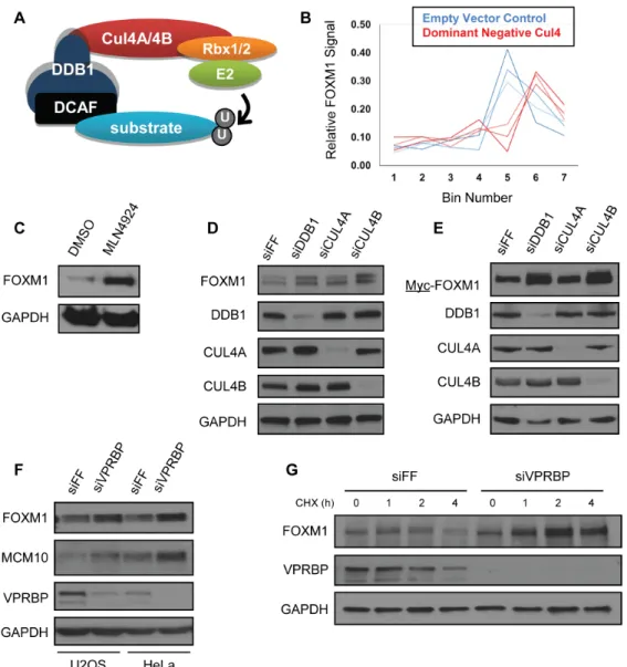
ligase family in humans (6). CRL assembly is based on a common molecular scaffold and relies on a cullin backbone that simultaneously engages substrates and E2 ubiquitin-conjugating enzymes. Cullin4-based ligases (CRL4) use either of two highly related cullin proteins, CUL4A or CUL4B, that bind to the triple-β-propeller protein DDB1 and simultaneously recruit specific proteins to the enzyme complex for ubiquitylation (Figure 2.1A) (226). Human CUL4A and CUL4B are highly similar (75% amino acid similarity), however CUL4B has an amino-terminal extension and localizes exclusively to the nucleus, whereas CUL4A is both nuclear and
cytoplasmic (227). Importantly, CRL4 function has been linked to chromatin regulation, cell cycle, viral infection, and the DNA damage response (226).
More than 50 CRL4 substrate receptors, termed DCAFs or DWD proteins (DDB1 and CUL4 Associated Factors; DDB1 binding WD40 proteins), have been identified (26, 228, 229). VPRBP/DCAF1 is a nucleus-localized CRL4 substrate receptor named for its ability to bind the HIV accessory protein Vpr (and Vpx) following viral infection (36). Ectopic Vpr expression in human cells triggers a G2 arrest that is dependent on CRL4VPRBP (47). Significantly, VPRBP associates with chromatin only during G2/M phase of the cell cycle (230). Knockout of VPRBP in mice causes embryonic death prior to embryonic day 7.5 (E7.5), and conditional inactivation of VPRBP in mouse cells or depletion using RNA interference (RNAi) in human cells, produces cell cycle defects (230). Despite its importance in cell cycle control and development,
transcriptional regulation and cancer (233). Here, we describe a role for VPRBP in controlling both the degradation and activation of FOXM1.
Results
FOXM1 Stability is Regulated by CRL4VPRBP
Using a fluorescence-based genetic reporter system termed global protein stability Profiling (GPS) we previously searched for substrates of the CUL4-based Cullin-RING ligase (CRL4) (6). The GPS expression system relies on a viral vector that expresses a bicistronic mRNA encoding both DsRed and an enhanced green fluorescent protein-open reading frame (EGFP-ORF) fusion protein. Using this system, we infer relative changes in the stability of EGFP-ORF fusions by examining the ratio between EGFP and DsRed fluorescence using flow cytometry. DsRed normalizes for expression of the reporter cassette on a single cell basis. Using this system, we screened a pooled library of 293T cell expressing more than 13,000 individual EGFP-ORF fusion proteins (one ORF per cell). A schematic overview of the GPS screening system is described in detail elsewhere (6, 234).
stability for EGFP-ORFs (6). For example, a shift in the probe distribution to higher-numbered bins suggests that cells expressing a particular EGFP-ORF showed an increase in their
EGFP/DsRed ratio, indicative of an increase in the stability of the EGFP-ORF fusion protein. Three of the four probes corresponding to FOXM1 showed a shifted distribution after dominant-negative CUL4 treatment, suggesting that EGFP-FOXM1 was stabilized by CRL4 inactivation (Figure 2.1B). These three probes showed a highly consistent distribution across bins suggesting that they accurately report the distribution of EGFP-FOXM1 expressing cells and that FOXM1 stability is regulated by CRL4.
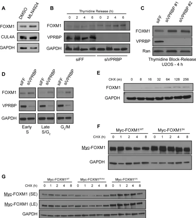
The two best-studied CRL4 substrate receptors implicated in cell cycle control are CDT2 and VPRBP. CDT2 engages substrates through a degron embedded within a PCNA-interacting peptide motif (PIP-box) (236). However, FOXM1 lacks a PIP-box. We therefore depleted VPRBP using siRNA and measured FOXM1 abundance by immunoblot. Depletion of VPRBP increased endogenous FOXM1, as well as the known substrate MCM10, in both HeLa and U2OS cells (Figure 2.1F).
FOXM1 is degraded in G1 phase by the anaphase-promoting complex/cyclosome-CDH1 (APC/CCDH1) ubiquitin ligase (153, 237). To determine if VPRBP regulates FOXM1 at other times during the cell cycle and to rule out the possibility that differences in FOXM1 abundance where due to changes in cell cycle progression, we treated U2OS cells with siRNA targeting VPRBP and then synchronized cells using thymidine in accordance with a previously established protocol (238). Cells were harvested 0, 2, 4 and 6 h after release and analyzed by immunoblot. FOXM1 was consistently increased in the VPRBP-depleted cells (Figure 2.2B), suggesting that VPRBP regulates FOXM1 degradation and that the increase in FOXM1 abundance is independent of gross cell cycle changes. Two independent siRNA oligonucleotides targeting VPRBP produced a similar increase in FOXM1 levels in S-phase synchronized cells (Figure 2.2C). Similarly, we synchronized HCT116 cells in early S-phase, late S-phase/G2 and G2/M using thymidine, thymidine block and release, and nocodazole, respectively. FOXM1 was increased in synchronized, VPRBP-depleted cells, indicating that VPRBP controls FOXM1 independent of its effect on the cell cycle (Figure 2.2D).
This is a reproducible result observed across several experiments. The increase in FOXM1 is evident in otherwise untreated cells after the addition of CHX (Figure 2.2E). While the mechanism underlying this phenomenon remains unclear, it has long been appreciated that some proteins can increase following treatment with CHX (239).
FOXM1 Interaction and Ubiquitylation by VPRBP
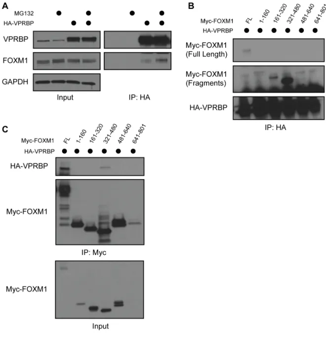
We determined if VPRBP binds to FOXM1 in 293T cells transiently transfected with Myc-FOXM1 and hemagglutinin-tagged VPRBP (HA-VPRBP) to examine the possibility that FOXM1 is a direct CRL4VPRBP substrate. Cells were treated with the proteasome inhibitor MG132 for 4 h prior to lysis and co-immunoprecipitation (co-IP) to promote the interaction between E3 ligase and substrate (240, 241). Following anti-HA-VPRBP IP, we detected an interaction with FOXM1, and this interaction was enhanced by treatment with MG132 (Figure 2.3A and Figure 2.4A). The interaction was also detectable when Myc-FOXM1 was
immunoprecipitated (Figure 2.4C). In addition, we immunoprecipitated endogenous FOXM1 from 293T cell lysates and detected endogenous VPRBP (Figure 2.3B). Finally, we determined if FOXM1 and VPRBP can directly interact. We immobilized bacterially purified hexahistidine-tagged FOXM1 (FOXM1-6HIS) on nickel-agarose beads and mixed them with purified, [35S]methionine-labeled VPRBP produced in vitro using purified transcription and translation machinery. VPRBP bound to beads that had immobilized FOXM1-6HIS but was observed only in the flow through of control beads, strongly suggestive of a direct interaction (Figure 2.3C).
isolated on nickel-agarose. VPRBP expression enhanced the ubiquitylation of endogenous FOXM1, measured by immunoblotting for FOXM1 (Figure 2.3D). Likewise, VPRBP significantly increased the ubiquitylation of ectopically expressed Myc-FOXM1 (Figure 2.3E). Therefore, VPRBP regulates the abundance, stability and ubiquitylation of FOXM1.
To identify the domain in FOXM1 that interacts with VPRBP we synthesized a series of constructs encoding 160 amino acid (aa) fragments of the FOXM1 protein spanning the length of its largest known open reading frame. We tested their ability to interact by expressing full-length HA-VPRBP and Myc-FOXM1 fragments in 293T cells and analyzing precipitates after anti-HA IP. A fragment of FOXM1 spanning amino acids 321-480 (FOXM1321-480) interacted most strongly with VPRBP (Figure 2.4B). The same Myc-FOXM1321-480 fragment bound to VPRBP when Myc was precipitated (Figure 2.4C). We conclude that the interaction between VPRBP and FOXM1 is dependent on the region of FOXM1 spanning amino acids 321-480.
To identify the degron sequence motif in FOXM1 we examined molecular and structural data of a known VPRBP substrate. SAMHD1 is targeted by CRL4VPRBP after HIV infection, and a ternary complex between VPRBP, SAMHD1 and the viral accessory protein Vpx has been crystalized (Figure 2.3F) (42). Importantly, the amino acids in SAMHD1 that mediate binding to VPRBP and degradation have been mapped (41, 42). A segment between amino acids 615 and 625 in SAMHD1 contributes significantly to VPRBP binding and several residues in this region are critical for its degradation. We looked for matching sequences in FOXM1 between residues 321 and 480 and identified a region of similarity between residues 414 and 422 (Figure 2.3F). We synthesized three mutant versions of the same 321-480 region, making amino acid
AA (aa 418 and 419), and K to A (aa 422). We tested the ability of each version of the fragment to bind HA-VPRBP by co-IP following treatment with proteasome inhibitors to normalize protein levels across IPs. We found that alanine substitutions at residues 418 and 419 (RI to AA) and 422 (K to A) impaired binding to VPRBP (Figure 2.3G). Notably, K622 in SAMHD1, corresponding to K422 in FOXM1, directly contacts VPRBP in the crystal structure and is required for SAMHD1 degradation (42).
We next tested the stability of these fragments by CHX chase. Significantly, fragments that showed reduced binding also had higher basal expression (note these experiments were not done in proteasome inhibitors) and increased stability relative to the wild type (WT) fragment when expressed in 293T cells (Figure 2.3H). We observed a similar increase in the stability of full-length FOXM1 when amino acids in the putative degron motif were changed (RVRIAPK to AAAAAPA; the half-life increased from 1.0 to 1.5 h). Minor changes, specifically K422A, similarly extended the half-life (Figure 2.2F and 2.2G). These data are suggestive that this motif sequence, R-I/V-X-X-(X)-K, represents a putative VPRBP degron. Similar sequences were found in established substrates TET2 and MCM10 (Figure 2.3F) (231, 232).
VPRBP is a FOXM1 Activator
coactivator MELK (174). VPRBP activated the reporter to a greater extent than MELK when transfected in equal amounts (Figure 2.5B). Furthermore, VPRBP activates the 6xDB reporter in the absence of ectopically expressed FOXM1 (Figure 2.5B). We also measured the effect of VPRBP on an unrelated luciferase reporter controlled by the oxidative stress response transcription factor Nrf2. FOXM1 and VPRBP expression did not affect the Nrf2 luciferase reporter, whereas strong activation was achieved by Nrf2 expression (Figure 2.6A).
We next expressed the 6xDB reporter with increasing amounts of VPRBP and analyzed luciferase activity, cell cycle dynamics and protein abundance all in the same experiment (Figure 2.5C). VPRBP activated reporter activity at both 24 and 48 h post-transfection in a dose-dependent manner (Figure 2.5C.1). Importantly, the abundance of FOXM1 remained unchanged at both time points and concentrations of VPRBP (Figure 2.5C.2). This demonstrates that the change in reporter activity is not due to changes in the overall level of FOXM1. It also suggests that VPRBP does not activate FOXM1 by triggering its degradation, as is the case for Myc activation by SKP2 (219,
220). We examined the cell cycle using propidium iodide staining and analysis by flow cytometry and found no significant changes at either time point relative to controls (Figure 2.5C.3). We therefore conclude that VPRBP activates FOXM1, and that activation is independent of FOXM1 abundance, and is not due to gross changes in cell cycle dynamics.
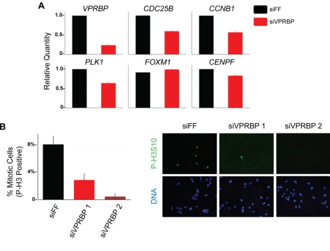
VPRBP depleted cells, including CDC25B, CCNB1, PLK1 and CENPF (Figure 2.7A). The expression of VPRBP was reduced by siRNA treatment as expected; however, the level of FOXM1 mRNA was unchanged. We performed a similar experiment by treating cells with siRNA targeting FF, FOXM1, and VPRBP, and then blocking cells in mitosis with nocodazole. Mitotic cells were specifically isolated by shake-off and we found that the expression of CDC25B, PLK1 and CCNB1
were reduced to a similar extent in both FOXM1 and VPRBP-depleted cells relative to controls (Figure 2.6B). We therefore conclude that VPRBP activates the transcription of FOXM1 target genes during G2/M phase of the cell cycle.
FOXM1 has been implicated in mitotic entry and progression (211, 212). We next examined the role of VPRBP in mitotic entry since its depletion reduces the expression of several FOXM1 target genes. U2OS cells were treated with either control or VPRBP siRNAs and after 72 h were fixed and processed for phospho-histone H3 (P-H3) immunostaining to mark mitotic cells. Imaging was performed and the percent of P-H3 positive cells was determined. 8% of cells were in mitosis in control depleted populations (Figure 2.7B). Depletion with two independent VPRBP siRNAs significantly reduced the percentage of P-H3 positive mitotic cells, to 2.8% and 0.5% (Figure 2.7B). Thus, VPRBP depletion blocks the accumulation of mitotic cell cycle genes and prevents M-phase entry, consistent with a role for VPRBP in activating FOXM1.
extent to the FOXM1 targets CENPF and CDK1 (Figure 2.8A). Thus, FOXM1 and VPRBP co-localize to FOXM1 target promoters.
Myc and ERα are activated by ubiquitylation via their respective ligases (219, 220, 242). We sought to gain similar mechanistic insight into the regulation of FOXM1 by VPRBP. We determined if two independent versions of VPRBP that cannot bind DDB1 (N909 and RARA) are able to activate FOXM1 reporter activity (231). The 6xDB reporter activity was increased using an amino-terminal fragment of VPRBP (N909) which cannot bind to the DDB1/CUL4 complex (Figure 2.8B). Similar results were obtained with a mutant version of VPRBP (RARA) that is also impaired in DDB1/CUL4 binding (Figure 2.8B) (231). Further, the response of 6xDB to VPRBPWT, VPRBPN909 and VPRBPRARA is dose dependent (Figure 2.6C). We conclude that VPRBP activation of FOXM1 is independent of the CRL4 complex. These data also suggest that FOXM1 activation by VPRBP is not due to a change in the abundance or stability of a secondary, unknown CRL4VPRBP substrate.
VPRBP Protein is Upregulated in High-Grade Serous Ovarian Tumors
The FOXM1 gene expression signature is upregulated in ~90% of high-grade serous ovarian cancers (HGSOC) (214). However, FOXM1 mRNA is only overexpressed in ~12% of tumors. To analyze the expression of VPRBP in HGSOC tumors we obtained surgically resected ovaries from HGSOC patients that had been histologically confirmed as serous ovarian cancer. As controls we examined ovaries from women who underwent oophorectomy for reasons other than gynecological malignancy. FOXM1 levels were not elevated in HGSOC tumors relative to controls. Remarkably, VPRBP protein was upregulated in seven out of nine HGSOC tumors tested relative to control ovaries where VPRBP levels were low or undetectable (Figure 2.9A). Further, the well-established FOXM1 target cyclin B was expressed in six of the seven tumors where VPRBP was increased. VPRBP mRNA is not overexpressed in HGSOC based on genomic analysis, suggesting that post-transcriptional mechanisms account for its overexpression (214).
Discussion
Ubiquitylation has long been implicated as a key regulator of transcription and chromatin regulation in human cells. Ubiquitin was identified due to its conjugation to the core histone H2A, and only later was identified by Hershko and colleagues as part of intracellular protein degradation system (243). The yeast α-2 transcriptional repressor was one of the first identified
aspects of cellular physiology, beside protein degradation, illustrates the complex and sometimes paradoxical role the ubiquitin machinery can play in signal transduction.
These multiple functions are evident in the role of ubiquitin signaling in transcriptional regulation, where transcription factors and coactivators can be activated by the ubiquitin machinery. For example, the transcriptional activation domain (TAD) of VP16 is activated in yeast by the SCF substrate receptor F-box protein, MET30 (222). Moreover, fusion of ubiquitin directly to VP16 restores its activity in the absence of MET30 without effecting VP16-TAD stability, providing an example of ubiquitin-dependent, degradation-independent transcriptional activation (222). There are also well-established examples of ubiquitin- and
proteasome-dependent transcriptional activation in human cells. First is the Myc transcription factor. Myc degradation is controlled by the SCFSKP2 E3 ubiquitin ligase and SKP2 also promotes Myc transcriptional activation (219, 220). Consistently, a lysine-less version of Myc, which cannot be ubiquitylated and degraded with normal kinetics, still binds to the coactivator Max and localizes to target gene promoters but is unable to fully activate gene expression (221). Similarly, ERa is ubiquitylated and degraded in response to ligand (estrogen) and this is required for its full activation (223).
suggest that FOXM1 activation by VPRBP is ubiquitin- and degradation- independent. The ability of VPRBP to activate FOXM1 independently of CRL4 binding provides a clear demonstration of E3 ligase substrate receptor repurposing. Moreover, it suggests the possibility that the activity of other CRL substrate receptors can be context dependent and dynamically altered by controlling their association with ubiquitin machinery.
We hypothesize that VPRBP acts as a rheostat to control FOXM1 degradation and activation (Figure 2.9B). FOXM1 abundance is tightly controlled throughout the cell cycle by ubiquitination. By tempering FOXM1 accumulation, VPRBP could impair its activation. Late in the cell cycle, before mitosis, FOXM1 activity increases, contributing significantly to the expression of genes involved in mitotic entry and progression (156). At this time, the partial dissociation of VPRBP from DDB1 and CUL4 (Figure 2.8) would enhance its ability to activate FOXM1 (216). Consistent with this prediction, VPRBP partially dissociates from CRL4 in mitosis and a mutant version of VPRBP that is impaired for DDB1 binding can still activate FOXM1. Importantly, since VPRBP depletion increases FOXM1 protein levels in mitotic cells, it is unlikely that VPRBP acts through a switch-like mechanism, whereby it is either targeting FOXM1 for degradation of promoting its activation. In a rheostat model, VPRBP could tune the output of FOXM1 by controlling both its activation and its degradation with the relative role of each in a given cell determined by the extent of CRL4VPRBP disassembly. It is interesting that VPRBP is unique among CRL4 substrate receptors in that it is likely stoichiometrically bound to CRL4 (230,
245). In addition, VPRBP association with chromatin is tightly cell cycle regulated and occurs only at the time when FOXM1 is activated (G2/M).
to DDB1/CUL4. Alternatively, modification of DDB1 or CUL4 could regulate their association with VPRBP. Modifications that affect FOXM1-VPRBP binding cannot be ruled out, although they did not change in mitosis in our experiments. It is interesting that CHK2 phosphorylates FOXM1 in the same region as the one that we mapped as being important for VPRBP binding (165) and CHK2 has been linked to mitotic progression (246). Dissecting the signaling pathways that control the relationship and interactions between VPRBP, CRL4 and FOXM1 is an important area of future study.
FOXM1 is activated in variety of human malignancies. Specifically, its transcriptional signature is upregulated in HGSOC, serous uterine cancer and basal-like breast cancer. (190, 214,
Materials and Methods
Immunoblot Analysis
Cell extracts were prepared by lysis in ice-cold NETN buffer (20 mM Tris pH 8.0, 100 mM NaCl, 0.5 mM EDTA, 0.5% NP40, 1 mM 4-(2-aminoethyl)-benzenesulfonyl fluoride (AEBSF), 10 μg/ml leupeptin, 2 μg/ml aprotonin, 2 μg/ml pepstatin A). Cell were lysed on ice for 15 minutes and then centrifuged at 14,000 rpm for 15 minutes at 4°C. Supernatant was collected and protein concentrations were determined by Bradford assay.
Tumor specimens were sampled from patients undergoing surgery for HGSOC at the University of North Carolina at Chapel Hill. The protocol was reviewed, and exemption granted by the Institutional Review Board at the University. To extract protein from tissues, normal ovarian tissues and ovarian tumors were homogenized in ice-cold tissue homogenizing buffer (10 mM HEPES (pH 7.4), 50 mM β-glycerophosphate, 1% Triton X-100, 10% glycerol, 2 mM EDTA, 2 mM EGTA, 1 mM dithiothreitol (DTT), 10 mM NaF, 1 mM Na3VO4, 10 μg/ml leupeptin, 2 μg/ml aprotonin, 2 μg/ml pepstatin A, and 1 mM AEBSF) using TissueLyser II (Qiagen). The homogenates were placed on ice for 15 minutes and then centrifuged at 14,000 rpm for 15 minutes at 4°C. Supernatant was collected, and protein concentrations were determined by Bradford assay.
conjugated to horseradish peroxidase (HRP) were purchased from Jackson ImmunoResearch Laboratories.
Immunoprecipitation
Briefly, soluble protein extracts were prepared from 293T cells transiently transfected with HA-VPRBP and Myc-FOXM1 (wild type or mutants). Cell were lysed in NETN as described above. Precipitation was performed by rotating 50 μl of EZview™ Red Anti-HA Affinity Gel or EZview™ Red Anti-c-Myc Affinity Gel (Sigma) with 2 mg of soluble, clarified lysate overnight at 4 °C. The affinity resin was recovered by centrifugation at 1,000 × g for 1 minute and washed 3 times with ice-cold lysis buffer. Precipitates were eluted in SDS-PAGE buffer and analyzed by immunoblot.
RT-qPCR
Total RNA was isolated with an RNeasy Plus mini Kit (Qiagen) and then reverse transcribed with a SuperScript® III First-Strand Synthesis System (Invitrogen). The resulting complementary DNA was used for quantitative PCR with iTaq™ Universal SYBR® Green Supermix (Bio-Rad). Data was normalized to GAPDH. Real-time PCR and data collection were done with a QuantStudio 6 Flex Real-Time PCR System and Software (Applied Biosystems). All of the primers sequences used for RT-qPCR analysis, ChIP and site directed mutagenesis are described in Table 2.2.
Luciferase Reporter Assays
in combination with different constructs (FOXM1, MELK and VPRBP) using PolyJet Plus transfection reagent (SignaGen Laboratories). The MELK expression vector was a kind gift from Lee Graves (University of North Carolina). The Nrf2 expression vector and Nrf2 luciferase reporter were gifts from Ben Major (University of North Carolina). VPRBP expression vectors were a kind gift from Yue Xiong (University of North Carolina). Cells were routinely collected at 48 h post-transfection. Luciferase activity was measured using the luciferase reporter assay system (Promega). Experiments were performed in three technical replicates each. Statistical differences were determined using Student's t test.
Flow Cytometry
For cell cycle profiling, trypsinized cells were washed with phosphate-buffered saline (PBS), fixed in cold, 70% ethanol and stored overnight at −20°C. DNA was stained for 30 minutes in 25 μg/ml propidium iodide and 100 μg/ml RNase A. Samples were analyzed using CyAn flow cytometer (Beckman Coulter) and FlowJo X software.
In Vivo Ubiquitination Assay