THE EFFECTS OF PROLONGED SITTING ON CEREBRAL PERFUSION AND EXECUTIVE FUNCTION
Quentin Willey
A thesis submitted to the faculty at the University of North Carolina at Chapel Hill in partial fulfillment of the requirements for the degree of Masters of Arts in the Department of
Exercise and Sports Science (Exercise Physiology).
Chapel Hill 2018
ABSTRACT
Quentin Willey: The Effects of Prolonged Sitting on Cerebral Perfusion and Executive Function
(Under the direction of Lee Stoner)
The study purpose was to determine if prolonged (3-hr) sitting impaired (a) cerebral perfusion and executive function, (b) systemic vascular function, and (c) if heel raise exercises prevent impairments.
Subjects (n=20) participated in a control (CON) and experimental heel-raise (HEEL) study. Near Infra-red Spectroscopy was used to measure cerebral perfusion and venous pooling in the legs. A Stroop Task was used to assess executive function. Vascular health was measured using pulse wave velocity and pulse wave analysis.
Cerebral perfusion and Stroop was not significantly changed. However, venous pooling did occur in the legs (p<0.05) and systemic vascular health was negatively affected (p<0.05) in both days.
ACKNOWLEDGEMENTS
TABLE OF CONTENTS
LIST OF TABLES………..viii
LIST OF FIGURES………..ix
LIST OF ABBREVIATIONS……….………..x
CHAPTER 1: Introduction………1
Purpose………3
Research Question……….3
Research Hypothesis………..4
Significance of Study……….4
CHAPTER 2: Literature Review Introduction………6
Sedentary Behavior………7
Executive Function………7
Chronic Associations……….8
Acute Associations……….8
Mechanisms………9
Blood Flow………10
Vascular Function………11
Autonomic Nervous System………11
Brain-derived Neurotropic Factor……….12
Chronic Sedentary Behavior and Mechanisms……….12
CHAPTER 3: METHODS
Subjects……….19
Study Design……….………..………..20
Pre-Assessment……….20
Familiarization………...………..20
Visit 2 & 3...…….……….21
Measurements…………..………22
Executive Function………..22
Cerebral Perfusion.………..………22
Venous Pooling in Lower Extremities.………...24
Vascular Health….………...24
Power Calculation.…….………..………..………..26
Statistical Analysis……….………..26
CHAPTER 4: RESULTS Subjects……….29
Executive Function………...29
Cerebral Perfusion………...29
Venous Pooling……….30
Vascular Health………33
CHAPTER 5: DISCUSSION Summary of Findings……….……….37
Executive Function………...37
Cerebral Perfusion and Calf Pooling……….38
Arterial Stiffness and PWV..………...40
Central Hemodynamics and Augmentation Index………...40
Strengths and Limitations..……….41
Implications…..………….………...………42
APPENDIX A………...44
APPENDIX B………...49
APPENDIX C………...54
APPENDIX D………...56
APPENDIX E………...57
APPENDIX F………...58
APPENDIX G………..59
LIST OF TABLES
Table 1 – Subjects Characteristics……….29
Table 2 – Stroop Completion Time………29
Table 3 – Normalized NIRS Forehead Data Trend………..30
Table 4 – Normalized NIRS Calf Data Trend..……….31
Table 5 – Normalized NIRS Pre-Post……….31
Table 6.1 – Seated Cardiovascular Measurement Trend……….34
LIST OF FIGURES
Figure 1 – Mechanism Pathway……….………….10
Figure 2 – Scheme of the Cardiovascular Control Mechanisms……….14
Figure 3 – Aortic Pulse Wave Analysis………...………...25
Figure 4 – tHb Calf Trend………...32
LIST OF ABBREVIATIONS
AIx – Augmentation Index
APL – Applied Physiology Laboratory AS – Arterial Stiffness
BF – Blood Flow BP – Blood Pressure
cBP – Central Blood Pressure CO – Cardiac Output
CV – Cardiovascular
CVD – Cardiovascular Disease FMD – Flow Mediated Dilation Hb – Hemoglobin
MAP – Mean Arterial Pressure NIRS – Near-infrared Spectroscopy NO – Nitric Oxide
Chapter 1. Introduction
Prolonged sitting may pose a public health risk through its effects on the vascular system and may lead to a reduced ability to process cognitive tasks, thereby negatively effecting the work or school day. Recent evidence indicates that sitting for three hours results in decreased blood flow (BF) to the legs and subsequent local (leg) vascular dysfunction36-38. This previous work did demonstrate that short bouts of walking106 or fidgeting38 prevented the decline in BF and shear stress, and subsequently local (lower extremity) vascular dysfunction. However, it is currently unknown whether (1) prolonged sitting effects the brain’s executive function, or whether (2) sitting impairs perfusion of the prefrontal cortex of the brain.
Executive function (EF) is required to turn sensory input into an actionable output and is necessary to carry out simple daily tasks. This EF may rely on cerebral perfusion to the prefrontal cortex of the brain. Previous works have shown that EF declines with chronic
sedentary behavior,16-20 however, few studies examining prolonged sitting have been conducted. Of the studies that have examined prolonged sitting, there is evidence suggesting that EF may have declined after a period of prolonged sitting; though they lacked objective evidence as the data consisted of questionnaires and the mechanisms were not investigated.90 Arguably, EF may become impaired as a function of decreased perfusion to the frontal cortex. 68 While the
Cerebral perfusion may become impaired as a function of blood pooling in the lower extremities during a bout of prolonged sitting. For example, when an individual stands up with locked knees for too long, blood pools in the calf as gravity pulls down and the muscles of the legs are not being repeatedly relaxed and contracted. This can result in fainting and light-headedness from a lack of adequate blood supply. Though similar mechanisms occur while sitting, it is unknown how brain function is effected by a bout of prolonged sitting. Indeed, one of the previous studies examining the effects of prolonged sitting reported increased circumference of the calf indicating an accumulation of blood in the lower limbs.37 The effects of hydrostatic pressure may be compounded by decreased muscle activity, which would normally act as a venous “pump”.42 Blood is then more easily pooled in the lower extremities resulting in less venous return from these regions which may lead to fluctuations along the systemic vascular tree.
Decreased blood flow – and subsequent shear stress – throughout the vascular system may also result in impaired vascular function, including the cerebrovascular. Impaired vascular function may compromise the ability to regulate blood flow to the brain. Though it is unknown how impaired vascular function is linked to brain blood flow, studies have found that a 3-hour sitting intervention is sufficient to impose changes in vascular function.36-38,106. Therefore, a 3-hour sitting protocol will likely be enough time to identify if there is a potential link between changes in vasculature and cerebral perfusion. To determine underlying mechanisms that may link vascular health and cerebral perfusion, vascular measurements that estimate what is happening at the level of the aorta may be helpful as it is thought that stiffness, pressure, and waveforms in the aorta may be an accurate representation of what is happening throughout the peripheral vascular tree.66
and ensure adequate venous return. Maintaining the flow of blood may thereby prevent decrements to local vascular health and prevent fluctuations in cerebral perfusion leaving executive function unaltered. If intermittent calf contractions caused by raising the heels up and down prevent blood from pooling in the legs and thereby maintain adequate cerebral perfusion, then heel raise exercises may be a viable solution to prevent deleterious effects of prolonged sitting on EF.
Purpose
The purpose of the current study is to examine the acute effects of sitting on executive function and the associated mechanisms in the cardiovasculature. Findings from this study may identify a simple strategy such as performing calf raises while being seated for offsetting the negative consequences of sitting and may contribute to public health policy pertaining to sedentary behavior.
Research Questions Primary
1. Does prolonged (3 hr) sitting impair: (a) measures of cerebral perfusion and (b) executive function?
2. Do intermittent heel raise exercises, to engage the muscle pump, prevent venous pooling in the lower extremities and thereby prevent changes in (a) cerebral perfusion and (b) executive function?
3. Are changes in executive function associated with cerebral perfusion? Secondary
2. Are changes in calf muscle perfusion associated with changes in (a) cerebral perfusion and (b) executive function?
Research Hypothesis Primary
1. Prolonged (3 hr) sitting does impair (a) measures of cerebral perfusion and (b) executive function.
2. Intermittent heel raise exercises, to engage the muscle pump, prevents venous pooling in the lower extremities and thereby prevent changes in (a) cerebral perfusion and (b) executive function.
3. Changes in executive function are associated with cerebral perfusion. Secondary
1. Changes in systemic vascular measures are associated with changes in (a) cerebral perfusion and (b) executive function.
2. Changes in calf muscle perfusion are associated with changes in (a) cerebral perfusion and (b) executive function.
Significance of Study
Chapter 2. Literature Review Introduction
Sedentary Behavior
Although, driving and working are often productive and can make defining sedentarism murky when individuals are still carrying out tasks. However, sedentary behavior is defined as any waking behavior characterized by an energy expenditure ≤1.5 METs while in a sitting or reclining posture.2 Sedentary behavior such as prolonged sitting may be effecting a large portion of the population because of occupation, transportation and leisure activity. For example, one study reported that an average of 77% of occupational time was spent sitting.11 Also, another study with a population of 6,329 from the US showed that 54.9 percent of their waking time was spent in sedentary behavior.9 This is further supported by multiple national surveys where respondents reported an average of 7 hours/day sitting at work.12 Though survey responses typically underestimate their sitting time, these conservative data still present a large problem. For example, the US Department of Labor conducted a survey in 2015 where an average 3 hours was dedicated to TV watching and was spent as leisurely, sedentary time.13 These reports may be shocking, but according to the American Heart Association, there has been an 83% increase in sedentary jobs since 1950.10 Therefore, it should be no surprise that sedentary behavior effects a large population in westernized societies.
Executive Function
It is important to note here that sedentary workers are still working. However, the
executive function would decrease the work capacity of individuals in sedentary jobs ranging from students to full-time employed persons.
Chronic Associations
The deleterious effects of chronic sedentary behavior and inactivity have been well documented by many studies. It is clear that sedentary behavior increases risk factors for all-cause mortality and is positively associated with cardiometabolic diseases.3-8 Other negative effects of sedentary behavior, like declines in Executive function, have become of recent interest. For example, recent evidence suggests that chronic sedentary behavior is associated with
declines in EF16,17 and is true for both young18 and old19,20 populations. In addition, individuals with various diseases and mood disorders are negatively affected regarding cognitive measures by sedentary behavior.21-25 As expected, chronic sedentary behavior is bad for human health and executive function is no exemption.
Acute Associations
In contrast to chronic behaviors, less is known about the effects of acute behaviors concerning Executive Function. However, it is well understood that Executive Function improves with acute bouts of physical activity and exercise.26-31 For this reason, interest has mounted whether the opposite might be true; that acute bouts of inactivity, or sedentary behavior, could lead to declines in Executive Function. How the duration of an acute bout is defined may be up for debate; however, several studies could help narrow the definition of acute. For
physiological dysfunctions are seen in as little as 2-3 hours.35-37,40 However, the same
complications may not occur in less 2-3 hours as one study showed no significant changes in Executive Function over the course of 1 hour of sedentary behavior.33 In short, the exact definition of an acute bout has not been well defined, though any longer than 2-3 hours may likely have deleterious effects including Executive Function.
Mechanisms
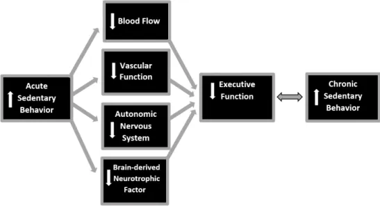
The evidence presented thus far provides a foundation for research concern regarding the effect of chronic, and more particularly, acute sedentary behavior on Executive Function. One of the aims of the current research study is to discover if Executive Function declines with acute sedentary behavior such as prolonged sitting of 3 hours. If declines in Executive Function are found following an acute bout of prolonged sitting, this study could provide potential
Figure 1. Mechanism pathway for declines in EF due to acute sedentary behavior like Prolonged sitting.
Blood Flow
The purpose of this section is to briefly discuss and define each of the four mechanisms in column 2 of Figure 1 which will then be discussed in the context of chronic (section 2.5.1) and acute (section 2.5.2) bouts of sedentary behavior followed by a discussion of how these
of time and is mostly unchanged from one day to another, resistance to blood flow is mostly attributed to changes in vessel diameter. In this way, blood flow is connected to changes in vessel diameter and function.
Vascular Function
How well the vessel diameter changes in response to different stimuli is known as vascular function. The vessel’s responses are largely dependent on the second inner-most lining of the vessel walls known as the endothelium and on smooth muscles which receive input from the nervous system. First, the endothelium is made up of endothelial cells that release nitric oxide (NO) in response to shear stress in the vessel. Shear stress is the force produced by blood moving along the endothelial layer and is proportional to blood flow. As blood flow and shear stress increase, NO is released and the vessel dilates to keep blood flow and shear stress at healthy levels. This is one way that vascular function is maintained.
Autonomic Nervous System
effected by sympathetic and parasympathetic tone. Heart rate (HR), contractility, rate of relaxation and rate of conduction are mainly controlled by the autonomic nervous system and these factors can be described in part by Heart Rate Variability (HRV). Why this information is important will be further discussed in section 2.6.
Brain-derived Neurotrophic Factor
This last portion will discuss Brain-derived Neurotrophic Factor (BDNF) which is a growth factor protein related to the Nerve Growth Factor and are found in the brain and periphery. BDNF has been shown to increase in acute bouts of exercise and mediates
improvements in EF.28 For this reason, it may be of interest to explore how sedentary behavior effects BDNF in chronic and acute bouts of sedentary behavior.
Chronic Sedentary Behavior and Mechanisms
As discussed previously, EF declines in response to chronic sedentary behavior. The mechanisms contributing to this phenomenon are not fully understood, however evidence
regarding BF, VF, the ANS and BDNF may provide potential explanations. First, the effects that chronic sedentary behavior has on BF and VF may be of greatest interest. As individual’s age, their vascular health, including cerebrovascular health, has been shown to decline;59 however, physical inactivity further exacerbates their decline. As discussed previously, strong evidence links sedentary behavior to cardiovascular disease and is the leading cause of death.4-8
independent of age and metabolic diseases60 making it a meaningful outcome when considering the effects of chronic sedentary behavior. For example, as individuals are sedentary over time, AS will increase. Increasing AS will decrease blood flow due to an added amount of resistance which will ultimately result in elevated blood pressures and vascular dysfunction. This would lead to less blood reaching working brain tissue and may be a potential mechanism as to why EF declines with chronic sedentary behavior.
This may be the case because as VF is already declining with age and sedentary behavior is intensifying the same effects. However, the negative effects from sedentary behavior appear to be controllable and have positive effects on EF and VF. For instance, evidence has shown that as older individuals make the effort to substantially reduce their blood pressure, EF is maintained over time. Furthermore, older adults with lower sedentary time and higher levels of physical activity have been shown to have better vascular function.57 In fact, the physiology of vessels seems to change with chronic sedentary behavior as seen in mice models where the endothelial layer and surrounding layers thickened, stiffened and became dysfunctional.61 Whether these effects are found in cerebrovascular regions is less understood, but systemic effects may be reaching the brain thereby decreasing EF.
then be effected which will keep the sympathetic tone at an elevated state. Figure 2 may be helpful in making connections between the ANS and responses in the body. Well-being in the ANS and sympathetic tone can be measured by HRV and will be further discussed in section 2.6. However, it is important to note here that as the sympathetic tone increases and remains
increased, the ANS becomes dysfunctional and the body is less able to respond to stress
appropriately.47,48 The combination of factors contributing to ANS dysfunction may be another potential explanation for EF decline with chronic sedentary behavior.
Figure 2. Scheme of the cardiovascular control mechanisms responsible for the main periodic fluctuations in heart rate.49
a role in43 as BDNF has also been shown to mediate the effects of physical activity on EF.52 However, chronic sedentary behavior may have a negative effect on EF through the
dysregulation of insulin and cytokines that in-turn may dysregulate BDNF. Finding a study evidencing this mechanism would be rare, although the recent findings mentioned here are provoking in context of the link between insulin and cytokines to the regulation of BDNF. If BDNF is decreased with chronic bouts of sedentary behavior, this might stimulate a new research avenue previously untapped.
Acute Sedentary Behavior and Mechanisms
The effects of acute sedentary behavior on EF is relatively recent area of research interest. However, there is a large body of evidence to support that the proposed mechanisms in column 2 of Figure 1 do occur. Whether or not these mechanisms are the same mechanisms that could lead to declines in EF has not been fully investigated. Although, when considering the all the evidence together in context of one another, there is reason to believe that EF could be effected by acute bouts of sedentary behavior. Discussed below are the findings related to acute sedentary behavior starting with BF and ending with BDNF similar to section 2.3.1.
to slow moving blood. Blood then becomes more viscous and resistance continues to increase.41 As discussed previously, increased resistance will reduce BF and thus BF decreases in the lower extremities.
Second, the decreases in BF cause decreased amounts of shear stress which results in vascular dysfunction. Endothelial cells have been shown to become dysfunctional in response to a bout of prolonged sitting lasting 3 hours.36-38 The combination of decreased BF and VF results in a decrease in venous return. According to the Frank Starling Law of the Heart, less blood filling the heart results in weaker contractions and a lower stroke volume (SV). Drops in venous pressure, such as when a person stands up too fast and becomes light-headed, has obvious effects on EF. This happens because there is temporarily less blood being ejected from the heart,
meaning less blood reaches and perfuses brain tissue causing temporary executive dysfunction. Therefore, if VF is compromised, there is reason to believe that there could be a connection between prolonged sitting of 3 hours and EF as one study has shown.32
Finally, BDNF could be used to better understand effects of sedentary behavior on EF and in light of current research, might be more useful in studying acute bouts versus chronic. This is because most of what is known about the associations between BDNF and physical activity come from acute bouts of exercise or physical activity.50-52 As mentioned in section 3.1, BDNF is regulated by insulin and cytokines. These can be altered in acute bouts of sedentary behavior and may contribute to BDNF declines leading to diminishing EF.
Implications
Sedentary behavior is known to be hazardous to health as has been demonstrated throughout this review. However, it is important that organizations with the ability to impact large populations recognize the evidence and findings regarding sedentary behaviors such as prolonged sitting. A recent example of this happening is the American Medical Association announced in their June 2013 annual meeting that sitting down for too long is hazardous to health. In addition, more audiences are recognizing that simply meeting physical activity guidelines each day does not prevent the negative effects of prolonged sedentary behavior.15 Mounting evidence of this nature should promote change in policy and guidelines in corporations and public health initiatives.
Chapter 3. Methods Subjects
A relatively homogenous cohort of 20 young (19–35 year) and healthy but sedentary participants was recruited to participate in the study. To be eligible, subjects could not have been meeting current American College of Sports Medicine activity guidelines to be characterized as sedentary. However, because subjects had no other cardiovascular risk factors outside of activity levels, they were considered to be a homogenous, healthy sample. Their sedentary behavior more appropriately represents populations that are frequently sitting for long periods of time. Further reason for selecting sedentary subjects is explained in the discussion. Because this is the first study of its kind and because elderly and diseased populations have different vascular sensitivity, a young, healthy, homogenous population is ideal.
It should also be noted that the effects of estrogen levels on vascular measurement may cause differing levels of vessel reactivity to changes in blood flow and effect cognitive
Study Design
This study was a randomized crossover design with two experimental conditions (control [CON] and heel raises [HEEL]). The experimental conditions took place on separate days and were preceded by a familiarization session. Each experimental testing session was separated by no more than 7 days. Data collection began between 6:00 am-10:00 AM in the Applied
Physiology Laboratory (APL). Prerequisite to participation, subjects took part in an overnight fast while abstaining from alcohol and exercise 24 hours prior to experiment. In addition, they were not allowed caffeine or other supplements the morning of the visit. All subjects were emailed, texted, or personally contacted the day before testing to be reminded of test visit. Pre-assessment
Prior to subject participation, ethical approval was obtained through the IRB and the Office of Human Research Ethics at the University of North Carolina-Chapel Hill. All subjects reported to the APL to fill out general health questionnaires (Appendix A) and gave informed consent (Appendix B).
Familiarization
arrival, subjects were required to confirm protein bar ingestion and were reminded via email or text message prior to testing day to consume the bar 2 hours prior to arrival.
Visit 2 & 3
negative effects of prolonged sitting. After 180 minutes, subjects were transferred back to the supine position to conclude with PWA and PWV.
Blood volume may change throughout the course of the sitting protocol due to filtration of the blood in the kidneys and insensible water loss through perspiration and respiration. This can cause between 100-250 ml of water loss in a period of 3 hours. For this reason, and at the request of the IRB, water intake was monitored during both testing sessions and subjects were given 40 mL of water every 30 minutes. In addition, subjects were instructed to refrain from using the restroom during the study because standing and walking to the restroom would alter CV mechanisms. There were no instances of subjects getting up to use the restroom at any point during the study.
Measurements Executive Function:
To assess if EF was affected by prolonged sitting and perhaps declines in cerebral perfusion, the Stroop Word-Color Task (Stroop) and was administered on a laptop computer.31 For both the word and color portion of the Stroop Task, 32 iterations were performed and time till completion was used as the primary outcome. Familiarization consisted of a minimum of seven tasks, or until total completion time plateaued for both the word and color portions of the Stroop as has been done previously.80 Stroop has been widely used for the testing of EF and is considered a valid and reliable test including young, healthy populations.69-71
Cerebral Perfusion:
several non-invasive measures and other works’ have shown its effectiveness in measuring oxygen uptake and delivery in the brain.93 For this reason, a NIRS probe was placed on the forehead to measure relative changes in total hemoglobin as has been described in previous works’31,79 and as shown in Appendix C. By placing a NIRS probe on the forehead, over the prefrontal cortex, the total amount of Hb (tHb), and thereby perfused blood, was measured over the course of the study. In addition, the total saturation index (TSI) was measured, which is an index represented as a percentage of oxygenated Hb (O2Hb) versus deoxygenated (HHb) found in the tissue of interest.
The deepest probe depth where the infrared light would travel the furthest into the tissue (or T3, meaning the third infrared light emitter) for tHb measurement was determined to be the best measurement output for capturing Hb within the prefrontal cortex. TSI, however, uses an average saturation of all three NIRS light infra-red-light sources and produces a single percentage making probe placement very important. For this reason, the tHb and TSI were measured with careful consideration to avoid any large vessels and ultrasound was utilized to verify probe placement. Snap-shots of ultrasound capture can be found in Appendix D.
each bout of heel raise exercise. Because the live NIRS data feed rises and falls slightly with each heartbeat and breath, selecting a single data point is less accurate. A 30 second average has been previously used in studies utilizing NIRS and was determined appropriate for this study.107
Venous Pooling in Lower Extremities:
To measure the amount of blood pooling in the lower extremities, the muscle belly of the medial gastrocnemius was found using ultrasound to avoid large vessels. A NIRS probe was placed on the medial gastrocnemius of the right leg as shown in Appendix C and blood pooling was monitored using NIRS set to a pathlength correction factors for the calf (Appendix F). To test if cerebral perfusion and calf perfusion changed as a result of prolonged sitting, tHb and TSI were the dependent variables and were first normalized relative to baseline by using change scores instead of absolute outputs.
Vascular Health:
To obtain a comprehensive overview of systemic vascular health, and to see if changes may be related to possible declines in cerebral perfusion and EF, several non-invasive methods were used, including PWV and PWA. Both measurement techniques have high reliability, validity and provide quantified data for AS and VF.64,65 If AS is shown to increase, there is reason to believe that the burden on the heart will increase.101 Any general relationships between systemic vasculature, cerebral perfusion and EF may be helpful in determining underlying mechanisms for the effects of prolonged sitting.
by the use of a large, hand-made caliper so as to avoid measuring the contours of the body and strictly measure the length of the descending aorta (Appendix E).
PWA was obtained by using oscillometric pressure waveforms recorded on the left arm using a brachial cuff following standard manufacturer guidelines.62 An aortic pressure waveform was generated using a validated transfer function based off of the pressure waveform derived at the arm.99 The AIx is the augmentation pressure (AP), expressed as a percentage of central pulse pressure. AP is defined as the peak systolic pressure minus the pressure at the inflection point (Fig. 1). The aortic wave can be separated (bottom panel) into its forward (Pf) and backward (Pb) waves
Fig. 3 Aortic pulse wave analysis. Using the generated aortic pressure waveform (top panel), the augmentation index (AIx) is calculated by expressing augmentation pressure (AP) as a percentage of the central pulse pressure (cPP). The AP is the additional pressure added to the forward wave by the reflected wave and is defined as the maximum central systolic
pressure minus the pressure at the inflection point. Using a
physiologic flow waveform (middle panel), the aortic wave can be separated (bottom panel) into its forward (Pf) and
after assuming a triangular flow wave, and reflection magnitude (RM) is given by Pb/Pf.84,96-98 AIx@75 is simply a measure of AIx after being corrected for heart rate deviations. However, because they responded nearly identically, only AIx will be discussed moving forward.
Power Calculation
Sample size calculations were based on the primary central vascular health outcomes, aortic pulse wave velocity (PWV). While the effects of prolonged sitting on central vascular health have not been investigated, previous studies have reported that prolonged sitting reduces leg vascular health between 57-80%.36-38,73 Based on a PWV of 6.6 m/s, which is expected for healthy participants <30 y (Reference Values for Arterial Stiffness’ Collaboration 2009), a 57% decrease in PWV would be 3.8 m/s. For the current study, it was opted to sample based on a conservative change score of 1 m/s. We also opted to use a conservative typical error of 1 m/s.74 Using magnitude-based inference, to estimate the sample size required to detect the smallest detrimental (or beneficial) effect in a cross-over study, with the maximum chances of a type 1 and 2 error set at 5% (i.e. very unlikely), approximately 12 participants are required. However, over-sampling became necessary here because there is no current representative study involving executive function to use as a reference for sample size.
Statistical Analysis
All statistical analyses were performed using SPSS 21. First, to test the hypothesis that prolonged sitting results in decreased executive function, data were analyzed using general linear modelling with repeated measures for the 2x3 ANOVA crossover design using the two
To test the hypothesis that cerebral perfusion trends downwards and calf perfusion trends upwards over the course of a prolonged sitting protocol the same statistical model used for executive function was repeated to test perfusion changes in the prefontal cortex and the calf. Secondary outcomes were also tested using the same model as previously described.
To test the hypothesis that prolonged sitting results in reduced cerebral perfusion, before and after data was analyzed using general linear modelling with a repeated measures 2x2
ANOVA crossover design using the two conditions (CON and HEEL) and two time-points (pre- and post) as the independent variables. This same design was also used to test the hypothesis that vascular health measures would significantly become worse as a result of prolonged sitting.
To test the hypothesis that EF, cerebral perfusion and vascular function are related, Pearson’s correlation tests were used to first assess if changes in EF were associated with changes in cerebral perfusion. A Pearson coefficient (r) would be evaluated by using r values of 0.1-0.3 being a small association, 0.3-0.5 being medium, and 0.5-1.0 being large. Since it
hypothesized that EF will decline as cerebral perfusion declines, we would expect these values to be negative but of the same magnitude from interpretation.
Then, to test secondary hypotheses, Pearson’s correlation tests will again be performed to assess if important vascular health variables such as PWV, Central Blood Pressures, and
Lastly, partial eta squared effect sizes were considered in the statistical interpretation of all relevant significant (p<0.05) outcomes where ƞ2 = .02, 0.13 and 0.26 represent a small, medium and large effect respectively. partial eta squared effect sizes were used because partial eta squared is the variance explained by a given variable of the variance remaining after
Chapter 4. Results Subjects
There was some ethnic diversity, though predominantly Caucasian (n=13). The remaining subjects were African American (n=4), Hispanic (n=1), Middle-eastern (n=1), and Asian (n=1).
Executive Function
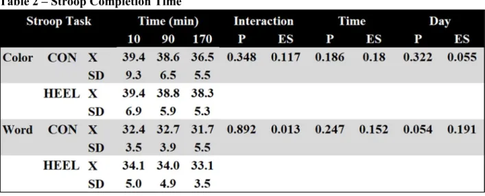
There were no interaction effects for executive function as measured by the use of a Stroop Color and Word task. In addition, there was no effect of day on Stroop scores (Table 2).
Table 2 – Stroop Completion Time
Table 1. Subject Characteristics
Age X 21.7
SD 8.6
BMI X 25.7
SD 5.3
Sex (% Female) X 70
SD N/A
Con Control, Exp Experimental, X mean, SD standard deviation, P p-value, ES effect size * represents statistically significant main effect (p<0.05).
Cerebral Perfusion
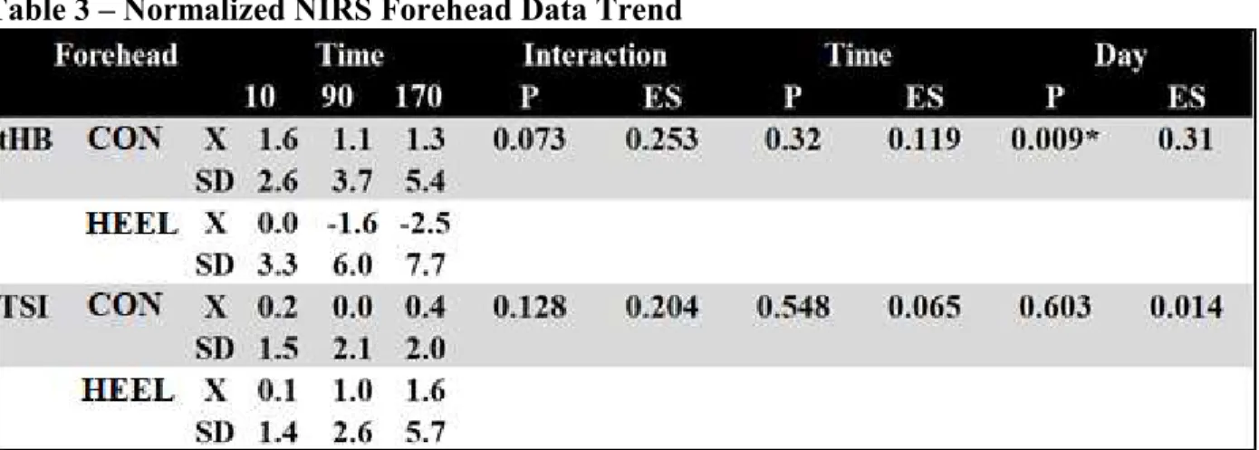
There were no interaction effects for cerebral perfusion (Table 3). Also, cerebral perfusion was not shown to significantly change over the course of the bout of sitting and we fail to reject the null hypothesis that prolonged sitting does not result in decreased cerebral perfusion. However, tHb in the HEEL day was found to be significantly (p=0.009) different from the control day with a mean difference of 2.685 ± 0.919, [CI @95% 0.76, 4.609]. This, and a large effect size, may partially explain the trend (p=0.073) towards significant interaction effects. Although there was some effect of day, because tHb changed in the opposite direction, we fail to reject the null hypothesis that calf raises prevent changes in cerebral perfusion.
Table 3 – Normalized NIRS Forehead Data Trend
Venous Pooling in Lower Extremities
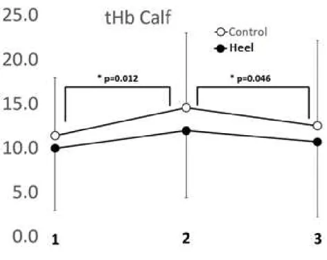
There was no interaction effect for tHb or TSI in the calf. However, blood was shown to accumulate in the calf on both days as there were significant (p=0.002) within-day differences found between the first and second (p=0.012) as well as the second and third (p=0.046) time points for tHb in the calf for CON and HEEL with a mean increase of 2.577 ± 0.922 [CI 95% 0.646,
Con Control, Exp Experimental, X mean, SD standard deviation, P p-value, ES effect size * represents statistically significant main effect (p<0.05).
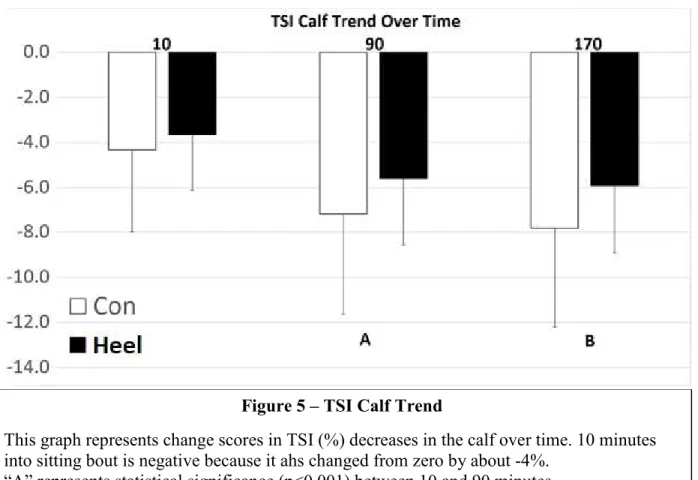
4.507] and 1.655 ± .774 [CI 95% 0.36, 3.275] respectively. In addition, within-day TSI for CON and HEEL was found to be statistically different (p<0.001) from first to second (p<0.001) and first to third time point (p<0.001) with a mean decline of 2.399% ± 0.402% [CI 95% 1.557, 3.24] and 2.863% ± 0.473% [CI 95% 1.872, 3.854] respectively(Table 4, Figures 2-3).
Table 4 – Normalized NIRS Calf Data Trend
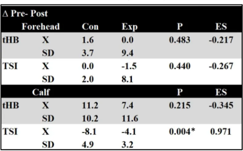
In addition, the effect size suggests a large effect of time on both tHb and TSI. For this reason, we fail to reject the null hypothesis that calf raises prevent venous pooling. However, CON declined significantly more than the HEEL from pre- to post and the effect size suggests a large effect of time on TSI (Table 5). These trend data can be found in.
Table 5 – Normalized NIRS Pre-Post
Table 4 represents mean change scores over time points 10, 90, 170 mins into sitting for Forehead and Calf. tHb total hemoglobin, TSI total saturation index
* represents statistically significant main effect (p<0.05).
** represents statistical trend toward a significant main effect (p<0.010).
Table 5 represents change scores between the beginning of the sit trial and end.
* represents statistically significant main effect (p<0.05).
** represents statistical trend toward a significant main effect (p<0.010).
Figure 4 – tHb Calf Trend
Vascular Health
There were no interaction effects of any vascular measures over the course of the three time points (10,90,170 mins) (Table 6.1) nor from pre-post sitting (Table 6.2). Although,
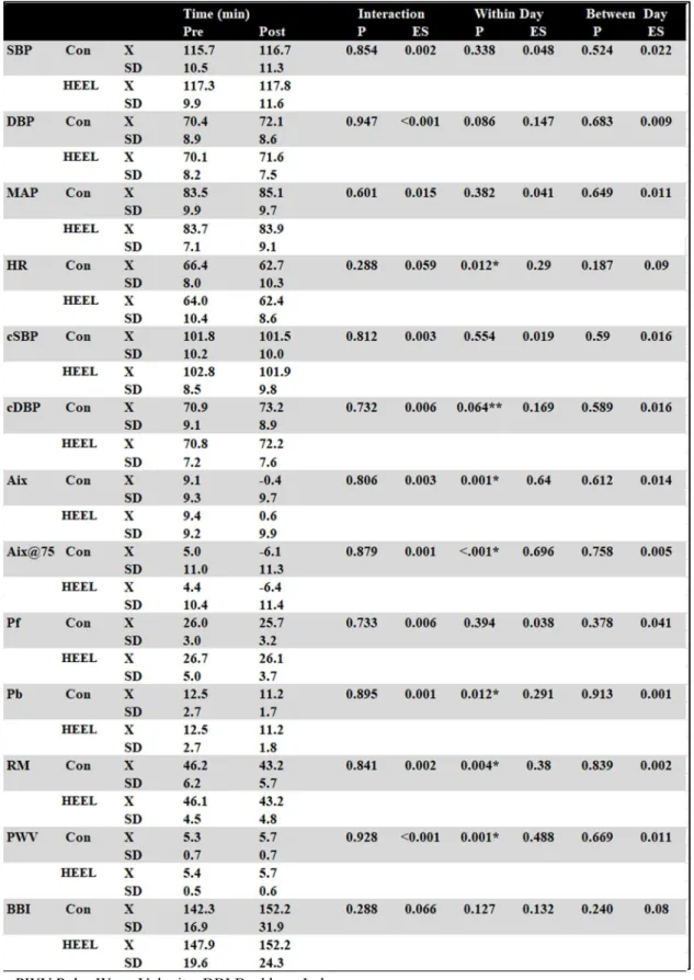
Augmentation Index (AIx) declined over time on each day from 10 minutes in to 90 minutes in by an average of 9.638 ± 1.863 (p<0.001, [CI 95% 5.739, 13.536]) and from 10 to 170 minutes in by 8.236 ± 2.224 (p=0.001, [CI 95% 3.608, 12.917].) The same effect was seen from pre- to post trial with a decrease of 9.14 ± 1.574 (p<0.001, [CI 95@ 5.846, 12.434]). Effect sizes for both pre-post and trend over the course of the study indicates large effects of time on AIx.
Figure 5 – TSI Calf Trend
This graph represents change scores in TSI (%) decreases in the calf over time. 10 minutes into sitting bout is negative because it ahs changed from zero by about -4%.
Table 6.1 – Seated Cardiovascular Measurement Trend
Con control, Heel experiemental, SBP systolic blood pressure, DBP diastolic blood pressure, MAP mean arterial pressure, HR heart rate, cSBP central systolic blood pressure, cDBP central diastolic blood pressure, AIx augmentation index, AIx@75 augmentation index @ 75bpm, Pf pressure wave forwards, Pb pressure wave backwards, HF high Frequency, LF low frequency.
Table 6.2 Supine Cardiovascular Measurement Pre-Post
Next, the reflected pressure wave represented by Pb decreased from the first (10 min) and second (90 min) time point on average by 1.45 ± 0.363 (p<0.001, [CI 95% 0.69, 2.21]) and from the first to third (170 min) time point on average by 1.388 ± 0.385, (p<0.002, [CI 95% 0.0.583, 2.192]). Similar to other cardiovascular measures, Pb also decreased within day from pre- to post by 1.3 ± 0.466 (p=0.012, [CI 95% 0.325, 2.275]). Other vascular measures that are representative of hemodynamic changes such as HR, AIx@75, and RM were also found to be significant. These data are presented in Table 6.1 where the trend over three time points (10,90,170 min) were tested. Then, in Table 6.2, data points from before and after the sitting protocol was undertaken are presented as pre-post averages along with their associated ANOVA results.
Finally, PWV increased in the control and HEEL days with an average increase of 0.3 ± 0.075, [95% CI 0.143,0.457]. In addition, the effect size proposes a large effect of time. However, increases in the control day were not significantly different from increases in HEEL.
From these data, our secondary hypotheses cannot be clearly tested because there were no detriments to EF or cerebral perfusion, and calf raises did not seem to have any preventative effect warranting tests of association with systemic vascular measures.
Associations
Chapter 5. Discussion Summary of Findings
We aimed to provide evidence as to 1) whether or not prolonged sitting resulted in declines in EF and cerebral perfusion, and 2) and if change in cerebral perfusion were found, whether they we associated with blood pooling in the calf, and/or impaired vascular function. It was also hypothesized that doing a set of 10 seated calf raises every 10 minutes could be a minimally effective dose to attenuate negative effects that may arise due to prolonged sitting. Several considerations are discussed below to interpret the results of this study.
Executive Function
in many studies as a measure of executive function.69-71 Even so, it may be too simple to generalize to more complicated tasks in the typical work or school day. Also, the Word portion of the stroop is easier and thus more resistant to change, and though the main effect of day trended towards significance, the effect size of this trend (ƞ2 = 0.191) is medium, and is small relative to the effect size of significant outcomes in this study such as the effect sizes of the NIRS trend data and AIx in both the pre-post and trend data. For these reasons, it should be interpreted that Stroop scores remained mostly unchanged and the null hypothesis is failed to be rejected.
Cerebral Perfusion and Calf Pooling
Previous research from Restaino et al. showed that shear stress was reduced in the popliteal artery after 3 hours of sitting. Shear stress is the frictional force that is caused by blood cells passing over vessel walls and is indicative of adequate blood flow. When shear stress is decreased and stays low, blood flow has likely been disrupted in some way. This may have been caused in this previous study because of increased pooling in the microvasculature of the lower extremities as it was shown that calf circumference increased with an acute bout of sitting.37 The findings from the current study are consistent with findings from this previous work and may lend further clarity regarding the effects on the microvasculature during a bout of prolonged sitting.
deoxygenated Hb because as blood accumulates or pools in the veins and venules in the muscle belly, the oxygen is offloaded from O2Hb into the tissue and deoxygenated Hb then remains in the area. Though NIRS was the only device used, the evidence is overwhelming that venous pooling does occur as represented by increases in tHb and declines in TSI.
It was hypothesized that blood pooling in the legs would lead to a decrease in cerebral perfusion and that heel raises would prevent blood from pooling and thereby prevent decreases in brain blood flow and cerebral perfusion. This is because the muscle contraction of the calf normally does generate enough venous return to prevent blood pooling such as in walking. Although blood was expected to pool in the calf as has been shown to occur before,37,38 it was not expected that HEEL would also express significant blood pooling. However, because HEEL demonstrated significant blood pooling in the calf and there were no main effects of day on tHb, venous return was likely not significantly different between days. Though it cannot be determined for certain why this occurred, one possibility is that calf raises increased oxygen demand in the lower extremities prompting increased BF and may help to explain these unexpected findings.
time may be a potent variable when considering the effects of prolonged sitting on BF in the lower extremities.
Arterial Stiffness and PWV
Though it is not entirely clear how vascular dysfunction acutely effects cerebral perfusion and EF, it is clear that AS did increase as a result of prolonged sitting and may be one critical consequence that could be contributing to long term detriments of sedentary behavior. Likewise, AS was likely increased at the level of the aorta represented by increases in PWV, though aortic stiffness was not accurately represented by decreases in AIx. In addition, PWV was measured 15 minutes after having returned to the supine position and with the large effect of time on PWV, prolonged sitting likely has effects that may not be quickly reversed. Nonetheless, it should be noted that these repeated increases in AS at the aorta as a result of prolonged sitting may be placing additional burden on the heart while sitting.
Central Hemodynamics and Augmentation Index
changes in AIx are likely not healthy regardless of directionality. Though these large changes cannot explain what occurred in the brain in this study, it should be understood that acute vascular dysfunction from prolonged sitting could be leading to poor chronic outcomes.
Strengths and Limitations
Three considerations regarding subjects need to be discussed here including BMI, age, and sex. First, the subjects’ average BMI crossed into the overweight category and being overweight is an independent risk factor for CVD,81 subjects were normotensive and free of disease. By using sedentary, overweight subjects, this study more closely represents the population of interest as it has been shown that only 21.7% of Americans meet the ACSM guidelines83 and sedentary individuals are often overweight or obese.82 Second, age is also thought to be an independent risk factor for CVD and for this reason, participants were all young and relatively homogenous with respect to age. Lastly, the majority of subjects were female which should be considered a strength making sample more homogenous. Additionally, most studies around this topic have used predominantly male participants, which makes this study increasingly novel.36-38
Implications
Moving forward, the data presented here further support the increasing interest and body of evidence that being sedentary for even a few hours negatively effects venous return from the legs and subsequently, negatively effects AS and arterial wave reflections. However, it is still unclear how these changes in cardiovascular health effect other areas such as brain function. If nothing else, the repeated bouts of negative responses to prolonged sitting may summate and largely contribute to the known risks accompanied by chronic inactivity. Though our hypotheses regarding cerebral perfusion and EF were not correct, it is clear that chronic inactivity has been linked to poor cognitive outcomes and there are still questions to be answered.
Chapter 6. Conclusion
Department of Exercise and Sport Science
Medical History Questionnaire Screening
Subject:__________________________ Telephone:______________
Address:________________________________________________________________
Email:___________________________________ Age:______________________
Patient History
1. How would you describe your general health at present? YES NO 2. Excellent______ Good_______ Fair______ Poor______
3. Do you have any health problems at the present time? _____ _____ 4. If yes, please describe:
5. Have you ever been told you have heart trouble? _____ _____ 6. If yes, please describe:
7. Do you ever get pain in your chest? _____ _____
8. Do you ever feel light-headed or have you ever fainted? _____ _____ 9. If yes, please describe:
10. Have you ever been told that you have high blood pressure? _____ _____ 11. If yes, please describe:
12. Have you ever had difficulty breathing at rest or with exertion? _____ _____
13. If yes, please describe:
14. Have you ever been treated for infectious mononucleosis, hepatitis, pneumonia, or another infectious disease during the past year? _____ _____ 15. If yes, name the disease:
18. Have you ever experienced heat stroke or heat exhaustion? _____ _____ 19. If yes, when?
20. Are you now taking any pills, medications, or supplements? _____ _____ 21. If yes, please list:
22. Have you had any recent (within 1 year) difficulties with your:
a. Feet _____ _____
b. Legs _____ _____
c. Back _____ _____
Menstrual Cycle
23. What was the start date of your most recent menstrual cycle? __________ Family History
24. Has anyone in your family (grandparent, father, mother, and/or sibling) experienced any of the following?
a. Sudden death _____ _____
b. Cardiac disease _____ _____
c. Marfan’s syndrome _____ _____
Bone and Joint History
25. Have you ever been treated for Osgood-Schlatter’s disease? _____ _____ 26. Have you ever had any injury to your neck involving nerves or
vertebrae? _____ _____
27. Do you experience pain in your back? _____ _____
28. Have you ever had an injury to your back? _____ _____
29. If yes, did you seek the advice of a doctor? _____ _____
30. Have you ever been told that you injured the ligaments or cartilage of either knee joint? _____ _____
31. Do you think you have a trick knee? _____ _____
32. Do you have a pin, screw, or plate anywhere in your body as the result of bone or joint surgery that presently limits your physical capacity? _____ _____ 33. If yes, indicate where:
Activity History
35. During your adolescent years (age 13-18) would you say you were:
Very active ____ Quite active____ Moderately active____ Seldom active____
36. Did you participate in:
a. Intramural high school sports? _____ _____
b. Community sponsored sports? _____ _____
c. Varsity high school sports? _____ _____
d. Active family recreation? _____ _____
37. Since leaving high school, how active have you been?
Very active ____ Quite active____ Active____ Inactive____
38. Have you previous participated in strength training _____ _____ 39. Do you participate in any moderate to vigorous activity at present?_____ _____ 40. If yes, please list:
Activity Frequency Duration Intensity
41. Whom shall we notify in case of emergency? Name:
Phone: (Home) (Work)
Appendix B University of North Carolina at Chapel Hill
Consent to Participate in a Research Study Adult Participants
Consent Form Version Date: ______________ IRB Study # 16-3051
Title of Study: Effects of Prolonged Sitting on Cerebral Perfusion and Executive Function Principal Investigator: Quentin Willey
Principal Investigator Department: Exercise and Sport Science Principal Investigator Phone number: (919) 962-0396
Principal Investigator Email Address: [email protected] Co-Investigators: Erik Hanson, Claudio Battaglini, William Evans
Faculty Advisor: Lee Stoner
Faculty Advisor Contact Information: (919) 962-0534
_________________________________________________________________ What are some general things you should know about research studies?
You are being asked to take part in a research study. To join the study is voluntary.
You may choose not to participate, or you may withdraw your consent to be in the study, for any reason, without penalty.
Research studies are designed to obtain new knowledge. This new information may help people in the future. You may not receive any direct benefit from being in the research study. There also may be risks to being in research studies. Deciding not to be in the study or leaving the study before it is done will not affect your relationship with the researcher, your health care provider, or the University of North Carolina-Chapel Hill. If you are a patient with an illness, you do not have to be in the research study in order to receive health care.
Details about this study are discussed below. It is important that you understand this information so that you can make an informed choice about being in this research study.
The purpose of the current study is to examine the acute effects of prolonged sitting on
cardiovascular health and cognition. Findings from this study may identify a simple strategy for offsetting the negative consequences of sitting, and may contribute to public health policy pertaining to sedentary behavior.
You are being asked to be in this study because you are between the ages of 18-35, and are not engaging in 90 minutes of moderate intensity activity or 30 minutes of vigorous activity per week.
Are there any reasons you should not be in this study? You should not be in this study if you are/have:
• Diabetes • Heart Disease • Atherosclerosis • Arrhythmias
• Taking medications known to affect cardiovascular function • Smoking cigarettes
• Pregnant
How many people will take part in this study?
There will be approximately 20 people in this research study at UNC-Chapel Hill. How long will your part in this study last?
Should you wish to participate in the study, you will be required to attend Fetzer Hall for three visits. These visits will include an initial 30 minute visit to familiarize you, followed by two additional 4 hour visits.
What will happen if you take part in the study?
During visit one, participants will report to the UNC EXSS Laboratory where we will discuss the study with you. You will be screened for participation in the study which will include a medical history questionnaire. You will be fitted for a small probe and a chair. You will also take a cognitive test 7 to 10 times or until your scores normalize. Lastly, you will sit while measurements are taken during which you perform several sets of 10 calf raises.
two and three will be the same procedure, but you will be doing 10 calf raises to a metronome every 10 minutes in one of these visits.
What are the possible benefits from being in this study?
Research is designed to benefit society by gaining new knowledge. You will not benefit personally from being in this research study.
What are the possible risks or discomforts involved from being in this study?
While in this study, blood will be collected. This requires an initial needle stick which may be uncomfortable and could cause bruising.
There may be uncommon or previously unknown risks. You should report any problems to the researcher.
A Urine Pregnancy test provided by the study will be obtained for all women of child-bearing potential.
What if we learn about new findings or information during the study?
You will be given any new information gained during the course of the study that might affect your willingness to continue your participation.
How will information about you be protected?
Your identity will be confidential and protected through the use of identification numbers. Additionally, all measurements will be collected in a private setting with access to the laboratory behind several secure doors. Identification numbers will be assigned to attached data and stored in a locked filing cabinet in the EORL, which only Quentin Willey and William Evans will have access to. Your identification number and associated data will only be accessible to the research team. All information uploaded to an external hard drive will be encrypted.
Participants will not be identified in any report or publication about this study. Although every effort will be made to keep research records private, there may be times when federal or state law requires the disclosure of such records, including personal information. This is very unlikely, but if disclosure is ever required, UNC-Chapel Hill will take steps allowable by law to protect the privacy of personal information. In some cases, your information in this research study could be reviewed by representatives of the University, research sponsors, or government agencies (for example, the FDA) for purposes such as quality control or safety.
What will happen if you are injured by this research?
emergency medical services will be contacted.
All research involves a chance that something bad might happen to you. This may include the risk of personal injury. In spite of all safety measures, you might develop a reaction or injury from being in this study. If such problems occur, the researchers will help you get medical care, but any costs for the medical care will be billed to you and/or your insurance company. The University of North Carolina at Chapel Hill has not set aside funds to pay you for any such reactions or injuries, or for the related medical care. You do not give up any of your legal rights by signing this form.
What if you want to stop before your part in the study is complete?
You can withdraw from this study at any time, without penalty. The investigators also have the right to stop your participation at any time. This could be because you have had an unexpected reaction, or have failed to follow instructions, or because the entire study has been stopped. Will you receive anything for being in this study?
You will be receiving measures of cardiovascular health reports for taking part in this study. Otherwise, there will be no compensation for study participation
Will it cost you anything to be in this study?
If you enroll in this study, you will not have any associated costs.
What if you have questions about this study?
You have the right to ask, and have answered, any questions you may have about this research. If you have questions about the study (including payments), complaints, concerns, or if a research-related injury occurs, you should contact the researchers listed on the first page of this form.
What if you have questions about your rights as a research participant?
Participant’s Agreement:
I have read the information provided above. I have asked all the questions I have at this time. I voluntarily agree to participate in this research study.
______________________________________________________ Signature of Research Participant
____________________ Date
______________________________________________________ Printed Name of Research Participant
______________________________________________________ Signature of Research Team Member Obtaining Consent
____________________ Date
______________________________________________________ Printed Name of Research Team Member Obtaining Consent
Appendix C NIRS Optode Positions
Cerebral:
• Positioned at FP1 and FP2.
• Place elatic band on head.
• Measure Nz to Lz (approx. 36cm).
• Mark distance of 10% upwards for Nz and Lz, these are Fpz and lz.
• Move elastic band onto 10% line.
• Measure circumference at 10% line (Fpz to lz, approx. 56 cm).
• Measure and mark 5% of total circumference to left and right of Fpz and mark.
• These are Fp1 (LEFT) and Fp2 (RIGHT).
*Position probe 1 on Fp1 and probe 2 on Fp2.
Gastrocnemius:
• Positioned bilaterally on the medial gastrocnemius belly.
• Ask patient to stand against bed and move on to tip toes (if possible)
• Identify outer edge of muscle. Identify muscle belly and mark with dot.
(This is just a preliminary identification to help with initial template placement.)
• Ask participant to sit on a bench or table high enough that their leg is relaxed and
suspended with approximately 90o between calf and thigh.
• Next, find the joint line between the femur and the tibia on the medial side. Follow that
joint line by palpation laterally towards the patella. Mark the point at which the joint line and the patella first intersect with one dot.
• Then, palpate the medial malleolus and find an approximate center and mark another
• Between the two dots described above, use a flexible meter stick to mark a straight line
at the level of the gastrocnemius that if continued would intersect with each dot (Image
A)
• The line just drawn will be used as a base for the Calf ROI Stencil. Place the edge of the
stencil on the line and make sure the slots of the stencil are parallel to the leg and on
top of the medial gastrocnemius muscle belly (Image B).
• Mark the bottom edge of the stencil by making a perpendicular line with the line already
marked on the participant’s leg. Then, measure and record the distance (cm) between the line at the bottom of the stencil to the dot placed in the center of the medial
malleolus. This distance will be used on this participant in future visits.
• Finally, there will be 10 slots labeled on the stencil to mark where the NIRS probe will be
secured. Two or three slots will need to be marked on the first visit in order to find the best NIRS placement. The best placement will be a relatively flat surface on the medial gastrocnemius.
• Note which slots were marked, which slot mark will be used, and on which side of the
mark the NIRS will be placed (+ = towards higher number or - towards lower number).
• Remove any slot markings not being used from skin.
*Position probe 3 on left and probe 4 on right.
Appendix D
Appendix E
The cartoon (below) is a
Appendix G Table 1. BF in Femoral Artery Sitting with Heel Raises
0 200 400 600 800
0:00 1:00 2:00 3:00 4:00
A N T E G R A D E S H E A R S T R E S S (D Y N E S /C M 3 ) Time (minutes)
ANTEGRADE SHEAR STRESS
CONTROL SHEAR STRESS CONT ANTE EXERCISE SHEAR STRESS EX ANT
CONTROL SHEAR STRESS CONT ANTE EXERCISE SHEAR STRESS EX ANT
-200 0 200 400 600 800
0 50 100 150 200 250 300
REFERENCES
1. University of eastern finland - brisk exercise linked to better arterial health already in childhood. (2017, Feb 06). PR Newswire Retrieved from
http://libproxy.lib.unc.edu/login?url=http://search.proquest.com/docview/1865132562?accou ntid=14244
2. http://www.nrcresearchpress.com.libproxy.lib.unc.edu/doi/pdf/10.1139/h2012-024. Date Accessed: February 6, 2017.
3. Hamilton MT, Healy GN, Dunstan DW, Zderic TW, Owen N. Too Little Exercise and Too Much Sitting: Inactivity Physiology and the Need for New Recommendations on Sedentary Behavior. Current cardiovascular risk reports. 2008;2(4):292-298. doi:10.1007/s12170-008-0054-8.
4. Dunstan, D. et al. 2011. Prolonged sitting: is it a distinct coronary heart disease risk factor? Current Opinion in Cardiology. 26, 5 (2011), 412.
5. Chomistek AK, Manson JE, Stefanick ML, Lu B, Sands-Lincoln M, Going SB, Garcia L, Allison MA, Sims ST, LaMonte MJ, Johnson KC, Eaton CB. Relationship of sedentary behavior and physical activity to incident cardiovascular disease: results from the Women’s Health Initiative. J Am Coll Cardiol. 2013; 61:2346–2354. [PubMed: 23583242]
6. Katzmarzyk PT, Church TS, Craig CL, Bouchard C. Sitting time and mortality from all causes, cardiovascular disease, and cancer. Med Sci Sports Exerc. 2009; 41:998–1005. [PubMed: 19346988]
7. Mokdad AH, Marks JS, Stroup DF. Gerberding JL. Actual causes of death in the United States, 2000. JAMA. 2004;291:1238–1245.
8. Van der Ploeg HP, Chey T, Korda RJ, Banks E, Bauman A. Sitting Time and All-Cause Mortality Risk in 222 497 Australian Adults. Arch Intern Med. 2012;172(6):494-500. doi:10.1001/archinternmed.2011.2174
9. Matthews CE, Chen KY, Freedson PS, et al. Amount of Time Spent in Sedentary Behaviors in the United States, 2003–2004. American journal of epidemiology. 2008;167(7):875-881. doi:10.1093/aje/kwm390.
11. Thorp AA, Healy GN, Winkler E, Clark BK, Gardiner PA, Owen N, Dunstan DW:
Prolonged sedentary time and physical activity in workplace and non-work contexts: a cross-sectional study of office, customer service and call centre employees. Int J Behav Nutr Phys Act 2012, 9:128.
12. http://www.ipsos-na.com/news-polls/pressrelease.aspx?id=4915. Date Accessed: February 7, 2017.
13. https://www.bls.gov/news.release/atus.nr0.htm. Date accessed: March 18, 2017. 14. http://www.ldonline.org/article/29122/. Date accessed: March 18, 2017.
15. Owen N, Healy GN, Matthews CE, Dunstan DW. Too Much Sitting: The Population-Health Science of Sedentary Behavior. Exercise and Sport Sciences Reviews. 2010;38(3):105-113. doi:10.1097/JES.0b013e3181e373a2.
16. Waldstein, S., Rice, S., Thayer, J., Najjar, S., Scuteri, A., and Zonderman, A. Pulse Pressure and Pulse Wave Velocity Are Related to Cognitive Decline in the Baltimore Longitudinal Study of Aging. Hypertension 51, 1 (2008), 99–104.
17. Lopez, O.L., Jagust, W.J., Dulberg, C., et al. Risk factors for mild cognitive impairment in the Cardiovascular Health Study Cognition Study: part 2. Archives of neurology 60, 10 (2003), 1394–9.
18. Haapala, E. A., Väistö, J., Lintu, N., Westgate, K., Ekelund, U., Anna-Maija Poikkeus, . . . Lakka, T. A. (2017). Physical activity and sedentary time in relation to academic
achievement in children. Journal of Science and Medicine in Sport, 20(6), 583-589. doi:http://dx.doi.org/10.1016/j.jsams.2016.11.003
19. Edwards, Meghan K. et al. Combined associations of sedentary behavior and
cardiorespiratory fitness on cognitive function among older adults. International Journal of Cardiology , Volume 229 , 71 – 74
20. Gallagher D, Kiss A, Lanctot K, . Depressive symptoms and cognitive decline: a longitudinal analysis of potentially modifiable risk factors in community dwelling older adults. J Affect Disord. 2015;190:235–240.
21. Jaffery, A., B.S., Edwards, M. K., M.S., & Loprinzi, P. D., PhD. (2017). Randomized control intervention evaluating the effects of acute exercise on depression and mood profile:
Solomon experimental design. Mayo Clinic Proceedings, 92(3), 480-481. Retrieved from http://libproxy.lib.unc.edu/login?url=http://search.proquest.com/docview/1874378277?accou ntid=14244
22. Asare M, Danquah SA. The relationship between physical activity, sedentary behaviour and mental health in Ghanaian adolescents. J Child Adolesc Psychiatr Ment Health Nurs.