R E S E A R C H A R T I C L E
Open Access
Ceratocystis cacaofunesta
genome analysis
reveals a large expansion of extracellular
phosphatidylinositol-specific
phospholipase-C genes (PI-PLC)
Eddy Patricia Lopez Molano
1†, Odalys García Cabrera
1†, Juliana Jose
1, Leandro Costa do Nascimento
2,
Marcelo Falsarella Carazzolle
1,2, Paulo José Pereira Lima Teixeira
1,7, Javier Correa Alvarez
3,
Ricardo Augusto Tiburcio
1, Paulo Massanari Tokimatu Filho
1, Gustavo Machado Alvares de Lima
4,
Rafael Victório Carvalho Guido
4, Thamy Lívia Ribeiro Corrêa
1, Adriana Franco Paes Leme
5, Piotr Mieczkowski
6and Gonçalo Amarante Guimarães Pereira
1*Abstract
Background:TheCeratocystisgenus harbors a large number of phytopathogenic fungi that cause xylem parenchyma degradation and vascular destruction on a broad range of economically important plants.Ceratocystis cacaofunestais a necrotrophic fungus responsible for lethal wilt disease in cacao. The aim of this work is to analyze the genome ofC. cacaofunestathrough a comparative approach with genomes of other Sordariomycetes in order to better understand the molecular basis of pathogenicity in theCeratocystisgenus.
Results:We present an analysis of theC. cacaofunestagenome focusing on secreted proteins that might constitute pathogenicity factors. Comparative genome analyses among fiveCeratocystidaceaespecies and 23 other Sordariomycetes fungi showed a strong reduction in gene content of theCeratocystisgenus. However, some gene families displayed a remarkable expansion, in particular, the Phosphatidylinositol specific phospholipases-C (PI-PLC) family. Also, evolutionary rate calculations suggest that the evolution process of this family was guided by positive selection. Interestingly, among the 82 PI-PLCs genes identified in theC. cacaofunestagenome, 70 genes encoding extracellular PI-PLCs are grouped in eight small scaffolds surrounded by transposon fragments and scars that could be involved in the rapid evolution of the PI-PLC family. Experimental secretome using LC–MS/MS validated 24% (86 proteins) of the total predicted secretome (342 proteins), including four PI-PLCs and other important pathogenicity factors.
Conclusion:Analysis of theCeratocystis cacaofunestagenome provides evidence that PI-PLCs may play a role in pathogenicity. Subsequent functional studies will be aimed at evaluating this hypothesis. The observed genetic arsenals, together with the analysis of the PI-PLC family shown in this work, reveal significant differences in theCeratocystisgenome compared to the classical vascular fungi,VerticilliumandFusarium. Altogether, our analyses provide new insights into the evolution and the molecular basis of plant pathogenicity.
Keywords:Phosphoinositide-specific phospholipases C (PI PLC),Ceratocystiswilt of cacao, Plant pathogen
* Correspondence:[email protected] †Equal contributors
1Genomic and Expression Laboratory, Department of Genetics, Evolution and
Bioagents, Institute of Biology, University of Campinas, Campinas, SP 13083-970, Brazil
Full list of author information is available at the end of the article
Background
Ceratocystis cacaofunesta (Phylum: Ascomycota; Class: Sordariomycetes) is the causal agent ofCeratocystis wilt of cacao (CWC), a disease responsible for significant losses suffered by the cacao industry in both Central and South America [1–3]. The genus Ceratocystis encom-passes numerous plant pathogens, including the sweet potato pathogen Ceratocystis fimbriata [4, 5], the plane tree pathogen Ceratocystis platani [6] and the mango pathogen Ceratocystis manginecans [7]. Pathogens of
Ceratocystis genus cause diverse diseases, such as root and tuber rot, canker stains and vascular wilt [8, 9] in a broad range of economically important crops around the world [5, 10]. CWC is a severe disease that begins with the fungus accessing the host tissue through wounds caused by beetles or by contaminated tools during prun-ing [11, 12]. Once inside the plant tissue, chlamydo-spores (aleurioconidia) germinate, probably triggered by exudates of the host plant [13]. On susceptible hosts, the fungus infects xylem parenchyma cells in a radial direc-tion, from where generated hyphae reach and invade the xylem vessels [14]. C. cacaofunesta produces atypical smaller conidia which can pass through the cell wall pits and are probably involved in the rapid and massive plant colonization by the fungus [15]. As a necrotrophic fun-gus,C. cacaofunesta causes plant cell death during host colonization [2]. It can reproduce asexually, through vegetative propagation and conidia formation, and sexually [2, 16]. Ceratocystis species are homothallic due to an unidirectional mating-type switching mechanism resulting in the production of both self-sterile and self-fertile isolates [16, 17]. The ascospores are discharged by a long-necked perithecium and dispersed by insects from the genus
Ambrosia [18, 19]. These insect vectors are attracted by volatiles compounds produced by the fungus [20].
Plant-fungal interaction studies have reported the forma-tion of polysaccharide gels, tyloses, and phenolic compounds in the vascular vessels of plants infected byCeratocystis spe-cies [15, 21]. CWC causes vascular obstruction that leads to wilting, vascular necrosis and tree death within a few weeks [2, 21]. However, in contrast to classical vascular fungi, such as Verticillium spp. and Fusarium spp., which infect only the plant vascular system causing wilt disease,Ceratocystis
species can colonize all stem tissues [14, 22, 23]. Proteins secreted by fungal pathogens have an active role in host tissue colonization and plant symptoms development.
Fungal secretomes are comprised of a diverse group of proteins involved in nutrient acquisition, self-protection, or manipulation of the biotic and abiotic factors [24]. Among the major classes of enzymes commonly found in fungal secretomes are carbohydrate-active enzymes, pro-teases, lipases, and oxidoreductases [24]. The specific composition of a fungal secretome is closely related to the microorganism lifestyle and its phylogenetic history [25].
For instance, through comparison of the genomes of three fungi that cause plant wilt disease (V. dhaliae,V. albo-atrum
andF. oxysporum), Klosterman and coworkers (2011) identi-fied a set of genes likely involved in niche adaptation. The identified genes enable pathogens to deal with osmolarity fluctuations and low content of nutrients in the xylem [23]. Also,Verticilliumspecies secrete a large number of carbohy-drate active enzymes with important roles in plant roots penetration, overcoming the plant defense responses and further colonization [23].
In pathogenic fungi, a wide variety of secreted proteins are considered pathogenicity and virulence factors because of their involvement in the disease development and modu-lation of the infection intensity, respectively [26]. Extracel-lular phospholipases are considered universal pathogenic factors in pathogenic fungi [27]. This denomination is due to their hydrolytic activity on membrane phospholipids of the host cell, causing its functional impairment or physical disruption, facilitating the invasion of the host tissues [28].
Phospholipases C (PLCs) hydrolyze glycerophospholi-pids at the phosphodiester bond, linking the glycerol backbone to the phosphate head group. The phosphate head group is also linked to a polar moiety [29]. Phos-phatidylinositol (PI)-specific phospholipase C (PI-PLCs) are PLCs that cleave glycerophospholipids containing phosphoinositides as a polar head [29]. PI-PLCs have been found both in prokaryotes and eukaryotes [30]. However, enzymes from each group differ greatly regard-ing their structural properties, specific phosphoinositide substrates, released products and putative functional role. The fact that PI-PLCs from prokaryotes and eukaryotes are so different but carry the same name has caused great misunderstanding within the scientific community.
Enzymes from the bacterial PI-PLC family (EC 4.6.1.13) are calcium-independent and contain a single domain [29, 31]. These proteins cleave phosphatidylinositol (PI), lyso-PI and glycosyl-PI (GPI) lipids present in cell membranes. In patho-genic bacteria likeStaphylococcus aureus, Clostridium, Bacil-lus cereus and Listeria monocytogenes PI-PLC are secreted proteins, being considered virulence factors [27, 30, 32]. On the other hand, eukaryotic PI-PLCs (EC 3.1.4.11) are proteins organized into several distinct domains, including PH, X, Y, and C2 [29, 30, 33]. In eukaryotes, these enzymes play a key role on cell metabolism through the regulation of cell prolifer-ation and differentiprolifer-ation [33]. Eukaryotic PI-PLCs can only hydrolyze phosphorylated inositide PI 4, 5- bisphosphate (PIP2) releasing diacylglycerol (DAG) and inositol 1, 4, 5-triphosphate (IP3). DAG and IP3 are important secondary messengers required to trigger signal transduction pathways through the activation of protein kinase and intracellular cal-cium release, respectively [30, 33].
pathogenicity and release of glycosyl-PI (GPI)-anchored surface proteins from target membranes [34–36]. For instance, the Saccharomyces cerevisiaegenome contains a single PI-PLC gene (Plc1p) which encodes a protein with sequence and domain arrangement similar to the delta isoforms of mammalian PI-PLC [23]. Plc1p is involved in nutritional and stress-related responses [34]. The plant pathogenCryphonectria parasitica, which causes chestnut blight disease, encodes cplc1, a multi-domain PI-PLC pro-tein associated with mycelial growth and morphology [35]. The genome of another plant pathogen,Magnaporthe ory-zae,codifies five PI-PLCs-encoding genes. The coded pro-teins are involved in signaling pathways with distinct roles in fungal development, conidiation and appressorium for-mation [36]. Higher numbers of secreted PI-PLCs have been identified in the genomes of Fusarium oxysporum
(15 genes) andMetharizium species (8 genes), with pos-sible involvement in fungal pathogenesis [22, 37]. With regards to protein organization, fungal PI-PLCs are still poorly studied.
In the present work, we analyzed the whole genome content of the cacao pathogenC. cacaofunesta,with par-ticular emphasis on its secretome. Comparative analysis between the gene families of theCeratocystisspecies and other Sordariomycetes shows a large expansion of extra-cellular PI-PLCs genes in the Ceratocystis genome and, remarkably, almost all PI-PLC genes are clustered in the same region of the genome. Our findings suggest that the evolution of pathogenicity in the genus Ceratocystis
correlates with the expansion of the PI-PLC family. Additionally, we performed the prediction of structure and homology modeling of Ceratocystis PI-PLCs which suggested that this family of proteins must have phospho-lipids hydrolytic activity. Finally, we discuss possible roles of PI-PLCs proteins in the context of CWC. Altogether, our analyses provide new insights into the evolutionary and genetic mechanisms ofCeratocystispathogenesis.
Results
Genome assembly and structure
C. cacaofunesta whole-genome shotgun sequences were generated using Illumina sequencing technology and assembled using Velvet [38]. The total length of the assembly was 30.5 Mb organized in 229 scaffolds larger than 1000 pb with N50 of 21 scaffolds (386 Kb size). The coverage of the genome was 369 fold: 166 fold sequencing coverage from 66.987.414 reads paired-end of 76 bp plus 203× fold coverage from 124.359.558 reads mate-pair of 50 bp. In total, 7382 gene models were predicted from the genome assembly. The overall C. cacaofunesta genomic statistics are summarized in Table 1.
The assembly size and number of predicted genes in
C. cacaofunesta are similar to those of the previously sequenced Ceratocystis genomes (Fig. 1). As described,
Ceratocystis species have smaller genome sizes and lower gene content when compared to other Sordariomycetes spe-cies. The genome size reduction in the Ceratocystis genus and in the closely related species Huntiella moniliformis
might be directly associated to the lower gene content.
Genome functional annotation
Gene models of C. cacaofunesta were annotated using AUTOFACT [39]. From the 7382 queries set, 6609 (89.5%) genes showed significant matches for gene descriptions (Additional file 1). Annotation of all the 6609 genes was performed using the Gene ontology (GO) and Kyoto Encyclopedia of Genes and Genomes (KEGG) data-bases to assign putative functions. From this total, 6007 genes were successfully annotated, yielding 3293 GO terms. The most abundant GO terms were related to Table 1Genome descriptive statistics forC. cacaofunesta
Genome information Ceratocystis cacaofunesta
Genome size Mb 30.5
Number of scaffolds 229
N50 Kb 386
% GC total 48.1
Number of gene models 7382
Genes with introns (%) 75.5
Assembly software Velvet
Genome Coverage 369X
Fig. 1Correlation of genomes sizes and number of genes ofCeratocystis
species compared with other Sordariomycetes genomes. List of species: CG,Chaetomium globosum; CM,Cordyceps militaris; CT,Chaetomium thermophilum; FG,Fusarium graminearum; FO,Fusarium oxysporum; FS,
Fusarium solani(Nectria haematococca); FV,Fusarium verticillioides; GG,
Glomerella graminicola(Colletotrichum graminicola); MC,Metarhizium acridum; MG,Magnaporthe grisea; MN,Metarhizium anisopliae(Metarhizium robertsii); NC,Neurospora crassa; NT,Neurospora tetrasperma; OP,
Ophiostoma piceae; PA,Podospora anserina; TA,Trichoderma atroviride; TR,
Trichoderma reesei; TT,Thielavia terrestris; TV,Trichoderma virens; VA,
organic cycling compound binding (1701), followed by heterocyclic compound binding (1699), ion binding (1471), and hydrolase activity (1031) (Additional file 2). Additionally, a total of 1411 genes were assigned to 115 KEGG metabolic pathways, and the following functions were highlighted based on the largest numbers of genes in these categories: biosynthesis of antibiotics (151), purine metabolism (128), pyrimidine metabolism (42), thiamine metabolism (41), oxidative phosphorylation (34), and pyruvate metabolism (32) (Additional file 2). This annota-tion was also performed for the type species of the Cerato-cystis genus, C. fimbriata. The results show that the C.
fimbriata proteome was distributed among GO and
KEGG categories in a pattern similar to C. cacaofunesta
(Additional file 2). Following sequence annotation, it was noted that Autofact pipeline could not assign a definition to about 10% of genes from both genomes (773 genes in
C. cacaofunesta and 744 genes in C. fimbriata). These genes were assigned as“No-Hits”(Additional file 3). From the C. cacaofunesta set of No-Hits, transcripts were detected for 630 genes based on RNA-seq data of in vitro-grown mycelia, indicating that the respective gene models correspond to actual genes (Additional file 3).
The predicted proteomes for C. cacaofunesta and C. fimbriata were assigned to homolog gene groups using OrthoMCL. Gene groups suggest that these genomes share about 92% (6700) of their genes, from which 86% (6282 genes) had orthologs inC. fimbriata, and 6% (558 genes) had paralogs in the C. fimbriata genome. These paralog genes resulted from gene duplication after the divergence of these species (Additional file 4). Single-copy orthologs were distributed into 6282 gene families. Interestingly, we identified 112 and 25 exclusive genes for C. cacaofunesta and C. fimbriata, respectively (Additional file 4). Some of these genes, and also the unique genes of C. cacaofunesta (541) and C.
fim-briata (600) that were not clustered in the Markov
CLustering algorithm (MCL) groups, may be associ-ated with the host relationship for each species.
Using the data base for automated carbohydrate-active enzyme annotation (dbCAN), we identified 275 pre-dicted genes that encode potential carbohydrate-active enzymes (CAZymes) in the C. cacaofunesta genome, corresponding to approximately 3.72% of the predicted proteome of this fungus. Members of the Ceratocystis genus andH. moniliformishave far fewer CAZymes than do any other Sordariomycetes analyzed so far. This reduction would be expected given the low gene content in the genome of these species. The calculated coeffi-cient of determination between proteome size and CAZyme content for the Ceratocystis species was 0.89, suggesting that the observed decrease is mostly related to the smaller size of the predicted proteome. As expected, the number and distribution of the
CAZymes families are very similar within Ceratocystis
species (Fig. 2).
Next, we compared the CAZyme repertories of Cera-tocystis species with their close relative,H. moniliformis, which has a saprophytic lifestyle. Interestingly, it was verified that, in general, H. moniliformis has fewer CAZymes (246) than the Ceratocystis species, which have 268 CAZymes on average. Also, the CAZymes rep-ertories of these species are very different. For instance, the Ceratocystis species have a greater number of en-zymes belonging to the CAZyme families AA4, CBM1, CBM20, GH18, GH16, GH32, GT8, CE13 and PL3, as compared to H. moniliformis. These CAZyme families are related to the conversion of phenolic compounds, cellulose-binding activity, granular starch-binding, chiti-nase, endo-1,3-β-glucachiti-nase, sucrose 1-fructosyltransferase, and pectinase functions, respectively. However, in CAZy families AA1, AA2, CE1, CE3, GH3, and GH43 - known as oxidases, esterases, β-glucosidases, and xylanases, respectively - the number of enzymes is smaller for Cera-tocystisspecies (Additional file 5).
Cellulolytic activities were measured on cultures of C. cacaofunesta,C. fimbriata and compared to Thielaviopsis paradoxa. Our results showed thatC. cacaofunestaandC. fimbriata display Carboxymethyl-cellulase (CMCase) and Filter Paper activities (FPase) when grown in avicel (cellu-lose crystalline containing significant fraction of amorphous cellulose) and xylan in 2:1 proportion (Additional file 5).
Identification of genes potentially involved in plant-pathogen interaction
In order to identify genes with potential roles in pathogen-icity in theCeratocystisgenome, three different approaches were applied: (i) given that key proteins involved in patho-genicity are usually secreted during the plant-pathogen interaction, the fungus secretome was predicted; (ii) the predicted secreted proteins were then validated using mass spectrometry, and (iii) a search for potential effectors among the predicted secreted proteins was conducted. Effectors were defined as small proteins that had fewer than 200 amino acids and high cysteine content (at least 4%).
The secretomes of C. cacaofunesta and C. fimbriata
GO functional term represented only 39% of the total pre-dicted proteins in the secretomes; another 12.5% were no-hits, and about 36% of the predicted secretome was anno-tated as a hypothetical proteins. Figure 3 provides an over-view of theC. cacaofunestaandC. fimbriatasecretomes. In general, the distribution of GO term categories in the C. fimbriata secretome is similar to that of C. cacaofunesta
(Additional file 6).
Mass spectrometry was used to validate the prediction of secreted proteins byC. cacaofunesta. The fungus was grown in a simulated xylem medium (SXM) and the supernatant was analyzed. The experiment identified 24% (86 proteins) of the total predicted secretome (342 proteins) (Additional file 6). This included 10 of the 43 No-hits predicted as secreted proteins.
By manual annotation, we identified several classes of proteins that were secreted by C. cacaofunesta and C. fimbriata. The most abundant class of proteins identi-fied was the PI-PLC proteins. According to the con-served domain database (CDD) description, these proteins are related to the catalytic domain of the recog-nized virulence factors of pathogenic bacteria PI-PLC proteins. Interestingly, in bacteria, PI-PLCs are related to the host cell membrane degradation [40]. Four (4) of the C. cacaofunesta PI-PLCs were found in the extracts of cultures of the fungus grown in the inducer SXM medium. Also, we identified important proteins involved
Fig. 3Overview of total secreted proteins inC. cacaofunestacompared withC. fimbriataclassification using GO terms and Blast functional annotation results
in the necrosis process: two proteins annotated as necrosis-inducing proteins (NPP1s) and one cerato-platanin protein (CP). Other proteins with potential roles in Ceratocystis -plant interactions are listed in Table 2. Similar results were obtained forC. fimbriata, including the high number of PI-PLC secreted proteins (Additional file 6).
Finally, following the aforementioned criteria, 85 pro-teins were identified as putative effectors in theC. cacao-funestaproteome. The functional annotation revealed that these proteins are related to: (i) glycoside hydrolase (GH) activities (alpha/beta-hydrolase, arabinan endo-1,5-alpha-L-arabinosidase, arabinogalactan, endo-1,4-beta-galactosi-dase, cellulose-growth-specific protein, covalently linked cell wall protein, endo-1,3(4)-beta-glucanase, endopolyga-lacturonase, proteins similar to mixed-linked glucanase, and a pectin lyase); (ii) proteins that could elicit plant responses (allergen Asp, cyanovirin-N domain protein, a protein similar to an expression library immunization antigen from Colletotrichum gloeosporioides, major aller-gen Asp, the Mmc protein); (iii) lipid metabolism (11 PI-PLCs, a phosphatidylinositol transfer protein, and a palmitoyl-protein thioesterase); (iv) proteins that could be involved in the resistance to plant responses such as oxi-dative stress (2og-fe oxygenase family protein, carbonic anhydrase, long chronological lifespan protein, Cu/Zn
superoxide dismutase, proline oxidase, and short-chain dehydrogenase) and (v) proteases (Additional file 6).
Comparative analyses reveal expansion of the PI-PLC family inCeratocystisgenomes
Gene families that had potentially undergone significant expansion or contraction in the genomes ofC. cacaofunesta
and C. fimbriata relative to other Sordariomycetes were identified using the CAFE program (Computational Ana-lysis of gene Family Evolution) [41]. The rates of gene gains and losses of several gene families, as well as the gene family size in the internal nodes of the phylogenetic tree using maximum parsimony, were estimated. A well-supported phylogenetic inference tree for Sordariomycetes was obtained (all internal branches had more than 90% bootstrap support) using both Bayesian and maximum like-lihood analyses (Fig. 4a, left), positioning C. cacaofunesta
andC. fimbriataspecies clustered withVerticillium alfafae, Verticillium dahliae, and Glomerella graminicola. Pro-teomes of all fungal species studied were grouped into 16,679 gene clusters shared by at least two species. The number of total gene clusters obtained and the exclusive clusters for each species are shown in Fig. 4a. The right part of Fig. 4a shows that the patterns of gain (lines in green) and loss (lines in red) of genes varied throughout the
Table 2Potential secreted virulence factors inC. cacaofunestaandC. fimbriatagenome Virulence Proteins (Annotation) C. cacaofunestaa C. fimbriataa Putative Role in Plants
Extracellular Serine-rich Protein 10 (1b) 23 Cell adhesion
GH 5,7, 11, 13, 16, 20, 28, 43, 53, 72, 61 25 (14b) – Cell wall degradation
GH 30,61,93,13,16,43 – 10 Cell wall degradation
Pectin Lyase 1b 3 Cell wall degradation
Ligninase H2 1 1 Cell wall degradation
Cellobiose Dehydrogenase 1b nd Cell wall degradation
PI-PLC 54 (4b) 53 Membrane degradation and hydrolysis of GPI-anchored proteins
Necrosis- and ethylene-inducing protein 2 precursor
2 2 Phytotoxic proteins
Ceratoplatanin 1b 1 Phytotoxic proteins/adhesion?
Glucan 1,3-beta-glucosidase 2 1 Polysaccharide gels degradation
Mixed-linked Glucanase 1 2 Polysaccharide gels degradation
Lacasse 1 – Defense against oxidative Stress
Catalase 1 – Defense against oxidative Stress
Multicopper Oxidase 1 (1*) 1 Defense against Oxidative Stress
AA4 2 nd Degradation of phenolics compounds
Aspartic Protease 1 – Proteolysis
Protease 4 – Proteolysis (inactivated cutinases)
Alkaline Serine Protease nd 1 Proteolysis
Potential Effectorsd 85 nd Suppress plant defense/Modulate plant physiology to fungal
benefit a
phylogeny, without an evident relationship to phylogenetic groups and species lifestyle. Most lineages showed more loss (thick red line) than gain (thin green line) of genes in the evolution process. The loss of genes was especially marked for the ancestral lineage ofCeratocystisin compari-son to common Sordariomycetes ancestors (Fig. 4a, right). Approximately 4000 genes were lost in the Ceratocystis
genus (Additional file 7). Figure 4b shows the phylogenetic inference for the Ceratocystis genus and H. moniliformis, which is described in the next section.
Gene families with differential evolutionary patterns for Ceratocystis species were further investigated. The top 10 protein families with significant expansion or retraction are listed in Table 3 (See Additional file 7).
The most expanded family is annotated as phospholipase-C, which corresponds to the most abundant protein class found in the predicted secretome: the PI-PLC protein family. The number of PI-PLCs increased significantly from two genes in Sordariomycetes to more than 44 copies in the
Ceratocystis genus ancestor. Notably, within the most expanded families in the Ceratocystisgenus, we found two other families of proteins with lysophospholipase activities
that are potentially involved in phospholipids metabolism. Investigation of the enrichment of GO terms within the expanded families ofCeratocystis(Additional file 7) confirms that most enriched categories are related to the lipid meta-bolic process, phosphoric diester hydrolase activity, phos-phoric ester hydrolase activity, and hydrolase acting on esters bonds. Other expanded families are related to phos-phorylation; the metabolism of pantothenate; hydrolase activity; and the metabolism of vitamins. Within retracted families are protein families involved in transport and the oxidation/reduction process (Additional file 7).
Comparative analyses withCeratocystisandH. moniliformisgenomes
A second scale of comparative analysis was performed between species of theCeratocystis genus andH. monili-formis. Predicted proteins ofC. cacaofunesta were used as a training set to predict proteins in the previously published draft genomes ofC. fimbriata,C. platani, and
grouped into 5077 gene clusters shared by at least two species. Among all clusters, 71% are shared by all species, and the other 29% have varied distributions among the
Ceratocystisgenus (Fig. 4b). Several gene families were sig-nificantly expanded and retracted within the Ceratocystis
genus (Fig. 4b). A large number of genes that also ap-peared in the ancestor of theH. moniliformislineage or in the ancestor of the Ceratocystis genus (3608 and 5077 gene clusters, respectively) was observed. Overall, Cerato-cystisspecies showed greater similarity, sharing 3608 gene clusters (Additional file 8). Within the expanded families found in the Ceratocystis species, more than 300 genes belong to PI-PLC family, contrasting with the small num-ber of these genes in the saprotrophic H. moniliformis
(only three). In Fig. 5, the total number of PI-PLC genes observed for each species (red dots) and hypothesized for ancestors in the evolution process within theCeratocystis
genus is shown. The different numbers in each node reflect the fact that the expansion of this family occurred in the ancestor of the Ceratocystis genus and continued expanding into these 4 species.
Characterization and genomic distribution ofCeratocystis PI-PLC genes
In the C. cacaofunesta genome, we identified 82 genes coding for proteins belonging to the PI-PLC family. The
sizes of the PI-PLC proteins ranged from 157 to 487 amino acids, with an average of 350 a.a. From this total, 58 PI-PLCs were predicted to be extracellular proteins.
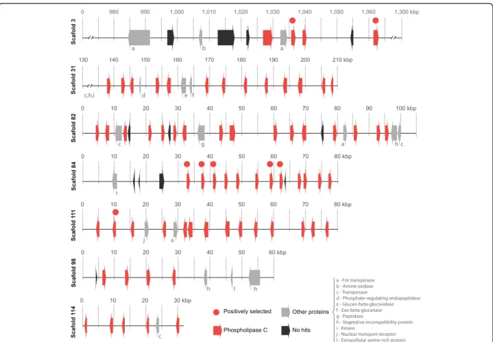
All of the 82 PI-PLC genes of C. cacaofunesta were found to be distributed throughout 15 scaffolds. However, 79% of these genes (65) were found in clusters larger than 4 genes concentrated in just 6 assembled scaffolds (31, 82, 84, 98, 111, 114) ranging from 31 kb to 210 kb (Fig. 6). It was observed that the PI-PLC genes that are located close together within a scaffold encode proteins of similar sizes and have the same pattern of orientation. PI-PLCs clusters are composed mainly of PI-PLCs interleaved by large regions with no predicted genes but with many traces of broken transposases, identified from blast searches of these intergenic regions against the NR-NCBI database. Scaffolds containing clusters of PI-PLCs featured high densities of transposable elements (TEs), with the terminal inverted repeats (TIRs) being the most abundant class (Additional file 9). Analyses of TEs content and classifica-tion overC. cacaofunestagenome showed that TIRs were the third most abundant type of TEs (18%), following long terminal repeats (LTRs), retrotransposons (27%) and“ un-known”type (25%). See Additional file 9 for complete TEs analysis.
About 40 other coding genes are distributed throughout these clusters (Fig. 6 and Additional file 10). Interestingly, Table 3Major expanded and retracted gene families in C. cacaofunesta genome
Families Ceratocystis
size family
Ceratocystisancestor size family
Difference Family Annotation
Family_230 76 1 75 Phosphatidylinositol-specific phospholipase c
Family_5 100 41 59 Vegetative incompatibility protein
Family_86 58 4 54 FOT5 transposase
Family_187 33 2 31 GAG-POL Protein
Family_50 38 7 31 GAG-POL Protein–retrotransposon
Family_167 28 4 24 Serine/threonine-protein kinase Sgk2
Family_130 25 4 21 Protein Kinase
Family_45 26 10 16 Kinesin Light Chain - Acyl transferase/acyl lysophospholipase
Family_442 16 1 15 MULE transposase domain
Family_349 15 2 13 Acyl transferase/acyl hydrolase/lysophospholipase
Family_23 3 16 −13 Major facilitator superfamily
Family_12 13 27 −14 ABC transporter
Family_11 8 23 −15 Phosphoethanolamine n-methyltransferase
Family_10 3 19 −16 Heterokaryon incompatibility
Family_13 8 25 −17 MFS general substrate transporter–Multidrug transporter
Family_15 2 19 −17 Heterokaryon incompatibility
Family_17 3 23 −20 Beta-ketoacyl synthase
Family_9 8 29 −21 Major facilitator superfamily
Family_8 12 34 −22 Major facilitator superfamily
the genes encoding proteins are related to noncellulolytic β-glucans hydrolysis (glucan 1,3-β-glucosidase and exocelular-β-1,3 glucanase); protein phosphorylation (kin-ase protein and serine/threonine kin(kin-ase protein); or returned as “No-hits”. No difference in GC content was observed in PI-PLC-rich regions in comparison to the rest of the genome (Additional file 10).
All 6 scaffolds shown in Fig. 6 are highly similar due to the conserved domains of the PI-PLC genes and the presence of other tandem repeats, such as TIRs. It must therefore be considered the strong possibility that the
genome assembler broke a single large genome region into different scaffolds because of the similarity and repeatability. This would mean that the PI-PLC gene clusters could be part of a single larger DNA fragment in C. cacaofunesta genome, with at least 560 kb. This region could actually be much longer, considering that other scaffolds contain a few PI-PLC genes. The same clustering pattern of PI-PLC genes was observed for otherCeratocystisspecies, but in many smaller scaffolds due to higher fragmentation of assemblies, suggesting that it could be an ancestral feature.
Fig. 5Evolution of PI-PLCs genes inCeratocystisspecies and the close relativeH. moniliformis. Numbers in the red dots displays the quantity of genes related to PI-PLC estimated using birth-death models by BadiRATE for each node
PI-PLCs gene family evolution
Because the expansion of PI-PLCs inCeratocystis species occurred in the genus ancestor and continued in the spe-cies’radiations and current lineages, we analyzed the evo-lution of this gene family inC. cacaofunestaand the other four members of theCeratocystisgenus. The phylogenetic hypothesis forCeratocystisPI-PLCs predicts that at least 2 protein clusters are composed by ortholog genes in the
Ceratocystisgenus (Fig. 7). The phylogeny is characterized
by one cluster of putative deep paralogs among all species that present a common ancestor before the divergence of
H. moniliformis and Ceratocystis, and a related star-like tree formed by 14 clusters of proteins distributed along
Ceratocystisspecies (Fig. 7; the complete phylogeny of the PLC gene family is shown in Additional file 11). PI-PLCs genes of theCeratocystisspecies showing a common ancestor with H. moniliformis are possibly the ones that retained ancestral characteristics. These ancestral-like
PLCs are composed of one group of orthologs among all
Ceratocystis and H. moniliformis, and another group of orthologs among allCeratocystiswith a single duplication inH. moniliformis. There is no evidence for the presence of signal peptides in the ancestral-like PI-PLC and a H. moniliformis PI-PLC proteins (s2.351), although a trans-membrane domain exists in one of them (Fig. 7).
The other 14 clusters of PI-PLCs are composed of pro-teins that have undergone duplications throughout the evolution of theCeratocystisgenus. The ortholog proteins among the fourCeratocystisstudied are organized into 42 derivate clusters (yellow dots in Fig. 7) and are distributed among many clusters of paralogs that duplicate within one or more species. Paralogs closely related in the tree are positioned in the same scaffold and often next to each other within at scaffold. Evidence of signal peptides was found in many of the derivate PI-PLC clusters (Fig. 7).
Models of evolution by positive selection were tested against models of neutral evolution using dN/dS likeli-hood ratios in codeml. In this analysis, 58 PI-PLCs from the Ceratocystisgenus showed significant values, indicat-ing that they might have evolved under positive selection (Fig. 7; Additional file 11). Among the putative positively selected proteins, some are the ancestral-like PI-PLC with no signal peptide, and many others are recently duplicated proteins with signal peptide, indicating that these newer proteins may be secreted (Fig. 7; Additional file 11).
Prediction of structure and homology modeling of CeratocystisPI-PLCs
An integrated approach including gene annotation, homologue identification, and comparative modeling was employed to construct a putative and reliable structural model of the product encoded by the gene s111.3 from the expanded gene family ofC. cacaofunesta. The PI-PLC encoded by the gene s111.3 was chosen for having been identified in both the predicted and experimental secre-tomes. The phosphatidylinositol-specific phospholipase C (PI-PLC, pfam ID PF00388.14) function of gene s111.3 product was predicted using the Web CD-Search Tool. The search indicated two well-conserved catalytic histi-dine residues. These histihisti-dine residues cleave the bond be-fore the phosphate, converting phosphatidylinositol into inositol and a lipid in the phosphodiesterase reaction. These data strongly suggested that the product encoded by gene s111.3 is a phospholipase C protein. Hence, the structural model of putative phospholipase C protein from
C. cacaofunestawas built using secondary structure opti-mized alignment.
A search for similar proteins was performed with the BLASTp algorithm. The search identified 31 proteins with available 3D structure related to phosphatidylinositol-specific phospholipase C. The template was selected using the following criteria: sequence identity, query cover
quality, resolution of crystallographic structure, presence of ligands and protein class. According to these features, phosphatidylinositol-specific phospholipase-C fromListeria monocytogenes(Protein Data Bank ID: 1AOD) was selected as the best candidate.
The best model comprised 332 residues and showed an RMSD value of 1.4 Å over Cαfor 369 equivalent residues of the L. monocytogenes putative homologue. The C. cacaofunestaPI-PLC model displays a typical TIM-barrel domain ((β/α)8-barrel) where the active site is located at the C-terminal end of theβ-barrel. InC. cacaofunestathe enzyme forms a deep cleft that is lined mainly by polar and charged amino acid side-chains. The PI-PLC model from C. cacaofunestaindicated that the active site shares 85% sequence identity with the template. The active site included amino acid residues His58, Asn59, Thr78, Asp93, Thr95, Asn107, Arg138, Gln140, Asp199, Ser227, His257, Thr259 and Ser261 (Fig. 8a). Next, we employed molecu-lar docking to verify whether the PI-PLC model from C. cacaofunestawould recognize inositol as substrate (Fig. 8b) [45]. The modeled binding modes indicate favorable polar interactions between inositol and active site residues (His58, Thr95, Arg138, Gln140 and Ser227) of the putative model. Moreover, the inositol binding mode to the PI-PLC model fromC. cacaofunestais very similar to the ex-perimental binding mode observed in the binary complex of PI-PLC fromL. monocytogenes[46]. Taken together, the presence of a predicted phosphoinositol binding domain, the conservation of catalytic site residues and the pre-dicted ability to recognize inositol as substrate suggest that the protein encoded by gene s111.3 is a phosphoinositol specific phospholipase C [47, 48].
Finally, In order to gain some insight into the conserva-tion of funcconserva-tion within the expanded PI-PLC gene family, we compared the sequence of the gene s111.3 with the other 75 PI-PLC-encoding genes present in the C. cacaofunesta
genome. The analysis generated a consensus sequence align-ment which indicates the most common residues at a given position (Additional file 12). According to this alignment, the catalytic residues are well-conserved throughout the gene family, suggesting a common function. Subsequently, we used the CD-Search Tool Web server to search for con-served domains in the 75 genes [49, 50]. In all cases, the PI-PLC catalytic domain was found with a high degree of confi-dence [51]. Therefore, our data strongly suggest that the analyzed genes from C. cacaofunestagenome encode pro-teins that can bind phosphatidylinositol molecules.
Discussion
C. cacaofunestawith focus on the identification of patho-genicity genes which might facilitate plant colonization and provide resistance against plant defense responses. More specifically, we describe the expansion and evolution of the PI-PLC protein family inC. cacaofunestagenome, as well as in the other three Ceratocystis genomes published to date [42–44]. We provide evidence that the expansion of this gene family correlates with the evolution of pathogen-icity in theCeratocystisgenus.
C. cacaofunestagenome,as well as the otherCeratocystis
genomes published so far, has a small size and low gene content when compared to other filamentous fungi (Fig. 1) [42–44]. The genome size reduction is proportional to the decrease in gene content for all species studied. This is also true, forHuntiella moniliformis,a related speciesthatalso belongs to Ceratocystidaceae family. Evolution analysis of gene families showed that Ceratocystis has the highest number of extinct proteins among the compared genomes. In addition, the Ceratocystis species have similar genome features and seem to share similar genetic content. Genome functional annotation and orthology results revealed high homology within the proteomes ofC. cacaofunestaandC. fimbriata (Additional file 4). These similarities are in accordance with the short evolutionary distance among these species. A marked phenotypic difference between these species is that C. cacaofunesta,unlike C. fimbriata,
has host specificity [1, 5]. Accordingly, slight differences between the proteomes of the two species were observed (Additional file 2) suggesting that this specialization might be due to some minor genetic variation. It has been described that in species with high genome similarity, host
specificity might be related to variations in a single locus or in clusters of closely linked loci [54].
Interestingly, differences in TEs content between these species were observed. Our results show a five-fold expan-sion of TEs in the C. cacaofunesta genome compared to the C. fimbriata genome, suggesting that TE expansions may play a significant role in the structure, adaptation, and evolution of theC. cacaofunestagenome (Additional file 9). TE expansion may be involved in gene duplication, and gene loss inactivation [55, 56], all of which have worked to increase the genetic variability between the two species. Similar expansions have been reported in other Ascomy-cota fungal pathogens, such as M. graminicola [57], and they appear to accelerate the evolution of genes related to pathogenicity and host range [58, 59]. Genome func-tional annotation for these two Ceratocystis species revealed pathways related to thiamine and biotin metabolism (Additional file 2). These vitamins have been previously reported as being essential for the sexual reproduction of C. fimbriata [60]. Also, important path-ways related to the biosynthesis of various terpenes and volatile compounds were found. The production of these compounds with fruity aromas has been reported for severalCeratocystisspecies [61].
In general, phytopathogenic fungi produce a variety of CAZymes during the plant colonization process. These enzymes are associated with plant cell wall degradation and perform crucial roles in the plant-pathogen interaction, being considered virulence factors [62]. Our results showed that theCeratocystisspecies present the lowest numbers of CAZymes, when compared with other Sordariomycetes, Fig. 8a and bCeratocystisPI-PLC molecular model.a. (Left) Catalytic active region is shown in red interacted directly with the ligand inositol or phosphoinositol in Green. (Right) Detailed view of the active site of phospholipases C specific for phosphoinositol crystallographic (in cyan) and modeled (red). The detailed residues refer to the modeled protein. The mutations in Gln140 and Ser227 are conservative and capable of promoting interactions with the inositol molecule, necessary for the maintenance of phosphodiesterase activity.bDetailed view of the active site showed amino acid residues interact directly with the ligand. Amino acid residues of the template (Crystallographic resolve protein in PDB code 1AOD) are showed in cyan and PI-PLCCeratocystis
including non-pathogenic species (Fig. 2). This difference was found to be related to the reduced gene content in the genomes of the Ceratocystis species, a trait that may be conserved within the genus. Nevertheless, the number of CAZymes observed for the pathogenicCeratocystisspecies is still higher than that of their relative H. moniliformis, a saprotrophic species. Moreover, the composition of detected CAZyme families differs between the two genera. This would be expected given their different lifestyles. Many phy-topathogenic fungi have even more CAZymes than do saprotrophic fungi, which are excellent degraders of plant biomass [63]. Furthermore, when compared toH. monilifor-mis, the Ceratocystis species have a higher number of CAZymes involved in the degradation of living plant tissues, such as pectinases. These enzymes are required for the deg-radation of pectin, a major component found between cells of living plant tissues [64]. Ceratocystis species have also a high number of genes classified in the AA4 CAZyme family, which contains vanillyl-alcohol oxidases that catalyze the conversion of a wide range of phenolic compounds. Histo-logical studies involving Ceratocystis infection showed the production of these compounds via the plant mechanism of defense againstCeratocystispathogens [21].
Another interesting fact discovered in the analyses of CAZymes is that there is a large difference when compar-ing theCeratocystisspecies with the classical or true vas-cular wilt pathogens (TVPs) ofVerticilliumandFusarium.
Traditionally, theCeratocystisspecies have been classified as vascular wilt pathogens due to the wilting disease they cause through the impairment of xylem vessels. However, recent reports showed differences related to the infected tissue; while TVPs infect xylem vessels,Ceratocystisinfect xylem parenchyma [21, 22]. Also, someCeratocystis spe-cies are able to infest other plant organs, suggesting that they are not true vascular pathogens (NTVPs) [14, 65]. This difference implies divergent pathogenesis strategies among TVPs and NTVPs. Ours analysis presents some molecular evidence that supports these differences. Klos-terman and coworkers [23] identified the expansion of polysaccharide lyase (PL) in Verticillium species, which have an enhanced capacity to degrade plant pectins. Meanwhile,Ceratocystis have fewer pectin lyases and do not present PL11 homologs, which according to the authors were present only in the wilt pathogens [23]. Additionally,C. cacaofunesta(andC. fimbriata) predicted proteome does not contain homologous to the specific wilt proteins involved in the maintenance of osmotic stability and the adaptation of TVP to their ecological-niche [23] (Additional file 13).
Otherwise, important virulence factors were found in the secretomes of C. cacaofunesta and C. fimbriata(Table 2), including a unique cerato-platanin (CP), a known phytotoxic protein. CP induces necrosis in tobacco leaves [66] and it has also been proposed that it may allow the attachment of
hyphae to hydrophobic surfaces during the formation of aerial structures [67]. Interestingly, Ceratocystis species do not have genes encoding class I and II hydrophobins sug-gesting that other proteins, such as CP, could be involved in cellular adhesion.
Additionally, two genes encoding proteins similar to NEP1-like proteins (NLPs) were identified. NLPs are classi-fied as Type 1NLP when contain two cysteine residues in the primary sequence and as Type 2NLP when there are four cysteine-residues [68]. We identified that C. cacaofunesta
andC. fimbriatahave a copy of each type of NLP. The first identified NLP (NEP1) was isolated from aF. oxysporum cul-ture filtrate. This protein was capable of producing necrosis and ethylene induction in the leaves of dicotyledonous plants [69]. Nowadays, NLPs are accepted as cytotoxic proteins, even though their mechanism of action is not well under-stood [70].
Among the CAZyme families detected in the secretome ofC. cacaofunesta, GH5, GH11, GH16, GH43 and GH61 are related to deconstruction of cellulose and hemicelluloses (Additional file 5 and Additional file 6). Thus, we measured the cellulolytic activity on cultures ofC. cacaofunestaandC. fimbriataand compared it to that ofT. paradoxa, a sugar-cane pathogen known as a cellulase producer [71]. Our results showed thatC. cacaofunestaandC. fimbriatahave cellulase activities when grown in conditions which reflect the cellulose:hemicellulose composition of the plant cell wall (Additional file 5). We also identified a single ligninase, sug-gesting that lignolytic activity is limited in these fungi, as described forOphiostomaspecies [72].
Other important virulence factors required for plant colonization by fungi are effectors. An effector is defined as any secreted molecule that modulates the interaction be-tween the pathogen and its host [73]. TheC. cacaofunesta
genome has a great variety of proteins with effector-like characteristics. Within this category we found proteins such as the allergen Asp and cyanovirin, which can elicit plant responses, and also proteins possibly involved in resistance to host-generated oxidative stress. Interestingly, many pro-teins with effector-like characteristics found in C. cacaofu-nesta did not match published sequences suggesting that these might be interesting targets for further studies.
The phylogenetic relationship within the Ceratocystis
genus was also studied. The obtained results showed that this genus diverged very recently, when compared to other Sordariomycetes genera, such as Fusariumand
phylogenetic investigations of the Latin American Clade ofCeratocystis,C. plataniandC. cacaofunestawere pro-posed as individual species butC. manginecanswas con-sidered a lineage withinC. fimbriatacomplex, implying that C. fimbriata would be a host generalist pathogen [74, 75]. As for many others pathogen where host speci-ficity drives genetic drift and individualisation of species, such species delimitations are difficult issues.
Despite the short evolutionary time for divergence, signifi-cant genomic structural changes occurred in theCeratocystis
ancestral lineage. First, the genome of the Ceratocystisand
Huntiella genera underwent a large reduction in size and gene number. A large proportion of genes was lost across gene families, many of which were involved in transport, detoxification and oxidation/reduction processes. Curiously, these processes are considered important for fungal plant pathogens, as they may play a crucial role in counteracting the oxidative stress generated by the host plant [58]. Despite the observed reduction in gene content, some gene families have shown a remarkable expansion in theCeratocystis gen-ome. This is especially true for the PI-PLC gene family (Fig. 5). Further analysis into the expansion and retraction of gene families showed that both the Ceratocystisand theH. moniliformis ancestors presented a large number of new genes. However, only theCeratocystisancestor displayed sig-nificant expansion of the PI-PLC gene family. The initial duplication of a PI-PLC enzyme, potentially surrounded by transposons, began a process of extensive transpositions in the genome, increasing the PI-PLC copy number in a short period of time. This expansion continued in the resulting
Ceratocystislineages (Fig. 5). All the transposition events and PI-PLC duplications created genomic clusters that might ac-tually be connected in a large single fragment or even a chromosome. This large sequence was divided into different scaffolds by assembly issues with repetitive regions (Fig. 6).
The rapid expansion of the PI-PLC gene family is also indicated by the star-like phylogenetic relationships among 14 PI-PLC clades, contrasting with the well-defined branching pattern from those of Huntiella and those that diverged within the 14 clades. Many PI-PLC genes that are closely related in gene phylogeny are also positioned near each other in genomic clusters, suggest-ing an evolution by duplications in tandem (Fig. 7). Pathogenic filamentous fungi present effector proteins that evolved on genomic regions that are not required for saprotrophic growth. These regions are called condi-tionally dispensable chromosomes (CDCs), in contrast to chromosomes whose gene content is essential and conserved across species [22]. CDCs have been associated to pathogenicity and host-range delineation in Lepto-sphaeria, Alternaria, and Fusarium species [76–78]. We are now working on obtaining the chromosomal map of
C. cacaofunestain order to analyze the possibility of the PI-PLC genes being located on CDCs.
Expansion of secreted PLC proteins is not a rare event in the genomes of fungal plant pathogens. For instances, the wilt pathogen Verticillium dahliae carries 19 genes encoding Patatin-like phospholipases which are likely involved in pathogen growth, lipid metabolism and sig-naling. Interestingly, 15 of these genes are located in V. dahliaelineage-specific regions (LS), which are enriched in repetitive sequences [23]. This PLC family expansion could be related to pathogenic properties required for the development of Verticillium wilt disease [23]. Fusarium oxysporumalso exhibits expansion of several protein fam-ilies related to lipid metabolism [22]. Specifically, eight PI-PLC encoding genes were described, all of which are located in LS regions [22]. Moreover, the entomopatho-genic fungus Metarhizium anisopliae presents 12 genes encoding secreted lipases, some of which are similar to PI-PLC and could be involve in pathogenicity [37]. Cerato-cystis species have a remarkable number of secreted PI-PLC genes, well above the average found in other fungi. Also, unlike the proteins from other fungi,Ceratocystis PI-PLCs show a significant similarity to the protein domain of pathogenic bacteria, which are known virulence factors. The extreme expansion in PI-PLC gene number and divergence in gene sequence, both in the ancestral genus of Ceratocystis and in current species, point toward a diversification process that might be adaptive for pathogen host infection. Considering thatHuntiellaspecies are not pathogenic, the genomic revolutions that expanded PI-PLC genes may have functioned as a pre-adaptation to the
Ceratocystis lineages that evolved as necrotrophic patho-gens. We obtained structural evidence that all of these newly duplicated proteins could be functional and that positive selection might be driving their evolution, with the accumulation of various non-synonymous substitu-tions in their sequences (Fig. 7). Pathogenicity-related pro-teins are expected to show high rates of non-synonymous substitutions. These changes are associated with the process of positive selection that drives the arms race estab-lished between the pathogen and its host [79]. Evidence of positive selection has been widely reported for plant patho-gens in virulence genes, especially for effectors [80].
Because the new non-synonymous substitutions diverge greatly among Ceratocystis that infect different hosts, we infer that different selective pressures may be acting on each lineage, possibly as a result of specific interactions between the pathogen and its host. Considering that C. manginecansmight be part ofC. fimbriataspecies complex [74, 75], differences in PI-PLC numbers and evidences of selection would not be a species-specific feature but a lineage specific one.
model, genes from the C. cacaofunesta PI-PLC family are likely able to bind phosphatidylinositol molecules. Our data indicates that these PI-PLC proteins have all the structural features to perform typical functions of bacterial PI-PLC: (i) catalyze the cleavage of phosphatidylinositol (PI) (or its phosphorylated derivatives) to produce DAG and the water-soluble head group phosphorylated myo-inositol [31]; and (ii) catalyze the release of proteins tethered to membranes by GPI-anchor proteins [81]. It is important to emphasize that PI-PLC acts on a substrate that does not occur in solu-tion, but is rather found in an aggregated state, as they are present in the cell membranes [82]. Therefore, PI-PLC from
C. cacaofunestamight be able to hydrolyze phospholipids in cell membranes, leading to their disruption or dysfunction, similarly to their bacterial homologues. Moreover, functional analysis showed that LmPI-PLC, unlike classical bacterial PI-PLCs, has a very low hydrolysis activity on GPI-anchored proteins [83]. This protein feature was associated to the lack of a small beta-strand (Vb), which it is presents in all bacter-ial PI-PLC [83]. Our analysis showed that Ceratocystis PI-PLCs contain the 8 amino acids region capable of forming the small beta-strand (Additional file 12). Therefore, Cerato-cystis PI-PLCs might also be able to release proteins an-chored to membranes by glycosylphosphatidylinositol (GPI). GPI-anchored proteins are ubiquitous and include enzymes, receptors, differentiation antigens and other bio-logically active proteins [84]. In fungi like Penicillium roqueforti, Paecilomyces variotii and Aspergillus niger, it has been found that GPI-anchored proteins are processed at the plasma membrane by a phosphatidylinositol-specific phospholipase C [85]. Many fungal cell wall pro-teins are covalently bound toβ-1,6-glucan via a remnant of a GPI anchor. These proteins are involved in important cellular processes, such as adhesion, invasion, biofilm for-mation, and flocculation [86–92]. In C. cacaofunesta, a total of 97 putative GPI-anchored proteins were identified. These were found to have similarly with various proteins groups, including chitin synthases, GH16, GH72, pepsin-like proteinases, Alpha-L-arabinofuranosidase, permeases and cysteine rich proteins (Additional file 14). Therefore,
C. cacaofunestaGPI-anchored proteins might be involved in fungal morphogenesis and invasion. On the other hand, GPI-anchored proteins in plants are involved in many processes [93]. For instance, Arabidopsis thaliana GPI-anchored proteins are related to cell wall deposition and remodeling, defense responses, and cell signaling [93].
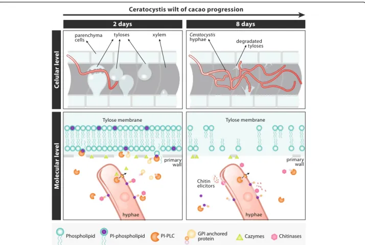
Based on the data obtained throughout this work and the available information of the histopathology of the C. cacaofunesta- cacao interaction, we suggest a model that would explain the possible roles of the PI-PLC family of proteins in the wilt disease context (Fig. 9). A histopatho-logical study of the interaction between C. cacaofunesta
and cacao showed plant responses toC. cacaofunesta in-fection in both resistant and susceptible plants [15]. Plant
responses included the discoloration of the primary walls of infected xylem vessels and the surrounding parenchyma cells; the mobilization of polyphenols in parenchyma cells, the translocation and accumulation of starch in the xylem and the production of gums and tyloses [15]. The main differences between genotypes were the intensities and kinetics of the appearance of those responses being more pronounced in the susceptible varieties [15]. In susceptible plants, the mycelium penetrated cells that were adjacent to the xylem and reached the xylem and parenchyma cells, overcoming structural resistance barriers such as tyloses. Tyloses are saclike structures that develop when turgor pressure causes parenchyma cell outgrowths through ves-sel parenchyma pit pairs into the lumen of xylem vesves-sels [94]. During tyloses formation, the primary wall compo-nent of the pit membrane is not pushed into the vessel. Instead, a fine protective layer containing pecto-cellulosic material is deposited between the protoplast and the pit membrane [94]. With regards to tyloses formation and
Ceratocystis wilt of cacao, Santos and coworkers (2013) observed that in susceptible plants, vessels containing the fungus were almost clear of tyloses. Nevertheless, adjacent vessels were completely occluded with tyloses formed in a matrix of gum 7 days after the inoculation [15]. Here, we hypothesize that PI-PLC could be involved in cellular dis-ruption and tyloses degradation aided by secreted proteins that degrade the cell wall. Also, PI-PLC might release pro-teins anchored to GPI, both in the fungal and in the plant cell walls, enhancing the process of tissue degradation and allowing the fungus to advance to regions more distant from the point of inoculation (Fig. 9). Collin & Parke (2008) suggested that Phytophthora ramorum produces enzymes able to degrade tyloses [95].C. cacaofunesta PI-PLCs would contribute to amplify both, the signals pro-duced by the pathogen and the host defense responses. We are currently gathering experimental data to support this model. In addition, necrosis in parenchyma cells may be assisted by other proteins reported in this work, such as different NEPs, cerato-platanin and the various types of CAZymes, which might assist in the degradation of the primary cell wall. All of the PI-PLCs hypothesized func-tions would contribute, together with the other secreted enzymes, to the development of wilt symptoms and the subsequent death of the host plant.
much greater than it is hypothesized here. Future investi-gations are aimed at elucidating experimentally the patho-genicity mechanisms related to PI-PLC.
The diverse arsenal of pathogenic genes present in C. cacaofunestaandC. fimbriatagenomes supports the idea that CWC is produced by the synergistic effect of toxin activity with parenchyma degradation and their fast growth and sporulation, with the consequent destruction of the plants vascular system, as reported in histopatho-logical studies [15]. All of these mechanisms might con-tribute to make ofCeratocystisspecies lethal pathogens.
Conclusions
Here, we presented the C. cacaofunesta whole-genome analysis. To our knowledge, this is the first work to report (i) the expansion of PI-PLC genes inC. cacaofunestaand other Ceratocystis species; (ii) PI-PLC genes distribution inCeratocystisgenome forming clusters in regions rich in TEs; (iii) their similarities with PI-PLC pathogenicity
crops, providing new perspectives for interference with plant-pathogen interactions.
Methods
Strain and nucleic acid isolation
The strain C1593 of Ceratocystis cacaofunesta used in this study was isolated from infected cacao trees of a cultivation located in the district of Uruçuca, Bahia, Brazil. The strain was generously donated by Dr. Tomas Harrington, from the Iowa State University Department of Plant Pathology. The methodology for nucleic acid isolation was previously described by Ambrosio and coworkers, 2013 [100].
Genome sequencing and sequence assembly
The genomic DNA was sequenced on a Genome Analyzer IIx platform (Illumina) at the University of North Carolina, Chapel Hill High-Throughput Sequencing Facility. A whole-genome shotgun strategy was used to produce 76-bp paired-end reads (400-bp insert size) and 50-bp mate-pair reads (3-kb insert size). The paired-end reads were assem-bled into longer contigs using Velvet version 1.0.12 [38], with an optimized k-mer size of 65. The pre-assemblies were used to construct scaffolds with the SSPACE program [101].
Gene prediction and annotation
The C. cacaofunestagenes were predicted by combining evidence retrieved from RNA-seq data and comparative gene finding. RNA-seq reads were obtained via in vitro laboratory experiments. RNA isolation, and RNAseq gen-eration protocols were previously described by Ambrosio and coworkers (2013) [100]. A second set of RNAseq data was generated in a parallel project for which a manuscript is in preparation. These data were used in this work to improve gene prediction. Mapping these reads into the assembled genome using STAR [102] produced splicing junction information, which were used in the self-training step of Genemark-ES [103] for the first prediction round. We used the BRAKER1 pipeline [104] with the splicing junctions’ information and the predictions from Genemark-ES to create a list of reliable genes. Next, we selected 1300 genes with Blastp [105] alignments (e-value cutoff of 1e−5) to proteins from close relative species obtained from a non-redundant (nr) database (NCBI), filtered by those with end-to-end alignment, gaps smaller than 15 bases, and at least 10 hits for each query. From the final list, 1000 genes were used as the training set in the gene predictor Augustus 3.1 [106], and the remaining 300 genes were used as the test set. We checked for the distribution of gene sizes in the training set to match the gene size distribution obtained from the first-round pre-diction of Genemark-ES. The prepre-dictions from Genemark and Augustus and also information from RNA-seq were combined by EVidenceModeler (EVM) [107], resulting in
the final prediction. For the otherCeratocystisspecies we analyzed, predictions were done with Augustus 3.1 using the training list of genes fromC. cacaofunesta.
The Autofact program [39] was used to perform auto-matic annotation of the C. cacaofunesta gene prediction. The main contribution of Autofact is its capacity to resume the annotation based on sequence similarity searches in several databases. For this, we used Blastp (e-value cut-off of 1e-5) to align the genes against protein databases, includ-ing the NR/NCBI, KEGG [108], UniRef90 [109], and CDD/ Pfam [110].
Protein general functional analyses
Protein classification of the C. cacaofunesta genome by gene ontology and the KEGG metabolic pathway was carried out using BLAST2GO tools [111].
Carbohydrate enzyme CAZy-families analysis
CAZymes content was predicted in 26 Sordariomycetes pro-teomes. Lists of all species and Data Bank access number of genome sequence are available in Additional file 15. The CAZyme analyses were performed using the HMMER3 and dbCAN HMMs databases, which are available online at the dbCAN homepage [112]. The amounts of CAZy enzymes in each CAZy category for each species were obtained from the dbCAN output.
Predicted secretome
Secreted proteins were identified by the prediction signal peptide/non-signal peptide using Signal P Version 4.1 [113]. The automatic annotation of the secretome was per-formed using the NR/NCBI, KEGG [108], and UniRef90 databases (BLASTp with an e-value cutoff of 1e-5) [114] and summarized using the AutoFACT program [39]. The CDD/Pfam database was used to identify the conserved domains. The Blast2GO program [111] was used to classify the gene ontology.
Secretome experimental identification using LC-MS/MS
from the liquid media by filtration through a paper filter (Whatmanno. 1) and centrifuged for 15 min at 2000 g.
The supernatants were concentrated 100-fold using (Vivaspin Concentration- GE) and quantified by Bradford assay. 295μL of protein from each sample was denatured, as described previously [116]. The resulting peptide solu-tion was dried in a SpeedVac concentrator and resuspended in 100μL of 0.1% formic acid. An aliquot of 4.5μL was sep-arated using C18 (75 μm × 100 mm) RP-nanoUPLC (nanoACQUITY, Waters) coupled with a Q-Tof Ultima mass spectrometer (Waters) with a nano-electrospray source. The flow rate was 600 nL/min, and the gradient was 2–90% acetonitrile in 0.1% formic acid over 45 min. The instrument was operated in the “top three” mode, in which one MS spectrum was acquired, followed by an MS/ MS analysis of the three most intense peaks [117].
The spectra were acquired using MassLynx v.4.1 soft-ware, and the raw data files were converted to a peak list format (mgf) using the Mascot Distiller v.2.3.2.0, 2009 soft-ware (Matrix Science Ltd.) without summing the scans, allowing for a label-free analysis. The files were then searched against theC. cacaofunestadatabase (7321 entries, 7269 proteins) using Mascot engine v.2.3.01 (Matrix Science Ltd.). Carbamidomethylation was used as a fixed modification, along with methionine oxidation as a variable modification, one missed trypsin cleavage, and a tolerance of 0.1 Da for both precursor and fragment ions. For the protein quantitation, the .dat files from the Mascot output were analyzed using Scaffold Q+ (version 3_00_03, Prote-ome Software), and quantitative values (normalized spectral counts) were obtained [118, 119]. For the endogenous pep-tide identification, methionine oxidation was set as a vari-able modification, with a tolerance of 0.1 Da for both the precursor and fragment ions. Only peptides with a mini-mum of 5 amino acid residues and statistical significance (p< 0.05) based on the Mascot scores were considered in the results.
Identification of potential effectors
Potential effectors were sought using the following parame-ters: small secreted proteins with cysteine-rich residues that are less than 200 amino acids in length and contain at least 4% cysteine residues, according to Klosterman et al. (2011) [23]. Additionally, GPI (glycosylphosphatidylinositol)-an-chored proteins were identified by the GPI-anchor attach-ment signal among the predicted secreted proteins using http://gpcr.biocomp.unibo.it/predgpi/ [120].
Comparative phylogenomics
Comparative analysis were carried out at two different scales: at a large scale, comparing C. cacaofunesta and
C. fimbriata to 21 other species from the Sordariomy-cetes class to identify Ceratocystis novelties; and at a small scale, comparing C. cacaofunesta to four other
species from the Ceratocystis genus to understand the evolution of novelties within the genus. List of all species and Data Bank access number of genome sequences are available in Additional file 15.
Genome sequences
In the large-scale analysis, we used the genome sequences of 23 Sordariomycetesspeciesavailable from the JGI (Joint Genome Institute) database. Accessions codes and refer-ences are available in Additional file 15. In the small-scale analysis, we used the genome sequences of 3Ceratocystis spe-cies (Table 1)—C. manginecans[GenBank: JJRZ01000000];C. fimbriata[GenBank: APWK00000000], andC. platani [Gen-Bank: LBBL01000012.1]—which are available from the Gen-Bank database of the National Center for Biotechnology Information (NCBI) (http://www.ncbi.nlm.nih.gov). The H. moniliformis data are available in the accession number JMSH00000000. The genome ofC. cacaofunestastrain 1593 was generated in the present study (see below) and has been deposited in DDBJ/EMBL/GenBank under the accession code PEJQ00000000.
Assignment of orthologous coding regions
The phylogenomic analyses assume that the sequences analyzed between species are homologs. The first step in predicting homologous regions was to cluster genes from all genomes of interest using a Markov clustering algorithm implemented in OrthoMCL [121], comprising possible orthologs and paralogs according to their func-tional groups. Then, all groups composed of only one gene for each species were isolated.
Sequences of multiple global alignments
Sequences within each orthologous gene families were aligned using the MAFFT 7 software [122] with iterative refinement methods using the weighted sum-of-pair (WSP) and consistency scores (G-INS-i), which implement a pipe-line combining the WSP score [123] and the COFFEE-like score [124] to evaluate the consistency between a multiple alignment and pairwise alignments [122]. We used the hmmalign tool of HHMER [125] to analyze the PI-PLC fam-ily, which is composed of much diversified proteins. This aligner uses a hidden Markov model (HMM) profile, ob-tained from a previous alignment of the protein family of interest, to guide the alignment of the domain regions. We downloaded the hmm profile for phospholipase-C in Pfam 27.0 protein database and used the HMMER hmmalign tool.
Expansion and retraction of gene families
process to model gene gain and loss across a phylogenetic tree, which we reconstructed using methods described below. We used the birth and death model in a maximum likelihood statistical approach to infer ancestral states for all tree nodes and calculate ap-value. The distribution of family sizes generated under this model provided a basis for assessing the significance of the observed family size differences among taxa.
Phylogenomic reconstruction
To reconstruct a phylogenetic hypothesis for Sordario-mycetes species that presents well-defined branch lengths, we used maximum likelihood methods, mented in RAxML [127], and Bayesian methods, imple-mented in MrBayes 3.2 [128], with the preselection of the nucleotide substitution model that best fits the mito-chondrial sequences used and keeping length parameter branches without restriction and relaxed relative to the molecular clock. The models were selected using Akaike’s (1974) criteria, as implemented in jModelTest2 [129]. Phylogenetic analysis for Ceratocystis species was carried out using a Bayesian analysis, as described below, involving gene beta-tubulin, which was successfully used in a previous broad phylogeny of Ceratocystis and related genera [5]. For themaximum likelihoodanalysis, the branch support was estimated using 1000 non-parametric bootstrap repetitions. The resulting tree was visualized and formatted using Figtree 1.3.1 [130]. For the Bayesian analysis, we used two independent rounds of Metropolis-coupled Markov chain Monte Carlo (MCMCMC), with a cold chain and three hot, each ana-lyzed by 1000,000 generations and sampled every 100 generations. The convergence of the chains was deter-mined by inspection through the TRACER 1.5 program [131]. The heating parameters of the chains and the number of generations could be adjusted over the ana-lysis. The list of Sordariomycetes and their genome data bank access numbers are in Additional file 15.
The phylogeny of the PI-PLC gene family was recon-structed using just the maximum likelihood method because the large amount individual genes requires a high computational processing time in Bayesian analysis. The protein alignments guided by HMM profiles had their highly variable ends trimmed using TrimAL [131]. The model of amino acid substitution that fits the data was selected using ProTest [129].
Evidence for positive selection inPI-PLC
In order to estimate the effect of natural selection on PI-PLC genes, the dN/dS ratio was estimated by maximum likelihood using the codon-based model of Goldman and Yang (1994), which was implemented using the codeml program in the PAML 4 package [132]. The divergence among Ceratocystis species from their most recent
common ancestor was considered to be proportional to the branch sizes in the phylogenetic reconstruction. The enrichment of higher dN/dS rates was tested statistically using the hypergeometric distribution.
Annotation and search for RIP-like signatures of transposable elements
TEs were identified and annotated from the genome of the fungiC. cacaofunesta; also, TEs were identified fromC. fim-briataCBS 114723 (Bioproject PRJNA67151) andM. ory-zae(Accession AACU00000000) using the REPET pipeline (http://urgi.versailles.inra.fr/index.php/urgi/Tools/REPET), which was optimized to better annotate nested and frag-mented TEs. Repeats were searched with BLASTER [133] for an all-by-all BLASTn genome comparison, clustered with Grouper, RECON, and PILER, and consensuses were built with the MAP Multiple Sequence Alignment program. The consensuses were classified with BLASTER 50 matches, using tBLASTx and BLASTx against the Repbase Update databank, and by identification of structural features such as long terminal repeats and terminal inverted repeats. The \resulting consensuses were used as input for the REPET annotation pipeline part, comprising the TE detection software BLASTER, RepeatMasker, and Censor [133–135]. Localizations of TEs were extracted from the gff3 files, and Blastn was used to find the number and size of TEs in theC. cacaofunestagenome. The TE consensuses and their annotated TE copies of the TIR transposon B120, satisfying strict quality criteria (longer than 400 bp in length and at least 80% identical), were aligned using clustalX [136]. This alignment was used for automated analysis of RIP inC. cacaofunestaTEs and for estimating dinucleotides using RIPCAL (http://www.sourceforge.net/projects/ripcal) [137]. RIPCAL output provides the numbers of transitions (Ti), transversions (Tv), and dinucleotide targets used in all possible transitions for each TE copy.
Prediction of structure and homology modeling of C. cacaofunesta PI-PLCs
The experimental data available in Web CD-Search Tool [138–140] was used to annotate function and retrieve domain information for the s111.3 of the expanded PI-PLC gene family ofC. cacaofunesta. Next, the search for homologous protein with available structural data was performed using BLASTp. Tertiary structure prediction of the protein encoded by gene s111.3 was performed by ViTaMIn4 program using Modeller’s spatial restraints algorithm [141–144]. The homolog phosphatidylinositol-specific phospholipase C proteins from Listeria
monocy-togenes (PDB ID, 1AOD) was used as templates. The