FATE1: CONTRIBUTOR TO TUMOR CELL FITNESS AND EXAMPLE OF AN ONCOGENE-ACTIVATED CANCER TESTIS ANTIGEN
Patrick J. Taus
A dissertation submitted to the faculty of the University of North Carolina at Chapel Hill in partial fulfillment of the requirements for the degree of Doctorate of Philosophy in the Department of Pharmacology, School of Medicine.
Chapel Hill 2016
Approved by:
Angelique Whitehurst John Sondek
Ian Davis
ABSTRACT
PATRICK J TAUS: FATE1: Contributor to Tumor Cell Fitness and Example of an Oncogene-activated Cancer Testis Antigen
(Under the direction of Angelique Whitehurst)
E3-ligase. We found further in vitro and clinical data that supports the hypothesis that FATE1 and RNF183 form a functionally relevant complex within tumors.
ACKNOWLEDGEMENTS
First and foremost I would like to thank my mentor, Dr. Angelique Whitehurst. Angelique has always been supportive of my current work and my future career as a physician-scientist. While writing this thesis I have been reminded of many things Angelique taught me, and I’m sure that will continue throughout my days as a scientist.
I would like to thank my committee including my chair, Dr. John Sondek, and members Dr. Ian Davis, Dr. Mohanish Deshmukh, and Dr. Lee Graves for the critical analysis they have given my project and their helpful insights at my committee meetings. I would also like to specifically thank Dr. Davis for serving as my clinical mentor while I was at UNC and Dr. Deshmukh for co-sponsoring my F30 grant application and helping to solve the administrative nightmare it became.
At UT-Southwestern I would like to thank Dr. James Amatruda for not only allowing me to round with him on the pediatric oncology service but also for his advice on the Ewing sarcoma chapter of my thesis. I would also like to thank Eunice Webb and Kathy Mercer for all their work keeping the department and lab running smoothly.
Macion, all of whom have made the lab a wonderful environment over the last two and a half years.
TABLE OF CONTENTS
LIST OF TABLES ... ix
LIST OF FIGURES ...x
LIST OF ABBREVIATIONS AND SYMBOLS ... xi
CHAPTER I: Introduction ...1
THE SEARCH FOR TUMOR ANTIGENS ...1
DISCOVERY OF CANCER TESTIS ANTIGENS ...2
CLASSIFICATION OF CTAS ...3
IMMUNOGENICITY OF CTA ANTIGENS ...5
ACTIVATION OF CANCER TESTES ANTIGENS ...7
FUNCTION OF CTAS WITHIN SPERMATOGENESIS...8
CTA-BASED THERAPEUTIC STRATEGIES ...9
FUNCTIONAL CONTRIBUTIONS OF CTAS TO TUMORIGENESIS ...11
SUMMARY OF DISSERTATION ...12
CHAPTER II: Materials and Methods ...16
CHAPTER III: FATE1 is a mitochondrial CTA that supports tumor cell viability ...26
INTRODUCTION ...26
RESULTS ...27
DISCUSSION ...31
CHAPTER IV: FATE1 modulates programmed cell death within tumor cells ...48
INTRODUCTION ...48
DISCUSSION ...54
CHAPTER V: EWS/FLI1 drives expression of multiple functional Cancer Testis Antigens ...66
INTRODUCTION ...66
RESULTS ...70
DISCUSSION ...75
CHAPTER VI: Conclusions and Future Directions ...87
SUMMARY ...87
FUTURE DIRECTIONS ...88
LIST OF TABLES
LIST OF FIGURES
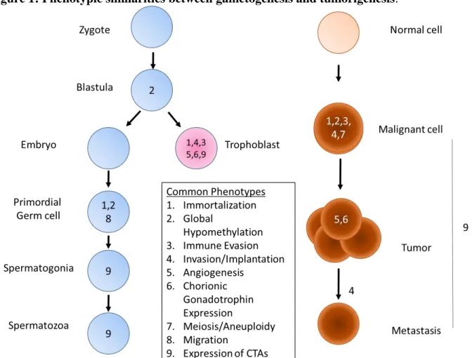
Figure 1: Phenotypic similarities between gametogenesis and tumorigenesis. ... 14
Figure 2: Cancer Testis Antigen presentation to cytotoxic T cells ... 15
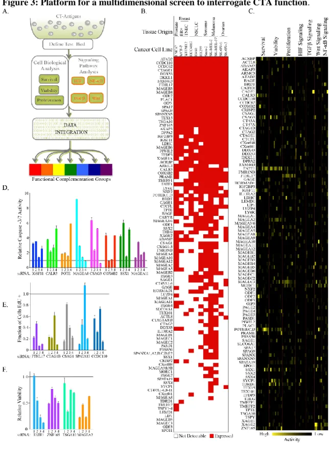
Figure 3: Platform for a multidimensional screen to interrogate CTA function. ... 36
Figure 4: FATE1 supports tumor cell viability ... 38
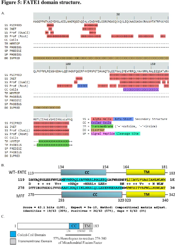
Figure 5: FATE1 domain structure. ... 40
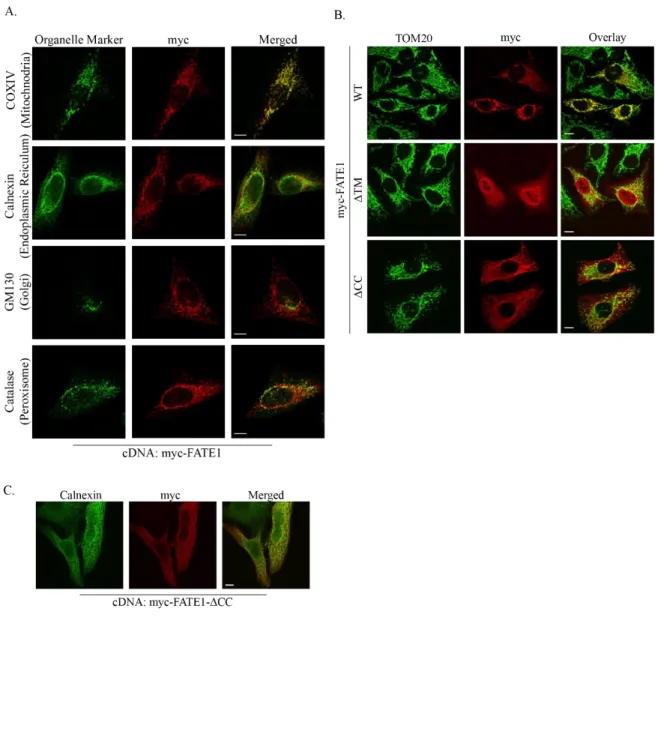
Figure 6: FATE1 is a mitochondrial protein. ... 42
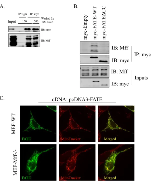
Figure 7: FATE1 interacts with Mitochondrial Fission Factor. ... 44
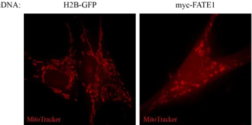
Figure 8: FATE1 alters mitochondrial morphology. ... 46
Figure 9: Overview of Bcl-2 family... 56
Figure 10: Loss of FATE1 induces programmed cell death. ... 57
Figure 11: FATE1 depletion engages the activity of Bcl-2 protein family. ... 58
Figure 12: FATE1 regulates BIK protein levels. ... 60
Figure 13: FATE1 interactor RNF183 possesses E3-ligase activity. ... 62
Figure 14: RNF183 impacts the stability of the apoptotic effector BIK. ... 64
Figure 15: FATE1 is a target of the oncogenic transcription factor EWS-FLI1. ... 78
Figure 16: EWS-FLI1 drives expression of FATE1. ... 80
Figure 17: FATE1 supports Ewing sarcoma cell viability... 82
Figure 18: EWS-FLI1 regulates a cohort of CTAs. ... 84
LIST OF ABBREVIATIONS AND SYMBOLS
Abbreviation/Symbol Definition
Δ Deletion
5-aza 5-aza-2'-deoxycytidine AIRE Autoimmune Regulator
AMPK Adenosine monophosphate-activated protein kinase APO Apo-ONE Homogenous Caspase-3/7 Assay
ATAD2 ATPase Family, AAA Domain Containing 2
BAGE B Melanoma Antigen
BH Bcl-2 Homology Domain
BORIS Brother of the Regulator of Imprinted Sites
CC Coiled-coil
CD Cluster of Differentiation
cDNA Complementary DNA
COX6BII Cytochrome c oxidase subunit 6BII CTA Cancer Testis Antigen
CTCFL CCCTC-Binding Factor Like
CTRL Control
EdU 5-ethynyl-2′-deoxyuridine ETS E26 transformation-specific EWS EWS RNA-Binding Protein 1
FATE1 Fetal and Adult Testis Expressed 1 FLI1 Friend Leukemia Virus Integration 1
GAGE G Antigen 1
HECT Homologous to the E6-APCarboxyl Terminus HIF Hypoxia Inducible Factor
HORMAD1 HORMA Domain Containing 1
HR Hazard Ratio
KM Kaplan-Meier
LDHC Lactate Dehydrogenase Isoform C
MAGE Melanoma Antigen
MARCH2 Membrane-Associated Ring Finger (C3HC4) 2 MARCH5 Membrane-Associated Ring Finger (C3HC4) 5
MFF Mitochondrial Fission Factor MHC Major Histocompatibility Complex mTEC Medullary Thymic Epithelial Cells
NF-κB
Nuclear Factor Kappa-Light-Chain-Enhancer of Activated B Cells
NSCLC Non-small Cell Lung Cancer
NUMA1 Nuclear Mitotic Apparatus Protein 1 NY-ESO-1 New York Esophageal Squamous Cell 1
PBMC Peripheral Blood Monocyte
SEREX Seriological Analysis of Expression cDNA Libraries siRNA Small Interfering RNA
SMAC Second Mitochondrial-derived Activator of Caspases SPATA19 Spermatogenesis Associated 19
SPO11 SPO11 Meiotic Protein Covalently Bound To DSB SSX Synovial Sarcoma, X Breakpoint
SYCE1 Synaptonemal Complex Central Element Protein 1 SYCP1 Synaptonemal Complex Protein 1
TCR T Cell Receptor
TET TLS, EWSR1, TAFII68
TEX15 Testis Expressed 15
TGFβ Transforming Growth Factor Beta
TM Transmembrane
TOM20 Translocase of Outer Membrane 20 TRIM Tripartitie Motif
Ub Ubiquitin
UPS Ubiquitin Proteasome System
CHAPTER I: INTRODUCTION
Similarities between cancers and germline tissues were first noted over one hundred years ago by British developmental biologist John Beard who proposed the Trophoblastic Theory of Cancer which proposed that cancer was the result of aberrantly localized trophoblast cells throughout that body [1]. Although the major tenets of this theory were disproven, numerous similarities between tumorigenesis and germ cell development were noted in the following century (Figure 1). In the last twenty-five years the similarities between tumors and germ cells have been demonstrated on a profound genetic level following the identification of genes highly expressed in those two entities. This group of genes, known collectively as Cancer Testis Antigens (CTAs), affords unique therapeutic opportunities through their immunologic potential and the potential novel insights they may functionally afford into tumor biology. As the work described herein focuses on Cancer Testis Antigens a review of their discovery, biology, and the current state of their therapeutic utility will be presented.
The Search for Tumor Antigens
Although dangerous, in many cases these injections led to shrinkage of the tumors presumably do to immune system activity. Later in the 20th century, a second line of evidence that tumors were vulnerable to immune destruction emerged. In 1943 Gross and colleagues found that mice in which tumors were chemically induced and then resected were able to reject the same tumor cells upon subsequent inoculation [2].
Several decades later in the 1970s the prominent role of T cells in tumor rejection was realized. This knowledge, coupled to the discovery of how to select and expand cytotoxic T lymphocyte clones in vitro would allowed for the identification of tumor antigens targeted by cytotoxic T cells through immunoscreening assays in the following decades [3-5]. In 1988 Boon and colleagues found that in vitro mutagenesis of a mouse tertacarcinoma cell line led to the expression of novel antigens that were the targets of cytotoxic T cells [2]. Using these T cell clones to screen cDNA libraries obtained from the vulnerable tumor cell clones the Boon group identified the first three tumor antigens, each being a mutated version of an ubiquitously expressed protein [6-8]. Subsequently, the first non-mutated antigen was identified using the parental cell line, this antigen was derived from a gene of unknown function, Trap1a [9]. The antigenicity of Trap1a’s protein product was due its restricted expression, the only normal tissues where it was detected were mouse spermatogonia cells and placental trophoblasts. Uniquely, both of these cell types lack major histocompatibility complex (MHC) class I molecules and therefore cannot present Trap1a-derived peptides to T cells [9].
Discovery of Cancer Testis Antigens
cells are screened against their own T cells to identify reactive T cell clones and the corresponding MHC Type I-loaded antigens they recognize. Using this method the Boon group discovered that expression of the gene MAGE-A1 rendered melanoma cells sensitive to destruction by autologous cytotoxic T-cells [10, 11]. It was subsequently discovered that among normal tissues the expression of MAGE-A1 was restricted to male germ cells and trophoblasts of the placenta [11]. Further analysis revealed that MAGE-A1 was a member of a large gene family consisting of over 40 proteins that contain a full or truncated MAGE homology domain [12]. The MAGE family can be divided into two categories: Group I MAGEs (-A, -B, -C) which are encoded on the X-chromosome and whose expression are restricted to the testis and Group II MAGEs (-D, -E, -F, -G, and –H) that are expressed throughout the body and are encoded for throughout the genome [12]. Autologous typing was used to identify additional CTAs including BAGE and GAGE which were identified using cytotoxic T-cells derived from the same patient in whom MAGE-A1 was discovered [13, 14]. In the mid-90s the identification of CTAs was further advanced by Serial analysis of cDNA expression libraries (SEREX), a technique in which cDNA expression libraries are screened with antibodies in lieu of T-cells [15]. Utilization of this technique led to the discovery of several CTAs including SSX, SCP1, and NY-ESO-1, the latter of which is a highly immunogenic CTA that is currently being targeted using multiple immunotherapy approaches [15-19].
Classification of CTAs
discovery, little is known about the immunogenic potential of many CTAs discovered solely through differential expression analysis. Large scale expression analysis also made clear that many CTAs are expressed at low but detectable levels in somatic tissues [20]. Based on these observations, the current criteria for designation as a CTA is as follows: a protein must be expressed within tumors, the testis and/or placenta, and no more than two somatic tissues [21]. CTAs can be further defined into three groups by the stringency of their expression profile: 1) testis restricted – found only in the testis, 2) testis-brain restrictive – expressed in both the testis and central nervous system, 3) testis selective – expressed in the testis and no more than two somatic tissues at lower levels than in the testis [20].
Immunogenicity of CTA antigens
Like all intracellular proteins CTAs are presented in MHC class I molecules on the cell surface, where, if recognized as foreign by circulating T cells, they can evoke an immune response (Figure 2). Initially, cancer testes antigens were discovered due to their antigenicity using autologous typing and SEREX techniques; however, with the recent reliance on gene expression analysis and large scale proteomic approaches to identify CTAs, the antigenicity of many newly identified CTAs is unknown. Which CTAs are immunogenic and why some CTAs can elicit varying degrees of immune responses amongst patients are critical questions whose answers will have significant repercussions on the use of CTAs as targets for immunotherapy. Here, the factors governing the antigenicity of CTAs and the current state of CTA-based immunotherapy will be discussed.
Another major contributor to the immune-privileged environment of the testis are Sertoli cells. Sertoli cells are somatic cells within the seminiferous tubule that support the development of male germ cells into spermatids [27]. Sertoli cells physically prevent infiltration of immune cells via specialized tight junctions that form the blood-testes barrier on the outside of the seminiferous tubules [28]. Although the blood-testes barrier is a significant barrier to immune cell infiltration into the seminiferous tubule it does not completely isolate the seminiferous tubule as ingress is possible through the straight tubules and the rete testis [25].
In addition to the physical barrier they create, Sertoli cell produce an immunosuppressive chemical environment within the testis. Multiple immunoregulatory cytokines are produced by Sertoli cells including transforming growth factor-β (TGF-β), activin A, FAS ligand, and inhibitors of both complement and granzyme B [25]. In addition to Sertoli cells, the Leydig cells of the seminiferous tubules also contribute to the immunosuppressive cytokine milieu through secretion of macrophage-migratory-inhibitor factor which can inhibit cell lysis mediated by cytotoxic T-cells and Natural Killer cells [29-31]. The resulting immunosignaling environment of the testes promotes a “type II” immunoregulatory, tolerant response in preference to “type I” cell-mediated immunity response [25].
Variability degree of tissue-specific gene expression within mTECs leading to varying levels of central tolerance has been reported for MART-1, a non-CTA melanoma antigen, and such variations may also contribute to the variation of CTA antigenicity [34].
Activation of Cancer Testes Antigens
The mechanism(s) by which CTAs are aberrantly activated within tumors is unclear but has major implications for their potential therapeutic exploitation. There is variation in CTA expression across tumor types. Some cancers such as bladder, non-small cell lung, and melanomas express numerous of CTAs, while others such as renal and colon express few [21]. CTAs also demonstrated a heterogeneous expression pattern within tumors themselves as immunohistochemistry has revealed focal expression patterns for several CTAs [24, 35]. Undoubtedly alterations in DNA methylation are major contributors to the aberrant expression of CTAs within tumors. Multiple studies have demonstrated activation of CTAs following treatment with DNA-Methyltransferase inhibitors such as 5-aza-2’-deoxycytidine (5-aza) [36, 37]. Interestingly, changes in DNA methylation may explain the focal nature of CTA expression within tumors as microdissection of ovarian tumors based on immunohistochemical staining for NY-ESO-1 demonstrated an inverse correlation between intratumoral NY-ESO-1 expression and DNA methylation [38]. Further epigenetic alterations such as histone post-translational modifications through inhibition of histone deacetylases or H3K9 methyltransferases, augment CTA expression following treatment with DNA methyltransferase inhibitors; however neither histone modification alone is sufficient to upregulate CTAs in these systems [39, 40].
studies have demonstrated differential activation of CTAs between tumor-derived and normal cell lines showing similar levels of demethylation [41]. Together these observations clearly demonstrate that mechanisms aside from DNA demethylation are required to activate specific CTAs. A few examples of such mechanisms have been previously described. For example expression of MAGE-A1 were found to be driven in large part by members of the ETS transcription factor family following promoter demethylation [42], while NY-ESO-1 activation is dependent on the binding of SP1 within its promoter [43]. Interestingly, SP1 is recruited to the NY-ESO-1 promoter by BORIS/CTCFL, which competes for NY-ESO-1 promoter occupancy with its paralog CTCF [43, 44]. BORIS, itself a CTA activated by hypomethylation, has been shown to activated a number of other CTAs in overexpression studies in both normal and transformed cellular contexts [45-49]; however, activation of BORIS/CTCFL alone is not sufficient to drive expression of CTAs in all cellular backgrounds [50, 51]. In total these data suggest the trans-acting factors are necessary and perhaps even sufficient to drive CTA-expression in the proper cellular backgrounds. Identification of such trans-acting factors may open therapeutic windows in the genetic backgrounds in which they are active.
Function of CTAs within spermatogenesis
The broad expression range of CTAs, throughout spermatogenesis from spermatogonia stem cells to implantation of the fertilized egg cell, as well as their genetic diversity suggests a vast array of functions for this group [22]. Numerous CTAs have been knockout in mouse models in efforts to characterize their roles in spermatogenesis. Many of these models develop normally aside from focal defects in male fertility, suggesting that while essential for
non-gametogenic tissues [22]. Not surprisingly, CTAs contribute significantly to the germ-cell specific processes of meiosis during the primary and secondary spermatocyte stages. The CTA SPO11 initiates homologous recombination during meiosis by introducing DNA double strand breaks through its transesterase activity [52]. Several CTAs: SYCE1, SYCP1, and HORMAD1, are members of the synaptonemal complex that ultimately resolves the DNA breaks via
homologous recombination; while another CTA, TEX15 is required for proper loading of repair proteins at the break sites [53-56].
Interestingly, several CTAs promote mitochondrial function, possibly to compensate for the high metabolic demands of spermatozoa motility [57]. These CTAs include COX6BII a sperm-specific isoform of a Complex IV subunit in the electron transport chain; LDHC, a sperm specific subunit of the lactate hydrogenase tetramer; and SPATA19, a mitochondrial adhesion protein that supports mitochondrial distribution, abundance, and respiratory capacity [58, 59]. Although examples have yet to be described, the functions of CTAs described above would potentially allow a tumor cell to counter increased burdens of DNA damage (Meiotic CTAs) or metabolic demand (Mitochondrial CTAs). As the functional spectrum of CTAs within the testis far exceeds the examples described here, their possible functional utility to tumors through isomorphic or neomorphic functions is immense.
CTA-based therapeutic strategies
results are encouraging as a recombinant NY-ESO-1 vaccine demonstrated a clinical benefit in patients with melanoma or ovarian cancer [60]. In addition to traditional vaccine approaches, recombinant NY-ESO-1 was used in the first study to elicit dendritic cell-induced T cell activity by fusing recombinant NY-ESO-1 to a monoclonal antibody specific for a dendritic cell receptor. In Phase I trials, this approach was well tolerated and six of eight patients who also received immune-checkpoint inhibitors had objective tumor regression [61]. In a murine model, vaccination with dendritic cells pre-loaded with a truncated form of BORIS inhibited tumor growth, metastasis, while increasing tumor infiltration by CD4 and CD8 T cells in a murine 4T1 mammary tumor implantation model, further demonstrating the potential use of CTA in non-traditional vaccination approaches [62].
Functional contributions of CTAs to tumorigenesis
To date the majority of research regrading CTAs focused on their potential roles as targets of immunotherapy and until the late 2000s functional studies into potential pro-tumorigenic roles for CTAs were few and far between. The first clues that CTAs may contribute to tumorigenesis include several studies around the turn of the century that showed overexpression of MAGE and GAGE genes contributed to tumor cell resistance to biological factors such as tumor-necrosis factor and FAS-ligand, chemotherapeutics such paclitaxel and doxorubicin, and γ-irradiation [67-69]. Further suggesting that CTAs functionally contribute to tumorigenesis were the results of an unbiased genome wide screen which identified a significantly enriched cohort of CTAs among genes that promote tumor cell resistance to the anti-mitotic chemotherapy drug paclitaxel [70]. Further studies demonstrated that one of the CTAs identified in that cohort, Acrosin Binding Protein, prevents disruption of microtubule dynamics and centrosomal clustering by negatively regulating levels of aberrantly expressed NUMA1 [71].
suppressor p53 leading to its degradation [74]. Further studies from the Potts lab showed that AMPK is degraded in a cancer-specific manner by a MAGE-A3/6-TRIM28 complex [75]. Although, several pro-tumorigenic roles for CTAs including the examples described here have been identified, no comprehensive approach has been taken to examine potential functional roles for the entire collection of CTAs.
Summary of Dissertation
EWS-FLI1, the oncogenic driver of the pediatric malignancy Ewing sarcoma. Using multiple cell viability assays we demonstrate that FATE1 is required for the short and long term viability of Ewing sarcoma cells. Additionally, by leveraging a previously published EWS-FLI1 ChIP dataset we nominate three additional CTAs: BORIS, MAGE-A4, and SPATA19, as potential EWS-FLI1 transcriptional targets, two of which, MAGE-A4 and SPATA19 are necessary for maintenance of Ewing sarcoma cell viability.
Figure 1: Phenotypic similarities between gametogenesis and tumorigenesis.
Figure 2: Cancer Testis Antigen presentation to cytotoxic T cells
CHAPTER II: MATERIALS AND METHODS
Cell lines. Cell lines were obtained from American Tissue Type Collection (ATCC) or John Minna (UT-Southwestern (UTSW)) except for: TC-32 (Children’s Oncology Group), SK-MEL-2 (National Cancer Institute); hMPro™ Mesenchymal Progenitor Cells (Aruna Biomedical); SK-MEL-37 and SK-OV-6 (Lloyd Old, Ludwig Institute); SUM159, SUM229, and SUM149 (Asterand); HuMEC (Charles Perou, University of North Carolina at Chapel Hill, (UNC)); HME50-hTERT, Fibroblasts (BJ) (Jerry Shay, UTSW); WHIM12 (Matthew Ellis, Baylor College of Medicine); ES-2 (Rolf Brekken, UTSW); PEO1 and U2OS (Michael White, UTSW); HEK293GP and HCC1806 (Gray Pearson, UTSW); RCC4 (William Kim, UNC); HCT116 (Cyrus Vaziri, UNC); HCT116-BAX-/-BAK-/- DKO (Bert Vogelstein, Johns Hopkins University); EWS502, EWS894, RD-ES, SK-ES-1, MHH-ES-1, A673, and SK-N-MC (Ian Davis, UNC). All cell lines were cultured in provider’s recommended medium. Because SK-OV-6 is on the ICLAC list of misidentified cell lines, Short Tandem Repeat (STR) profiling was used to validate the line used in this study [76, 77]. SK-OV-6 was used because it has previously been demonstrated to express a number of CTAs [78]. Cells were evaluated for mycoplasma contamination by DAPI stain for extra-nuclear DNA.
1:1000); Sigma: FATE1 (HPA034604; 1:2000), and RNF183 (SAB2106627; 1:1000); Cell Signaling Technology: Cleaved Caspase-3 (9661; 1:500), PARP1 (9532; 1:1000), and BCL-xL (2764; 1:5000); Abcam: FATE1 (ab111486; 1:1000); V5 (R960; 1:1000; Life Technologies); FLI1 (554266; 1:500; BD Biosciences). Antibodies for immunofluorescence were as follows: c-Myc (sc-40; 1:100; Santa Cruz Biotechnology), TOM20 (sc-11415; 1:500; Santa Cruz Biotechnology), GM130 (ab31561; 1:100; Abcam), Cytochrome c (556432; 1:200) and ; BD Biosciences), HA (MMS-101R; 1:100; Covance), Catalase (219010; 1:1000; Calbiochem), V5 (R960; 1:300; Life Technologies), Calnexin (ADI-SPA-860; 1:100; Enzo), COX IV (4850; 1:125; Cell Signaling Technology), and β-tubulin (T5293; 1:100; Sigma), and FLAG (F1804, Sigma).
Expression Plasmids and Mutagenesis. Full-length FATE1 cDNA in pRK5 (a gift from Michael White, UTSW), was subcloned into pCMV-myc (Clontech) and pcDNA3 (Thermo Fisher Scientific) between SalI and NotI, and EcoRI and NotI, respectively. Myc-FATE1 cDNA was from pCMV-myc-FATE1 was subcloned into pLPCX (Clontech) between BglII and NotI. Full-length RNF183 cDNA was obtained in pLX304 from the CCSB-Broad lentiviral ORF collection housed at UNC and cloned into pCMV-HA (Clontech) between SalI and NotI. Full-length BIK cDNA was obtained from pEGFP-BIK (Addgene plasmid #10952). HA-RNF183-C13A/C59A and BIK-L61G, which has limited toxicity as compared to wild-type[79], were generated using site-directed mutagenesis. BCL-xL cDNA (a gift from Mohanish Deshmukh, UNC) was cloned into pCMV-myc between SalI and NotI and then myc-BCL-xL cDNA was subcloned into pLPCX between BglII and NotI. eGFP cDNA (a gift from Michael White, UTSW) was subcloned into pLPCX between BglII and NotI. Viral packaging plasmids, pCMV-dr8.91 and pCMV-VSV-G, were a gift from Bill Hahn, Harvard). pRL-CMV (a gift from Deborah Chapman, University of Pittsburgh). EWS-FLII cDNA (a gift from Ian Davis, UNC) was cloned into pCMV-HA between SalI and NotI. 3xFLAG-tagged EWS-FLI1 cDNA in a Gateway Entry vector (a gift from James Amatruda, UTSW) was inserted into pLX304 via the Gateway LR clonase reaction.. pFATE1Luc was produced by amplifying a 1606 bp fragment -1634 to -29 bp upstream of FATE1 translational start site from HCT116 genomic DNA with KpnI and HindIII extensions. The insert was then ligated into pTA-Luc (Clontech) thereby removing minimal TA-promoter and placing the FATE1 promoter directly upstream of the luc
gene from Photinus pyralis. pFATE1ΔEF1-Luc was produced by amplifying the 1268 bps on the
siRNA screen and data processing. Transfection conditions for each cell line were optimized using the CTG assay and the formula: Transfection Efficiency = 1-(LuminescencesiUBB/LuminescencesiCTRL). A custom siGenome SMART pool siRNA library (Dharmacon/GE Healthcare Life Sciences) was purchased in 96-well plate format and resuspended as described [70]. siRNAs were diluted to 250 nM in serum free medium and 30 µL of this solution (8.3 pmoles of siRNA) was mixed in well with appropriate transfection reagents in 9.8 µL Opti-MEM® and incubated for 20 minutes. Then, 60 µL or 80 µL of cells in growth medium were added for cell biological and signaling assays respectively. Cell biological screens were performed 96 hours post plating using 20 µL CTG (ATP; viability), 90 uL APO (caspase-3/7 activity; survival), or the Click-iT® EdU Alexa Fluor® 488 Imaging Kit (DNA synthesis; proliferation) assay systems according to manufacturers’ protocols. CTG and APO assays were read with a Pherastar Plus or Pherastar FS (BMG Labtech) plate reader. siRNA pools with z-scores > 2 in the apoptosis screen or < -2 in the viability and proliferation screens were considered outliers. Only siRNA pools that exhibited statistically significant change (P ≤ 0.05 by unpaired Student’s t-test) were considered. For the HIF, NF-κB, Wnt, and TGFβ reporter screens, siRNA pools that reduced reporter activity by either > 60 % in a single cell line or > 30 % in more than 2 cell lines were considered positives. For the NF-κB, Wnt, and TGFβ signaling screens, both basal and ligand-induced values were considered.
transfected using FuGENE 6® or the calcium phosphate method[80]. All manufacturers’ protocols were followed.
Generation of stable cell lines. Cell lines stably expressing shRNAs were created via lentiviral-mediated gene transduction through co-transfection of HEK293T cells with viral expression and packing plasmids (pCMV-VSV-G and pCMV-dr8.91). For FATE1 studies, stable lines were created via retroviral-mediated gene transduction through co-transfection of HEK293GP cells with pLPCX expression plasmids and pCMV-VSV-G. Virus conditioned media was used to infect target cells in the presence of polybrene and stable populations were selected using appropriate antibiotics.
Luciferase assays. Indicated luciferase reporters (100 ng), Renilla reporter (pRL-CMV, 2 ng) and 100 ng indicated cDNAs were transfected into HEK293T using FuGENE 6®. Forty-eight hours later, luciferase activity was measured using the Dual-Glo® Luciferase Assay System.
Colony formation assay. At indicated incubation times following siRNA transfection, cells were replated at limiting dilution, fed twice a week and stained with Geimsa (Sigma).
Gene expression. RNA was isolated using or an RNA isolation kit (Sigma) and reverse transcribed using the High-Capacity cDNA Reverse Transfection Kit (Thermo Fisher Scientific) according to manufacturer’s instructions. An Applied Biosystems Real-Time PCR system and either Solaris™ (Dharmacon), SYBR® Green or TaqMan® Real-Time PCR (Thermo Fisher Scientific) gene expression assays were used. Gene expression assays were multiplexed with RPL27 as a control assays. Relative expression values were calculated using the comparative 2 -ΔΔCT
method [81].
Immunoblotting. Whole cell lysates were prepared in 2x Laemmli sample buffer and resolved using Sodium Dodecyl SulFATE1 Polyacrylamide Gel Electrophoresis (SDS-PAGE). Gels were transferred to Immobilon® PVDF (Millipore) or nitrocellulose (Bio-Rad Laboratories) membranes, blocked in tris-buffered saline containing 0.1 % Tween20 (TBST) and blocked in 5 % non-fat dry milk, bovine serum albumin (BSA), or Odyssey® (LI-COR Biosciences) blocking buffer followed by incubation with indicated primary antibodies for 1 hour or overnight. After washes in TBST, appropriate HRP-coupled secondary antibodies (Jackson Immunoresearch) or IRDye® antibodies (LI-COR Bioscience) were used for chemiluminescence or fluorescence detection (Odyssey®), respectively. Coomassie stain (Genlantis) was incubated with SDS-PAGE gels for 30 minutes followed by destain for 4 hours.
BSA, 0.1 % Tween-20 in 1X Phosphate Buffered Saline (PBS) (PBTA). Cells were incubated with primary antibodies for 1 hour followed by three washes in PBTA. Coverslips were then incubated with Alexa Fluor®-conjugated secondary antibodies (Thermo Fisher Scientific) for 30 minutes followed by 3 washes in PBTA and a wash in H2O. MitoTracker was added for 30 minutes prior to fixation. Prolong® Gold Antifade reagent with DAPI (Thermo Fisher Scientific) was used to mount slips on glass slides and images were acquired on either a Leica DM55000 B upright microscope or a Zeiss LSM510 confocal microscope.
Viability Assays. Cells were reverse transfected with RNAiMAX™ in Opti-MEM® with 50-100nM siRNA in 96 well format. 120 hours post-transfection (unless otherwise indicated), CTG was used to quantitate total ATP using a Pherastar Plus plate reader.
Immunoprecipitation. Unless otherwise indicated, cells where lysed on ice for 30 minutes in non-denaturing lysis buffer (NDLB): 50 mM HEPES pH 7.4, 1.0 % Triton X-100, 0.5 % Sodium Deoxycholate, 150 mM NaCl, 1 mM NaVO4, 25 mM β-Glycerophosphate, 1 mM EthyleneDiaminetetraacetic Acid (EDTA), 1 mM Ethylene Glycol Tetraacetic Acid (EGTA), plus protease-inhibitor cocktail (Sigma)). Lysates were clarified at 12,000g for 10 minutes. Then, 10% of each clarified lysate was set aside as an input loading control and the remainder was immunoprecipitated for 4 hours at 4 °C with antibodies coupled to Protein A/G beads. Unless otherwise indicated, beads were washed two times in high salt (350 mM NaCl) NDLB, once in NDLB, and then resuspended and boiled in 2X Laemmli sample buffer.
with pCMV-HA-RNF183 or pCMV-HA-RNF183-CC/AA. Twenty-four hours after transfection, cells were lysed on ice in non-denaturing lysis buffer (NDLB): 50 mM HEPES, pH = 8, 1.0 % Triton X-100, 0.5 % Deoxycholate, 150 mM NaCl, 1 mM NaVO4, 25 mM β-Glycerophosphate, 1 mM EDTA, 1 mM EGTA, plus protease-inhibitor cocktail, clarified at 12,000g for 10 minutes and then immunoprecipitated for 4 hours with anti-HA antibody (Covance) and Protein A/G beads (Life Technologies). Beads were then washed 3 times in NDLB with 350 mM NaCl and two times in ligase buffer: 50 mM Tris pH 7.5, 150 mM KCl, 1 mM MgCl2. After the final wash, beads were resuspended in ligase buffer containing 100 nM recombinant E1 (Enzo Life Sciences), 1 µM recombinant UbcH5b (Enzo Life Sciences), 5 µM ubiquitin from bovine erythrocytes (Sigma), plus or minus 5 mM Mg2+-ATP and incubated for 1.5 hours at 37 °C.
coomassie blue and those with GST-RNF183 bands were dialyzed overnight into protein storage buffer (50 mM Tris-HCl pH 7.7, 100 mM KCl, 10 % glycerol, 1 mM DTT).
In vitro ubiquitination assay. The Enzo® Ubiquitinylation kit was used to evaluate
GST-RNF183 for E3-ligase activity towards 6xHis-BIK (Abnova). Briefly, 100 nM ubiquitin
activating enzyme (E1), 100 nM ubiquitin conjugation enzyme UbcH5b (E2),1 mM recombinant GST-RNF183, 100 nM 6xHIS-BIK, and 5 mM bovine ubiquitin (Sigma) were incubated in 1 x Ligase Buffer (50 mM Tris-HCl pH 7.5, 150 mM KCl, 1 mM MgCl2, 1 mM DTT), with 5 mM Mg++-ATP. The reaction mixture was incubated at 37 °C for 90 minutes and then quenched by addition of 2x Laemelli buffer. Negative control reactions were performed in the absence of Mg++-ATP.
GREAT analysis. Genomic binding coordinated from Patel el al 2012 were converted to GRCh37 and subjected to GREAT 3.0.0 analysis using the basal plus extension gene regulator definition with the following parameters: Proximal- 5.0 kb upstream and 1.0 kb downstream, plus Distal up to 1000 kb.
Oncomine™ Analysis. For Oncomine™ BIK mRNA tumor/normal analysis, the follow studies were used: GSE165151, GSE25142, and The Cancer Genome Atlas (TCGA) Research Network (http://cancergenome.nih.gov/) [82-85].
CHAPTER III: FATE1 IS A MITOCHONDRIAL CTA THAT SUPPORTS TUMOR CELL VIABILITY1
Introduction
Recently, pro-tumorigenic functional roles for CTAs have begun to be defined; however, these studies have been confined to only a few CTAs or CTA gene families. Aside from several CTA gene families whose members have extensive homology, there is little sequence homology between CTAs. Additionally, the known functions of CTAs in spermatogenesis are quite diverse, impacting processes at each step of male cell differentiation up to and including implantation. The diversity of protein sequences and known functions within sperm suggests the spectrum of potential functions for CTAs in tumorigenesis may be quite large. To assess this in a comprehensive manner we interrogated the impact of expression of individual CTAs on tumor cell viability, survival, and proliferation, as well as five signaling modules implicated in tumorigenesis (NF-κB, HIF, TGF-β, Retinoic Acid, and Wnt). These assays were carried out in a panel of 11 tumor-derived cell lines from diverse cancer lineages chosen to provide maximal representation of annotated CTAs. The results of the viability and survival arms of this screen identified the uncharacterized CTA Fetal and Adult Testis Expressed 1 (FATE1) as a major contributor to tumor cell viability and therefore I will briefly summarize the previously published literature regrading FATE1.
1
FATE1
The FATE1 transcript was first identified in a 2001 study that sought to map transcripts with enriched testicular expression to the breakpoints of chromosome translocations in infertile men [87]. The same year, a follow up study cloned FATE1 from fetal tissue and identified its gene structure consisting of 5 exons spanning 7 kb on the X chromosome [88]. In 2003, FATE1 mRNA expression was detected in samples from patients with hepatocellular (66 %) and colorectal carcinomas (21 %). Of note, three patients with hepatocellular carcinomas positive for FATE1 expression had reactive sera against recombinant FATE1 protein indicating FATE1’s antigenic potential in vivo [89]. In 2005, immunohistochemical analyses detected FATE1 protein within the testis in spermatogonia, primary spermatocytes, and Sertoli cells but failed to detect its presence in other normal tissues [35]. No functional role for FATE1 within normal tissues or cancers had been described prior to following studies.
Results
Functional Analysis of CTAs
2 cell lines and 20 % were expressed in all 11 cell lines (Figure 3B).
To annotate tumorigenic CTAs, we individually depleted each CTA in each of the 11 cell lines and measured the consequences on viability, apoptosis and proliferation. In addition, we measured the consequences of CTA depletion on the Hypoxia Inducible Factor (HIF), Wnt, TGFβ, and Nuclear Factor Kappa-light-chain-enhancer of activated B cells (NF-κB) signaling pathways in a subset of cell lines using luciferase reporters. These pathways were chosen because they are classic tumorigenic signaling cascades that are also essential during development and therefore, we reasoned were most likely to be affected by CTAs in tumor cells. Importantly, each luciferase signaling reporter exhibited a broad dynamic range upon ligand-mediated stimulation in at least 5 testbed cell lines, providing an opportunity to examine CTA influence in multiple genetic backgrounds. Raw data from each screen were normalized to internal non-targeting controls and a z-score was calculated for all CTAs in each assay and cell line (Figure 3C).
Multiple CTAs are essential for tumor cell viability
were sufficient to recapitulate the activity of the FATE-targeted siRNA pool (Figure 3F). We also returned FATE1 as essential for viability in a previous genome-wide loss of function screen in H1155 cells, a NSCLC cell line [70]. Given the penetrance of siFATE-induced viability defects in our testbed cell lines, we further evaluated tumor cell dependency on FATE1 by expanding our analysis to additional cell lines derived from colorectal, ovarian, sarcoma, breast, cervical and NSCLC cancers. While all cell lines were sensitive to FATE1 depletion, we identified a subset (HCT116, WHIM12, U2OS, HeLa, ES-2, PEO1, SUM159, A549, LNCaP), which exhibited an almost complete loss of viability 120 hours post transfection with siFATE1 (Figure 4C). These observations corresponded with a potent loss in viability as assessed by colony formation assays in multiple tumorigenic backgrounds (Figure 4D). Looking at patient expression data we found that among colorectal cancer patients, a dataset chosen because of FATE1 significant impact on viability of the colorectal carcinoma cell line HCT116 cells, those patients with high expression of FATE1 had a significantly poorer outcome compared to those with low FATE1 expression, indicating FATE1 may play a clinically relevant role within tumors (Figure 4E).
FATE1 is a mitochondrial protein
transmembrane domains (TM), which exhibit 29 % identity and 55 % similarity to the corresponding regions of FATE1 (Figure 5B) [90]. MFF is a mitochondrial resident protein that functions during mitochondrial fission to recruit the mechanical effector of mitochondrial fission, Dynamin-related protein 1, to the mitochondrial outer membrane surface [91, 92]. Together these data suggest a domain map of FATE1 shown in Figure 5C.
Consistent with its homology to MFF’s mitochondrial-targeting transmembrane and coiled-coil domains, myc-FATE1 localized to the mitochondria with limited presence in other organelles (Figure 6A) [91]. We then assessed the requirement of FATE1’s putative coiled-coil and transmembrane domains on FATE1 localization. Both domains are required for FATE1 to localized to the mitochondria, deletion of the transmembrane domain localized FATE1 to the cytoplasm and nucleus and deletion of the coiled-coil domain localized FATE1 to the endoplasmic reticulum. (Figure 6B and C). Coiled-coil domains frequently mediate protein-protein interactions so we assessed a possible interaction between FATE1 and MFF. We found that the two protein interact within cells, and that this interaction requires FATE1’s coiled-coil domain (Figure 7A and B). We also assessed the requirement for MFF in localizing FATE1 to the mitochondria. Using mouse embryonic fibroblasts harboring a MFF-/- deletion we assessed the localization of wild-type FATE1 and found that it maintained its mitochondrial localization in the absence of MFF expression (Figure 7C).
8A). In H1299 cells FATE1 condensed the mitochondrial network into a perinuclear aggregate. Interestingly, this phenotype was also observed H1299 cells depleted of MFF, suggesting the two proteins may have opposing functions (Figure 8B). Additionally, the FATE1-induced perinuclear aggregation was attenuated when cells were depleted of the mitochondrial fusion-effector, Mitofusin 1 (MFN1) [93] (Figure 8B). Importantly, the H1299 cell lines used showed no differences in proliferation rates indicating the mitochondrial phenotypes were not due to alterations in cell cycle (Figure 8C).
Discussion
Intriguing correlative associations between gametogenesis and tumorigenesis have been noted for over 100 years (Figure 1). For example, tumor cells can produce trophoblastic hormones at sufficient levels to be used as a serum marker for tumor detection and recurrence [94]. Additionally, gene products whose expression is otherwise restricted to reproductive tissues are frequently re-expressed in a range of tumor types. However, despite widespread activation in tumors, a global investigation into the contribution of these genes to neoplastic behaviors has been lacking. Here, by integrating findings from a multi-faceted, comprehensive platform we find that CTAs engage divergent mechanisms in the tumorigenic regulatory network to promote cancer.
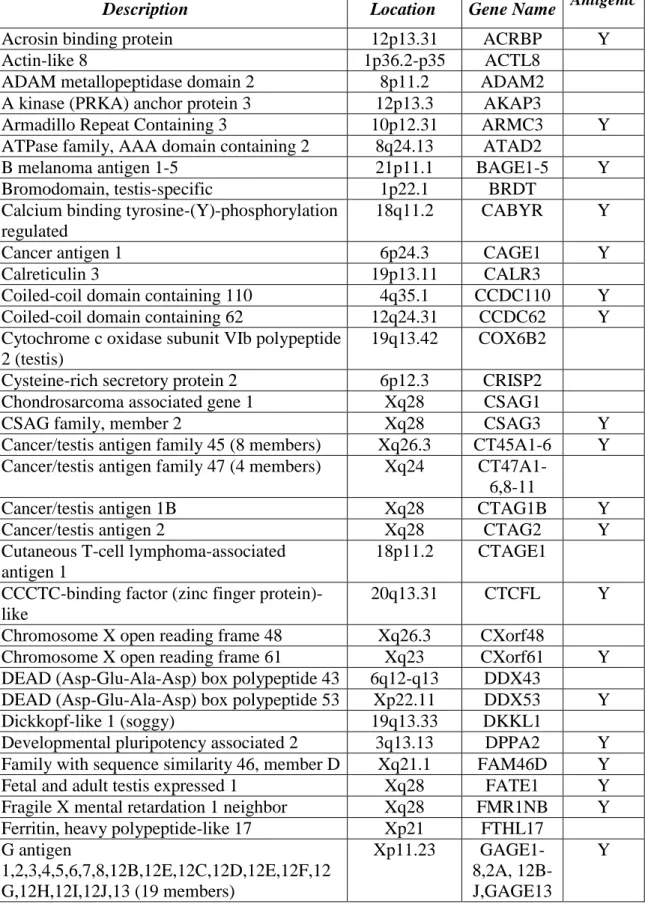
Table 1. Screened Cancer Testis Antigens
Description Location Gene Name Antigenic
Acrosin binding protein 12p13.31 ACRBP Y
Actin-like 8 1p36.2-p35 ACTL8
ADAM metallopeptidase domain 2 8p11.2 ADAM2
A kinase (PRKA) anchor protein 3 12p13.3 AKAP3
Armadillo Repeat Containing 3 10p12.31 ARMC3 Y
ATPase family, AAA domain containing 2 8q24.13 ATAD2
B melanoma antigen 1-5 21p11.1 BAGE1-5 Y
Bromodomain, testis-specific 1p22.1 BRDT
Calcium binding tyrosine-(Y)-phosphorylation regulated
18q11.2 CABYR Y
Cancer antigen 1 6p24.3 CAGE1 Y
Calreticulin 3 19p13.11 CALR3
Coiled-coil domain containing 110 4q35.1 CCDC110 Y
Coiled-coil domain containing 62 12q24.31 CCDC62 Y
Cytochrome c oxidase subunit VIb polypeptide 2 (testis)
19q13.42 COX6B2 Cysteine-rich secretory protein 2 6p12.3 CRISP2
Chondrosarcoma associated gene 1 Xq28 CSAG1
CSAG family, member 2 Xq28 CSAG3 Y
Cancer/testis antigen family 45 (8 members) Xq26.3 CT45A1-6 Y Cancer/testis antigen family 47 (4 members) Xq24
CT47A1-6,8-11
Cancer/testis antigen 1B Xq28 CTAG1B Y
Cancer/testis antigen 2 Xq28 CTAG2 Y
Cutaneous T-cell lymphoma-associated antigen 1
18p11.2 CTAGE1 CCCTC-binding factor (zinc finger
protein)-like
20q13.31 CTCFL Y
Chromosome X open reading frame 48 Xq26.3 CXorf48
Chromosome X open reading frame 61 Xq23 CXorf61 Y
DEAD (Asp-Glu-Ala-Asp) box polypeptide 43 6q12-q13 DDX43
DEAD (Asp-Glu-Ala-Asp) box polypeptide 53 Xp22.11 DDX53 Y
Dickkopf-like 1 (soggy) 19q13.33 DKKL1
Developmental pluripotency associated 2 3q13.13 DPPA2 Y Family with sequence similarity 46, member D Xq21.1 FAM46D Y
Fetal and adult testis expressed 1 Xq28 FATE1 Y
Fragile X mental retardation 1 neighbor Xq28 FMR1NB Y Ferritin, heavy polypeptide-like 17 Xp21 FTHL17
G antigen
1,2,3,4,5,6,7,8,12B,12E,12C,12D,12E,12F,12 G,12H,12I,12J,13 (19 members)
Xp11.23
GAGE1-8,2A, 12B-J,GAGE13
Table 1, continued. Screened Cancer Testis Antigens
Description Location Gene Name Antigenic
HORMA domain containing 1 1q21.3 HORMAD1 Y
Insulin-like Growth Factor 2 mRNA Binding Protein 3
7p11 IGF2BP3 Y
Immunoglobulin superfamily, member 11 3q13.32 IGSF11 Y Interleukin 13 receptor, alpha 2 Xq13.1-q28 IL13RA2
Lactate dehydrogenase C 11p15.1 LDHC
LEM domain containing 1 1q32.1 LEMD1
Lipase, member I 21q11.2 LIPI
Leucine zipper protein 4 Xq23 LUZP4 Y
Lymphocyte antigen 6 complex, locus K 8q24.3 LY6K Y
Melanoma antigen family A, 8 Xq28 MAGEA8
Melanoma antigen family A, 9 Xq28 MAGEA9 Y
Melanoma antigen family B, 1 Xp21.3 MAGEB1 Y
Melanoma antigen family B, 2 Xp21.3 MAGEB2 Y
Melanoma antigen family B, 3 Xp21.3 MAGEB3
Melanoma antigen family B, 4 Xp21.3 MAGEB4
Melanoma antigen family B, 5 Xp21.3 MAGEB5
Melanoma antigen family B, 6 Xp21.3 MAGEB6
Melanoma antigen family C, 1 Xq26 MAGEC1 Y
melanoma antigen family C, 2 Xq26 MAGEC2 Y
Melanoma antigen family C, 3 Xq26 MAGEC3
MORC family CW-type zinc finger 1 3q13 MORC1
Nuclear RNA export factor 2 Xq22.1 NXF2
Outer dense fiber of sperm tails 1 8q22.3 ODF1
Outer dense fiber of sperm tails 2 9q34.11 ODF2 Y
Outer dense fiber of sperm tails 3 11p15.5 ODF3 Y
Opa interacting protein 5 15q15.1 OIP5
P antigen family, member 2 (prostate associated)
Xp11.21 PAGE2 Y
P antigen family, member 4 (prostate associated)
Xp11.21 PAGE4 Y
P antigen family, member 5 (prostate associated)
Xp11.21 PAGE5
PAS domain containing 1 Xq28 PASD1 Y
Piwi-like 2 (Drosophila) 8p21.3 PIWIL2
Placenta-specific 1 Xq26 PLAC1 Y
POTE ankyrin domain family B,C,D (3 members)
8p11.1,15q11. 2,21q11.2
POTEB-D
Preferentially expressed antigen in melanoma 22q11.22 PRAME Y
Table 1, continued. Screened Cancer Testis Antigens
Description Location Gene Name Antigenic
Solute Carrier Organic Anion Transporter Family, Member 6A1
5q21.1 SLCO6A1 Y
Sperm autoantigenic protein 17 11q24.2 SPA17 Y
Sperm associated antigen 9 17q21.33 SPAG9 Y
SPANX family, member A1,A2,C,D, E2 (5 members)
Xq27.1 SPANX Y (B)
SPANX family, member N5 Xq27.1 SPANXN5
Spermatogenesis associated 19 11q25 SPATA19
SPO11 meiotic protein covalently bound to DSB homolog (S. cerevisiae)
20q13.31 SPO11
Synovial sarcoma, X breakpoint 1 Xp11.23 SSX1
Synovial sarcoma, X breakpoint 2 Xp11.23 SSX2 Y
synovial sarcoma, X breakpoint 4 Xp11.23 SSX4 Y
Testis Specific, 10 2q11.2 TSGA10 Y
Protease, Serine, 50 3p21.31 TSP50
Testis specific protein, Y-linked 1 (3 members) Yp11.2 TSPY1 X antigen family, member 1B, E (2 members) Xp11.22 XAGE1B;
XAGE1E
Y
X antigen family, member 2 Xp11.22 XAGE2
CHAPTER IV: FATE1 MODULATES PROGRAMMED CELL DEATH WITHIN TUMOR CELLS2
Introduction
A defining hallmark of malignant cells is the ability to resist signals arising from both internal (e.g. oncogenic stress, DNA damage) and external (e.g. hypoxia) sources that would otherwise activate the process of programmed cell death, apoptosis [98, 99] . Tumor cells employ a number of mechanisms to overcome these insults, including: upregulation of anti-apoptotic proteins, and loss or suppression of proteins that activate apoptosis (e.g. p53) or suppress pro-survival signaling (e.g. PTEN) [100, 101]. Targeting the dysregulated apoptotic pathway within tumors has proven effective in early phase clinic trials using navitoclax, an anti-apoptotic Bcl-2 family inhibitor; however, side effects including anemia and other cytopenias, infection, and gastrointestinal distress were common [102, 103]. The identification and targeting of tumor-specific mechanisms of deflecting cell death signals would significantly reduce such side effects while maintaining clinical efficacy.
Cancer Testes Antigens represent a cohort of genes that could deflect cell death in a nearly tumor-specific fashion. In our comprehensive analysis of CTAs (Chapter III) we found that depletion of multiple CTAs led to a significant cellular apoptotic activity within cells. The
2Elements of the work referenced in this chapter are published in: Kimberly E. Maxfield and Patrick J. Taus, Kathleen Corcoran,
studies presented here in Chapter IV will elaborate the role of FATE1 in antagonizing apoptosis. To begin I will discuss the regulation of the apoptotic pathway as mediated by members of the Bcl-2 protein family as well as the post-translational mechanisms that can alter levels of this protein family and in doing so alter cellular sensitivity to apoptosis itself.
Regulation of Apoptosis
Post-translational regulation of the apoptotic pathway
Apoptosis is regulated in part by the balance of pro- and anti-apoptotic Bcl-2 members. These levels can be controlled on transcriptional, translational, and post-translational levels [110-112]. For the purposes of the results discussed in this chapter, an overview of the current body of work concerning the post-translational regulation of Bcl-2 family members through the actions of the ubiquitin-proteasome systems (USP) will be presented at this time.
The UPS along with autophagy are the major mechanisms by which cells degrade intracellular proteins. While autophagy degrades proteins and macromolecules wholesale at an organelle level, the UPS targets individual proteins for degradation through conjugation of ubiquitin chains onto lysine residues within the target protein or onto a target protein’s free N-terminal α-amino group through the N-rule pathway [113, 114]. Tight ubiquitin-mediated degradation of numerous members of the Bcl-2 protein family is required to maintain cellular homeostasis [115]. The necessity of this regulation can be seen in the clinic as multiple Bcl-2 family members including BIK, NOXA, and BIM drive tumor cell death following treatment with Bortezomib a proteasome inhibitor which is used clinically under the name Velcade® for treatment of multiple myeloma and relapsed mantle cell lymphoma [116, 117].
HECT (Homologous to the E6-AP Carboxyl Terminus) domains that interact with E2 enzymes. RING domains facilitate the direct transfer of the charged ubiquitin from the E2 onto the target lysine while HECT domain-containing proteins form a thioester with the ubiquitin before transferring it to the substrate [118, 119].
E3-ligases are targeted to their substrates either directly or through additional binding partners that serve as scaffolding/adaptor proteins [118]. The activity of E3-ligases is also regulated by post-translation modifications of the substrates including phosphorylation (the most prominent), glycosylation and proline hydroxylation [113]. The activity of E3-ligases can also be regulated by the E3-ligase itself through self-ubiquitination which can occur in both substrate-independent, and substrate-dependent fashions [120]. Within the cell, the activity of E3-ligases is also regulated by the activity of deubiquitinases, proteases that cleave ubiquitin chains which serve to fine tune or even reverse ubiquitin modification on substrates and E3-ligases alike [121].
Results
Depletion of FATE1 activates programmed cell death
suppress apoptosis specifically in the transformed cellular environment irrespective of disease site.
siFATE1-induced apoptosis requires the core apoptotic machinery
Given the potent induction of the apoptotic pathway following depletion of FATE1 and FATE1’s localization to the mitochondria, a critical signaling hub in apoptotic signaling, we further analyzed the role of the key regulators of mitochondrial premeabilization siFATE1-induced death. Depletion of FATE1 in HCT116 cells siFATE1-induced cleavage of PARP1 and cytochrome c release, a phenotype that was absent in BAX/BAK null HCT116 cells (Figure 11A). Overexpression of the anti-apoptotic Bcl-2 family member, BCL-xL, also rescued cell death following siFATE1 (Figure 11B). Consistent with a general role in deflecting apoptosis, we found that cells overexpressing FATE1 exhibited attenuated PARP1 cleavage following staurosporine (STS) challenge (Figure 11C).
FATE1 regulates stability of the BH3-only tumor suppressor BIK
found upregulated in tumors compared to normal tissues (Figure 12B) [132-138]; however, BIK protein is expressed at low to undetectable levels indicating that tumor cells adapt mechanisms to prevent BIK protein accumulation [116, 117, 125, 139, 140]. These mechanisms likely consisted of augmented proteasome-mediated turnover as inhibition of this pathway or attenuation of BIK ubiquitination leads to its accumulation [134, 141]. Accumulation of BIK correlates with the induction of apoptosis following treatment with Velcade ® (Bortezomib) a proteasome inhibitor approved for clinical use [116, 117]. We verified that FATE1 and BIK interact within mammalian cell (Figure 12C) and found that upon depletion of FATE1, BIK protein was stabilized within cancer cells (Figure 12D). Significantly, depletion of BIK rescued siFATE1-induced apoptotic signaling (Figure 12E), leading us to hypothesize that FATE1 may be involved in the regulation of BIK stability.
Within a cell, targeted degradation of proteins is mediated by the ubiquitin-proteasome system which is initiated via the conjugation of poly-ubiquitin chains to proteins destine for destruction. Conjugation of ubiquitin to target molecules is carried out by E3-ligases. While FATE1 does not have any identifiable E3-ligase domains, yeast 2-hybrid studies have identified several FATE1 binding partners with potential E3 ligase activity [142, 143]. Among this cohort, depletion of RNF183 led to stabilization of BIK (Figure 13A and B). RNF183 is uncharacterized protein with a C3HC4-RING domain whose expression may be a biomarker for endometrial carcinoma in uterine aspirates (Figure 13C) [144]. We found that RNF183 localized to the endoplasmic reticulum (Figure 13D). We confirmed that FATE1 and RNF183 interact within mammalian cells and confirmed the RNF183 possess E3-ligase activity which is dependent on its canonical RING domain cysteine residues (Figure 13E and 13F) [118].
lacking ligase activity, reduced expression of exogenous BIK, indicating that RNF183’s E3-activity negatively regulates BIK protein accumulation (Figure 14A). We also verified RNF183’s ability to directly ubiquitinate BIK using a reconstituted in vitro system (Figure 14B). Given its potent effect on the stabilization of BIK, we evaluated the consequences of RNF183 depletion on cell viability. We observed a striking correlation between siFATE1 and siRNF183 sensitivity in a panel of tumor derived cell lines (Figure 14C and D). Furthermore, we found that BIK interacted with RNF183 along with FATE1 in intact cells (Figure 14E). These findings indicate that FATE1 and RNF183 may collaborate to restrain BIK protein levels and promote cellular escape from otherwise lethal apoptotic signaling.
We then examined a potential role for FATE1 and RNF183 cooperativity within patient tumors. In NSCLC, patients with tumors expressing high levels of both FATE1 and RNF183 were at highest risk for poor survival (hazard ratio (HR) = 2.80; p <0.0001) as compared to those with high expression of only FATE1 (HR = 1.73, p = 0.0124) or RNF183 (HR = 1.62, p = 0.0004) (Figure 14F). In a separate NSCLC data set, the majority of tumors with high FATE1 expression exhibited elevated RNF183 expression, and high expression of both genes predicted shortened overall survival time (HR = 2.529, p = 0.0004) (Figure 14G). The frequency of co-expression of RNF183 and FATE1 along with their correlation with poor outcome reinforces the notion that these proteins are functioning in human tumors to promote survival.
Discussion
physiological conditions. Prototypical examples include thwarting oncogene-induced pro-apoptotic signaling, DNA damage stress, toleration of proteotoxic stress due to aneuploidy, and buffering of oxidative stress resulting from altered mitochondrial function [145-147]. FATE1 may represent one mechanism by which tumor cells overcome these barriers by promoting the degradation of second messengers in this signaling system.
Figure 9: Overview of Bcl-2 family.
Figure 10: Loss of FATE1 induces programmed cell death.
CHAPTER V: EWS/FLI1 DRIVES EXPRESSION OF MULTIPLE FUNCTIONAL CANCER TESTIS ANTIGENS
Introduction
The mechanisms by which CTAs are aberrantly activated within cancers has not been fully elucidated. Although alterations in DNA methylation are a significant driver of CTA expression, rarely do these alterations lead to induction of the entire CTA group, arguing that in certain cases additional transcriptional factors are involved. A potential role for tumor-specific transcription factors driving the activation of CTAs has yet to be explored. In this Chapter, studies examining the role of EWS-FLI1, a chimeric transcription factor that underlies the molecular pathology of Ewing sarcoma, in activation of FATE1 will be presented. To begin, I will discuss Ewing sarcoma, including its underlying biology, clinical management, and the current state of ongoing efforts to improve outcome for patients with the disease.
Ewing sarcoma
under 15 % [150, 152, 153]
Molecular pathology of EWS-FLI1
Ewing sarcoma is driven by an oncogenic transcription factor resulting from a genomic translocation [154]. In roughly 90 % of cases this chimeric transcription factor contains the C-terminus of EWS, a TET family transcription factor, and the N-C-terminus of FLI1, a member of the ETS transcription factor family member [155]. The fusion, which is under control of the constitutively active EWS-promoter, couples the transactivation activation domain of EWS to the DNA-binding domain of FLI1 [154, 156, 157]. Within Ewing sarcoma cells the fusion is essential for proliferation and tumorigenesis and aside from its causative translocation, the genetic landscape of Ewing sarcoma tumors is nearly devoid of additional chromosomal abnormalities [158, 159]. The resulting chimeric transcription factor can up and down regulate genes and examples of each have been shown to contribute to the survival and proliferation of Ewing sarcoma cells and xenografts [155]. EWS-FLI1 can bind conventional ETS motifs but also acquires the ability to bind GGAA repeats [160]. The ability of EWS-FLI1 to bind GGAA repeats was discovered while investigating the regulation of NR0B1. These study found a series of 25 GGAA repeats with the in NR0B1 that were necessary and sufficient for EWS-FLI1 driven transcription of NR0B1 [160].
GGAA-repeats were found to be activated by EWS-FLI1 (as measured by enhanced p300 recruitment) while enhancers containing conical ETS motifs were repressed [162]. As microsatellite regions are known to be highly polymorphic, variations at such sites may underlie the ethnic differences in susceptibility to the disease [163, 164].
To date, the study of Ewing sarcoma oncogenesis has been hampered by a lack of biologically relevant model systems. Early studies demonstrated that immortalized murine fibroblasts (NIH3T3 cells) were transformed by expression of EWS-FLI1 [165]; however, recently the value of this as a model for Ewing sarcoma oncogenesis has come into question due to differences in expression profiles of these cells with tumor samples from patients and differential requirements of EWS-FLI1 regulated genes for cell survival in murine and human cell lines [166, 167]. A potential explanation for differences observed between mouse and human systems is the different microsatellite composition of each species’ genome [155]. These discrepancies suggest that production of a mouse model of Ewing sarcoma reflective of the human condition will be difficult. Also, the cell of origin of the diseases is unknown further adding to the difficultly of studying the oncogenesis of Ewing sarcoma. Several recent studies support mesenchymal progenitor cells as a strong candidate as the potential cell of origin for Ewing sarcoma; however while expression of EWS-FLI1 within mouse mesenchymal stem cells has induced transformation, transformation of human mesenchymal stem cells by EWS-FLI1 has yet to be demonstrated [168-170].
Current Treatment of Ewing sarcoma
consists of alternating courses of vincristine-cyclophosphamide-doxorubicin with ifosfamide-etoposide every three weeks for roughly one year [150]. Recently, an intensified protocol for patients presenting with localized disease in which chemotherapy was administered every two weeks increased event-free survival to 73 % (compared to 65 % in the standard arm, P = 0.48) [151]. Surgery is the first choice for local control of Ewing sarcoma tumors, however, radiation is considered for unresectable tumors, cases were potential functional morbidity from surgery is deemed too high or as adjuvant therapy in surgical cases in which negative margins are not obtained [171, 172]. Radiation in a preoperative setting in which clean margins are unlikely has led to tumor shrinkage and clear margins following subsequent surgery [173].
Ongoing clinical trials in Ewing sarcoma
Currently, on-going phase III clinical trials in the United States are testing the benefit of the addition of a second topoisomerase inhibitor, topotecan, to the current five drug backbone chemotherapeutic regimen (COG-AEWS1031) in patients with localized disease. A phase II study in patients with newly diagnosed Ewing sarcoma is also evaluating the efficacy of the addition of the a second topoisomerase inhibitor, irinotecan, as well as an additional alkylating drug, temozolomide, and the cardioprotecitve agent dexrazoxane to the current five drug backbone (NCI-2013-01094). A phase II trial is testing the efficacy of cabozantinib-s-malate, a receptor tyrosine kinase inhibitor, in patients with relapsed osteosarcoma or Ewing sarcoma (NCI-2014-01927).
(MAGE-A1, MAGE-A3, and NY-ESO-1) in children with relapse or refractory sarcomas (including Ewing sarcoma) found that the treatment was well tolerated and over half of the patients developed a response to the antigens over the course of treatment [174]. Preliminary in vitro
studies have also demonstrated the effectiveness of Natural Killer cells against Ewing sarcoma derived cell lines [175]. Approaches targeting the novel breakpoint region of the EWS-FLI1 fusion demonstrated that while the native peptide had poor loading onto HLA-A2.1, modified peptides with enhanced MHC loading could be used to raise CTLs that then demonstrated potent killing of tumor cells expressing endogenous EWS-FLI1 and could prolong survival of mice with Ewing sarcoma xenografts [176]. Unfortunately, although immune-mediated mechanisms are showing promise, Ewing sarcomas lose expression of MHC class I molecules during disease progression thereby limiting the temporal window in which immune therapies may be effective in treating the disease [177].
Results
FATE1 is a target of EWS-FLI1