Open Access
Review
Assisted reproductive technologies in rhesus macaques
Don P Wolf*
Address: Division of Reproductive Sciences, Oregon National Primate Research Center Beaverton, Oregon and Departments of Obstetrics/ Gynecology, Physiology and Pharmacology, Oregon Health and Science University, Portland Oregon, USA
Email: Don P Wolf* - [email protected] * Corresponding author
Abstract
The assisted reproductive technologies (ARTs) have been used in the production of rhesus monkey offspring at the Oregon National Primate Research Center (ONPRC) and that experience is summarized here. Additionally these technologies serve as a source of oocytes/embryos for monozygotic twinning, embryonic stem (ES) cell derivation and cloning. High fertilization efficiencies were realized with conventional insemination or following the use of intracytoplasmic sperm injection (ICSI) and approximately 50% of the resulting embryos grew in vitro to blastocysts. Both fresh and frozen sperm were employed in fertilization by ICSI and the resulting embryos could be low temperature stored for subsequent thawing and transfer when a synchronized recipient female was available or after shipment to another facility. Following the transfer of up to 3 embryos, an overall pregnancy rate of 30% was achieved with increasing rates dependent upon the number of embryos transferred. Singleton pregnancy outcomes following the transfer of ART produced embryos were similar to those observed in a control group of animals in the timed mated breeding colony at ONPRC. ICSI produced embryos were used in efforts to create monozygotic twins by blastomere separation or blastocyst splitting. While pregnancies were achieved following the transfer of demi-embryos, only one was a twin and it was lost to spontaneous abortion. ICSI produced embryos have also served as the source of blastocysts for the derivation of embryonic stem cells. These pluripotent cells hold potential for cell based therapies and we consider the monkey an important translational model in which to evaluate safety, efficacy and feasibility of regenerative medicine approaches based on the transplantation of stem cell-derived progeny. Finally, efforts to produce genetically-identical monkeys by nuclear transfer have been briefly summarized.
Review
Notable progress has been realized in the past 20 years in both the development and application of the ARTs in monkeys with advances in ovarian follicular stimulation, fertilization by intractyoplasmic sperm injection (ICSI), culture medium development for the production of blast-ocysts, low temperature storage of sperm and embryos and embryo manipulation including twinning, cloning and the derivation of embryonic stem cells. Unlike in
many fields where knowledge flows from non-human pri-mate (NHP) research to clinical application in humans, the development of the ARTs in monkeys historically postdates human application. The experience in NHP, however, while nowhere nearly as robust as that in humans has been associated with several landmark accomplishments and methods for establishing pregnan-cies are currently available as summarized by us in 2001 [1]. While a few reports in New World monkeys and Great
Published: 16 June 2004
Reproductive Biology and Endocrinology 2004, 2:37
Received: 23 March 2004 Accepted: 16 June 2004
This article is available from: http://www.rbej.com/content/2/1/37
Apes exist, most research efforts have focused on Old World macaques with the rhesus monkey the primary subject and that species is the focus of this review.
The first successful birth of a rhesus macaque following IVF was reported in 1984 by Bavister and coworkers [2] and the total number of infants born to date is approxi-mately one hundred. Development of the ARTs in NHPs has been impacted by resource availability, high expense and lack of compelling incentives to produce large num-bers of monkeys using this approach. The latter limitation has recently been eliminated by the need for Indian-ori-gin, rhesus macaques carrying the class 1 MHC allele, A*01 for HIV vaccine development research [3] (Figure
1). In addition to this demand for specific genotypes, many experimental paradigms could benefit from the availability of genetically-identical animals produced by monozygotic twinning or by somatic or ES cell cloning. Finally, development of cell-based therapies for the treat-ment of disease has gained motreat-mentum and a great deal of attention predicated on the availability of stem cells including those isolated from the blastocyst.
While application of the ARTs results in the production of embryos and pregnancies following transfer, information on pregnancy outcome is prerequisite to evaluating the impact of the ARTs on routine animal husbandry and more exotic applications such as the preservation of
Schematic presentation of our approach to producing mamu A*01 positive rhesus monkey offspring
Figure 1
endangered species. The monkey can also serve as a model for clinical application of the ARTs where fertilization by ICSI has been associated with an increased frequency of genetic problems that may be secondary to the inappro-priate expression of imprinted genes [4,5]. In nonprimate animals, a large offspring syndrome [6] has been associ-ated with in vitro manipulation of embryos and while this does not appear to be a problem in ART produced chil-dren, its possible existence in NHPs has not been evaluated.
This chapter will summarize the experience at ONPRC with the ARTs in the rhesus monkey building on two pre-vious reviews [1,7]. The methodology employed in the conduction of the ARTs in monkeys can also be found in these reviews. My objective is to disseminate the results of our extensive experience in the rhesus macaque in an effort to catalyze research using non-human primates.
Oocyte collection, fertilization and embryo
culture
Oocyte recovery from adult, cycling rhesus monkeys sub-jected to controlled ovarian stimulation has been described previously [1] and reviewed in the contribution by Drs. Stouffer and Zelinski-Wooten. The percentage of oocytes recovered at the mature, fertilizable MII stage var-ies as a function of the hCG to egg collection time between 27 and 36 hr [8]. Experiments requiring enucleation are ideally conducted immediately after 1st polar body extru-sion in which case a shorter time interval such as 27 hr is ideal. Oocytes that are recovered as metaphase I (MI) at the 27 hr interval can be monitored in vitro for release of the first polar body and used promptly thereafter. On the other hand, for routine fertilization, it is usually prefera-ble to recover the maximum number of oocytes at the MII stage, in which case the hCG to collection interval can be lengthened to 33–34 hr. The number of oocytes collected following controlled ovarian stimulation in our experi-ence is high, averaging 38 ± 2 [7]. Although not all are healthy, fertilizable oocytes, this number is considerably higher than that associated with the ARTs in women where a focus on the collection of approximately 10 high quality oocytes is becoming the norm [9]. This difference, along with the use of fixed length stimulation protocols, may be relevant to our efforts to improve ART outcomes in monkeys. A discussion of oocyte quantity and quality in the monkey can be found in the contribution by Drs. Stouffer and Zelinski-Wooten.
The conventional approach to embryo production using the ARTs is IVF and we reported a 61 ± 6% fertilization rate for 23 replicates involving 242 ova [7]. In our hands, complications associated with agglutination following sperm exposure to the capacitating agents, dibutyrylcyclic adenosine monophosphate and caffeine [10], are
signifi-cant and we have, therefore, adopted ICSI as our routine insemination procedure. Similar if not improved out-comes have been experienced with fresh sperm ICSI, 81 ± 3% for 61 replicates and 714 ova, however, we do not know if the use of ICSI creates problems with regards the transmission of genetic disease or the production of sub-optimal embryos. An evaluation would require the direct comparison of pregnancy rates obtained with IVF versus ICSI-produced embryos as well as long-term follow up on the offspring.
In general, conventional IVF is not feasible with frozen thawed sperm because of the rapid motility loss that occurs post-thaw [7]. However, control, fresh sperm ferti-lization rates can be achieved with cryopreserved sperm when ICSI is conducted 3 or more hours after sperm thaw-ing and cryoprotectant removal. This delay presumably reflects the need for a post-thaw recovery period and res-toration of the sperm's oocyte activating potential.
A complex medium, CMRL 1066 supplemented with serum, has historically been used to culture NHP embryos [11,12]. In early trials, coculture on buffalo rat liver cell monolayers in the presence of CMRL 1066 produced the highest blastocyst development rate [13]. However, while coculture may improve developmental potential, its com-plexity makes the specific requirements for development
in vitro difficult, if not impossible to study, not to mention the probable variability inherent in the coculture system. Attempts to culture embryos in a chemically defined medium (KSOM/AA) supplemented with serum resulted in improved, but inconsistent in vitro development rates [14]. Parenthetically, sequential media developed for human IVF such as G1/G2 have not produced satisfactory results in the monkey (unpublished). Recently, we initi-ated the use of HECM-9 in the absence of coculture [15-17] and reported a 76 ± 2% ICSI fertilization rate and a 47 ± 4% blastocyst formation rate [7]. Embryo culture in HECM-9 produced faster development than in CMRL/BRL or KSOM-AA with compact morulae present on day 3, cav-itating blastocysts on day 5 and expanded blastocysts on day 7 (Figure 2). The rates reported here for HECM-9 may closely approximate the in vivo rates although the latter are based on the recovery and staging of a limited number of embryos flushed from the uterus [see Figure 2; [12,14,18]]. A retrospective comparison of pregnancy and implantation rates for embryos produced in these in vitro
Embryo transfer
Parameters known to contribute to pregnancy and implantation rates following embryo transfer to synchro-nized recipients include the number of embryos trans-ferred, embryo quality and the method of transfer. When increasing numbers of embryos were transferred up to 3, a corresponding increase in pregnancy rate to 36% was realized and implantation rates when expressed per embryo were independent of the number of embryos transferred. However, these databases are not yet large enough to establish statistical significance [7]. Recently we experienced the first triplet pregnancy reported in rhesus monkeys that occurred following the transfer of 3 frozen-thawed embryos. The pregnancy terminated spontane-ously at 85 days possibly following intrauterine demise of the smallest member of the triplet set (Male, 0.067 kg; Female, 0.060 kg; Male,0.033 kg).
The ability to cryostore embryos and conduct transfers when recipients are available is essential to any wide-spread use of the ARTs in monkeys. In our experience, pregnancy and implantation rates for fresh and
frozen-thawed ICSI produced embryos were associated with sim-ilar outcomes at approximately 30%. However, while the average number of embryos transferred was comparable for fresh (2.3) and frozen (2.1) embryos, substantial embryo loss (35%) was associated with freeze-thawing.
The conventional approach to embryo transfer (ET) in rhesus monkeys involves mini-laparotomy with a preg-nancy rate of 27% (25/92) [7]. In order to avoid a surgical procedure, it would be ideal to conduct non-surgical ET with embryo deposition directly into the uterus, similar to the approach currently employed in women. However, the results to date in monkeys have been disappointing reflecting in part difficulty in passing the long tortuous cervix in this species. Laparoscopic ET with oviductal placement of embryos is the most recent variation devel-oped by Dr. John Fanton and the surgical team at ONPRC (Figure 3). Similar pregnancy and implantation results were obtained when mini-laparotomy was compared to laparoscopy and since laparoscopy avoids exposure of the animal to a major surgery, that technique is now preferred by us. Preliminary evidence suggests that embryos at any
Average time in days for ART-produced embryos to reach a given developmental stage
Figure 2
Average time in days for ART-produced embryos to reach a given developmental stage. Embryos were cultured in CMRL/BRL (yellow triangles), KSOM-AA supplemented with 10% fetal calf serum (pink squares) or HECM-9 with albumin (blue diamonds). M, morula; B, blastocyst; XB, expanded blastocyst. The results for CMRL/BRL coculture and KSOM-AA came from Weston and Wolf [14] and the embryos were produced by IVF while for culture in HECM-9, the results are from Wolf and coauthors [7] with the embryos produced by ISCI. Estimated in vivo developmental rates (open circles) have been included for reference purposes [12].
M
B
XB
0 2
4
6
8
10
stage of preimplantation development from 2-cell to expanded blastocyst can be transferred to the oviduct with pregnancy production.
The pregnancy success rate depends on the synchrony between the age of the transferred embryos, as measured by culture time in vitro, and the host endometrium, rela-tive to the predicted day of ovulation. Synchronous trans-fers appear to be optimal in women. However, evidence supports an asynchronous transfer in the rhesus monkey [19,20] with optimal timing for blastocyst (day 7/8) trans-fer into a day 4 uterine environment. This may reflect a retarded rate of development of embryos in vitro. In con-trast, younger cleavage stage embryos were optimally transferred into synchronized recipients with the highest success rates following the transfer of day 3 embryos into either day 2,3 or 4 recipients.
Finally, pregnancy success rate is likely also to be a func-tion of the psychological and physical health of the recip-ient. Thus, we have long suspected that imported Chinese rhesus monkeys are inferior to home grown, Indian ori-gin, rhesus macaques as recipients, because the imported animals may not be as physically healthy and are at increased risk for poor psychological health. However, a retrospective review of embryo transfer did not support a statistically significance difference between the two with 13 of 59 (22%) transfers resulting in pregnancy in Chi-nese rhesus recipients and 35 of 132 (26%) for Indian-ori-gin animals.
Oocyte/embryo manipulation
ART applications as described so far in this review, where ICSI has been used to compensate for poor motility in fro-zen-thawed sperm preparations, can also be extrapolated to the oocyte. Conventional IVF is a poor choice for ferti-lizing oocytes following extensive culture when zona hardening is a likely outcome. For instance, in cases were oocytes are matured in vitro. Another example is in the context of preserving fertility in young cancer survivors [21]. The strategy that we recently employed in the mon-key model involved heterotropic transplantation of fresh ovarian tissue followed by oocyte recovery from the grafts. ICSI was then employed to fertilize these oocytes and the transfer of the resulting embryos resulted in the produc-tion of a pregnancy.
Somatic cell cloning (SCC) is perhaps the ideal approach to producing genetically identical animals because the clone size is, in theory, infinite. Protocols for SCC have been developed for a number of mammalian species and each appears to require species unique modifications to succeed [22,23]. Cloned animal production rates are for the most part still very low except in widely studied spe-cies such as cattle where live birth rates in the 15–20%
Illustrations of the progressive stages in laparoscopic embryo transfer in rhesus monkeys
Figure 3
range are now realized [Andras Dinnyes, personal com-munication]. This increased rate in part reflects greater attention to fetal and neonatal monitoring with cloned pregnancies considered at high risk. The high fetal and neonatal wastage that is associated with SCC is thought to reflect incomplete reprogramming of the donor nucleus, perhaps secondary to the inappropriate expression of imprinted genes. Reprogramming is, of course, a bigger challenge when somatic cells are employed as the donor nuclei as opposed to relatively undifferentiated, embry-onic cells. In fact, cloning success was first achieved in sheep with embryonic blastomeres or cell lines derived from embryonic cells. In monkeys, we began efforts to produce animals by nuclear transfer before SCC was con-sidered feasible and were successful. Initially in 1997 [24], 3 pregnancies were achieved culminating in 2 live births in 9 embryo transfers. We repeated this success in 2002, with one term pregnancy in 11 embryo transfers, how-ever, the fetus was lost as a stillborn. The most likely cause of death was fetal asphyxia and since no gross abnormal-ities were observed, we concluded that fetal demise was unrelated to the nuclear transfer process [25]. A number of variables have now been studied in the context of SCC in monkeys, however, we have not yet been successful in achieving a clinical pregnancy. Reconstructed embryos often grow in vitro to the morula stage but seldom to the blastocyst and we have no independent confirmation that the embryonic genome is actived in these cloned embryos. Since oocyte enucleation to produce the cyto-plast, donor cell/nucleus fusion or injection to produce a reconstructed embryo, and chemical activation of devel-opment is common to both embryonic and SCC, we have concluded that the failure of SCC in the monkey is related to incomplete reprogramming of the somatic cell nucleus. This conclusion differs markedly from a recent report that cloning in primates [26] is precluded by the inadvertent removal of critical spindle related factors during enuclea-tion. Moreover, successful therapeutic cloning in humans, would also argue against the concept that SCC is impossi-ble in primates [27]. In order to follow up on our hypoth-esis that SCC is related to incomplete reprogramming, we have renewed our efforts to clone from monkey ES cells because these cells are relatively undifferentiated while still available in unlimited numbers supporting the
even-tual production of large numbers of genetically-identical, cloned animals.
In the absence of a successful protocol for SCC in mon-keys, the need for genetically identical animals can be, at least, partly satisfied by producing identical twins. How-ever, the twin rate in macaques is very low at 0.05% and is associated with poor outcomes [28]. Despite this low twinning rate in nature, when 2 ART produced embryos were transferred, the pregnancy rate was 25% (Table 1, top row) and the twinning rate was 20%. The latter statistic, with a different denominator, was derived when all preg-nancies produced following transfer of 2 or 3 embryos were considered. A 64% live birth rate was obtained. Therefore, it would seem reasonable, despite the reluc-tance of free-ranging animals to carry twins to term, that monozygotic twins could be produced following transfer of ART produced demi-embryos. Two approaches to monozygotic twinning; blastomere separation and blasto-cyst bisection have been considered [29]. Demi-embryo pairs were produced in high efficiency by both methods and shown to be capable of growth to blastocysts in vitro
similar to intact controls or able to re-expand following manipulation as in the case of blastocyst-derived, embryos. Differential cell counts indicated that demi-embryos contained one half the total number of cells of their intact counterparts with no change in the ratios of ICM/trophectoderm or ICM to total cells. However, when demi-embryo sets were transferred into recipients, the pregnancy rate was similar to that obtained following the transfer of two intact embryos (Table 1, bottom row) but no term twin pregnancies were realized. Despite these dis-appointing preliminary results, we are continuing efforts to produce monozygotic twins by these approaches.
Primate embryonic stem cell lines were first generated in rhesus monkeys and marmosets in 1995 [30] and 1996 [31] respectively, followed by the human in 1998 [32] and the cynomolgus macaque in 2001 [33]. A parthenog-enote ES cell is also available in the cynomolgus monkey [34]. Derivation of ES cell lines requires dissociation of ICM from trophoblast at the blastocyst stage with culture of the ICM on feeder layers of mitotically-inactivated, mouse embryonic fibroblasts (Figure 4). Initial success in the rhesus monkey by Thomson and colleagues [30] Table 1: Twin pregnancy rates and outcomes in rhesus monkeys following the transfer of intact or demi-embryos.
Pregnancy rate per transfer (%) Twinning rate per transfer (%) Live birth rate per twin pregnancy (%)
Intract-embryos 14/57 (25) 14/70 (20) 18/28 (64)
Demi-embryos 12/55 (27) 1/12 (8) 0/2 (0)
employed in vivo produced blastocysts recovered in uter-ine flushings. More recently, our group at ONPRC has developed a series of ES cell lines from in vitro produced, ICSI fertilized embryos. The availability of a stem cell bank comprised of 25 or so well characterized lines is cru-cial to future experimentation and an evaluation of ES cell line specificity. We envision the NHP as an essential trans-lational model required in efforts to bring ES cell-based therapies to clinical trials in human patients.
Pregnancy and pregnancy outcome
We recently reviewed the outcome of 87 ART-produced pregnancies, 14 of which were twin gestations [7]. A pop-ulation of pregnancies from the timed mated breeding colony was included as a gold standard for comparative purposes. In total, 234 of 257 (91%) clinical pregnancies resulted in live birth. In the TMB population, 14 cases of
fetal demise were recorded for a 92.5% live birth rate. This value compared favorably with a live birth rate for an indoor TMB rhesus colony at the California Primate Center of 83% (1106/1332) [28].
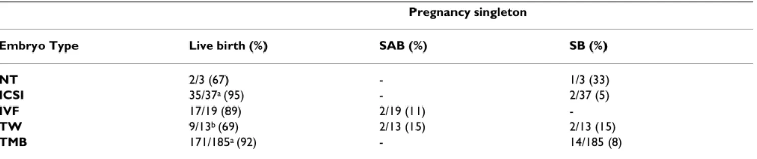
For ART produced, singleton pregnancies (nuclear trans-fer, demi-embryos, ICSI and IVF combined; Table 2) 63 of 72 resulted in a live birth for a rate of 87.5%, statistically identical to the TMB population. When ART produced pregnancies were evaluated separately, ICSI and IVF, gave a combined live birth rate of 92.8 % (52/56), while preg-nancy outcomes resulting from demi-embryo transfers were significantly lower at 69% (9/13). This impaired out-come with demi-embryos, although preliminary, suggests that despite appearances (high in vitro development rates to the blastocyst stage, ICM to total cell numbers similar
Schematic representation of the derivation of monkey embryonic stem cells
Figure 4
to intact controls), demi-embryos are not the same as their intact counterparts.
The sex ratios of ART produced pregnancies were the same as those for the TMB colony as were the gestational lengths and birth weights of singleton, live, term infants. The average twin birth weight was 0.37 kg with an average gestational age of only 150.7 days, in both cases signifi-cantly different from the corresponding singleton value. Growth rates based on animal weight for singleton and twin pregnancies are plotted in Figure 5. Offspring pro-duced in twin pregnancies were significantly lower in average weight until 1 year of age. Interestingly, despite fetal wastage in multiple pregnancies the outcome of twin pregnancies to date has resulted in the production of an increased number of viable infants with 15 twin pregnancies confirmed as clinically viable producing 18 live, term monkeys.
Conclusions
In the past 15 years, research into and practice of the ARTs in rhesus monkeys at ONPRC has resulted in a number of notable accomplishments. In 1988, the first was monkey produced following the transfer of an embryo that had been frozen, cryobanked and thawed after successful in vitro fertilization. This effort has also achieved the first twin pregnancies in rhesus monkeys, the first non-human primate infants produced by nuclear transfer of embry-onic cells, the first rhesus monkey infant born following the transfer of an ICSI-produced blastocyst employing a non-surgical procedure, the first monkey live birth result-ing from the transfer of a demi-embryo created by blast-omere separation at the 2-cell stage or blastocyst bisection and the first infants produced following laparoscopic embryo transfer. Recently we reported the outcomes of 87 pregnancies (7).
Based on the results summarized in this mini-review, the ARTs in non-human primates would now seem to be ready for widespread application in the propagation of selected, desirable genotypes. This is not to say that there is no longer room for protocol development. However, improvements may be slow in coming because the high cost and limited access to the NHP resource means that relatively few programs will be able to participate. Yet improvements in pregnancy rates should be anticipated based on extrapolation from the clinical experience in women where protocol development over the entire 20+ history of the technology has resulted in ever increasing pregnancy and implantation rates. The current expecta-tions include 50% pregnancy rates in patients under 35 and even higher levels when donor oocytes are used with implantation efficiencies of 0.5/embryo transferred [34-36]. How should we focus efforts in NHPs? This is a diffi-cult challenge but selection of higher, and perhaps fewer, quality oocytes following animal exposure to individual-ized ovarian stimulation protocols seems a good place to start. Although embryo development in HECM-9 was improved over that obtained on coculture or in KSOM-AA, it is likely that further improvements in embryo cul-ture that translate into higher pregnancy rates are possi-ble. Finally attention focused on embryo transfer is appropriate both from a technical perspective, for instance with regards to pH stability in the oviduct during the insufflation required for laparoscopic ET, as well as recipient selection based on parameters that have not yet been developed but might reasonably include size, age, parity, menstrual cycle regularity and systemic cortisol level as an indirect measure of psychological health. From a longer term perspective, it may be worthwhile to evalu-ate the possible recovery and maturation of ovarian oocytes from necropsy animals or perhaps even to derive gametes from ES cells as has been accomplished in the mouse [37,38].
Table 2: Live births and fetal loss for singleton pregnancies following application of the ARTs in rhesus macaques.
Pregnancy singleton
Embryo Type Live birth (%) SAB (%) SB (%)
NT 2/3 (67) - 1/3 (33)
ICSI 35/37a (95) - 2/37 (5)
IVF 17/19 (89) 2/19 (11)
-TW 9/13b (69) 2/13 (15) 2/13 (15)
TMB 171/185a (92) - 14/185 (8)
Before ending, a comment or two relative to cost effective-ness of the ARTs in NHP is appropriate. Clearly, if there is no other way to acquire specific animals i.e., they are
priceless, then this discussion is irrelevant. But consider acquiring rhesus macaques of either Indian-origin from local sources, if they are available at all, or Chinese-origin
Growth rates for ART-produced offspring
Figure 5
Growth rates for ART-produced offspring. The average weight in kilograms for singleton (closed diamonds) and twin (closed square) offspring are plotted versus time in years. Results are compared to a group of singleton infants produced in the timed mated breeding colony (closed triangle) at ONPRC. Significant differences were observed between the singleton and twin offspring at birth and up until 1 year of age. Results are from Wolf and coauthors [7].
0
0 .5
1
1 .5
2
2 .5
3
3 .5
4
4 .5
5
0
1
2
3
4
A nim a l A g e (y e a rs )
Average W
ei
ght
(
kgm
from importers. The cost of the former have been esti-mated at approximately $7,000 for a SPF, adult male and for the latter, approximately $5,000/yearling or adult (G. Heckman, personal communication). How much finan-cial sense does it make then to use the ARTs in the context of routine animal production? First off, the ARTs can't compete with open corral housing and animal production where infant costs reflect only per diem maintenance rates at approximately $2,000 [7]. When considering the TMB colony where animals are individually caged, per diem increases dramatically and the cost per animal may approach $5,000. For animal production by IVF or ICSI and based on 30% pregnancy rates, the estimated cost per animal is between $7,500 and $9,000. However, modest improvements in outcome to 50% pregnancy rates fol-lowing the transfer of 2 embryos can bring that cost down to $4,000. This is based on the expectation that a single controlled ovarian stimulation, oocyte collection, embryo production cycle can support 7–8 embryo transfers and 3–4 offspring. The use of frozen sperm ICSI, where a sin-gle ejaculate can provide adequate sperm for 100 oocyte cohorts, reduces the male costs to an insignificant level. Thus, the ARTs may soon be used extensively in the rou-tine propagation of valuable animals. These considera-tions assume that the technical support and the resources already are in place and are based on modest subsidies for ARTCore services at ONPRC. The logistics of providing such services to the wider NHP community could involve the use of one or more centralized facilities or a larger number of local units.
Acknowledgments
Appreciation is expressed to Drs. Richard Stouffer, Mary Zelinski-Wooten, Shoukhrat Mitalipov, John Fanton, Richard Yeoman, David Hess and Gwen McGinnis for their participation in the development of the ARTs in rhesus macaques at ONPRC. The competent technical support of S Thormahlen, C Ramsey, A Widmann-Browning, C Greenberg, C Gagliardi and M Sper-mann is acknowledged along with the surgical services of D Jacob, J Kelly and N Dewey. Ms Julianne White is recognized for her excellent adminis-trative support services. Serono Reproductive Biology Institute, a member of Serono, International, graciously provided the recombinant human gona-dotropins and Antide used in cited studies. Finally, recognition is made of the financial support provided by NIH grants funding ONPRC (RR00163), and the author (RR15199 and NS44330 to DPW).
References
1. Ouhibi N, Zelinski-Wooten MB, Thomson JA, Wolf DP: Assisted fertilization and nuclear transfer in nonhuman primates. In Assisted Fertilization and Nuclear Transfer in Mammals Edited by: Wolf DP, Zelinski-Wooten M. New Jersey: Humana Press Totowa; 2001:253-284.
2. Bavister BD, Boatman DE, Collins K, Dierschke DJ, Eisele SG: Birth of rhesus monkey infant after in vitro fertilization and non surgical embryo transfer. PNAS 1984, 81:2218-2222.
3. Cohen J: Vaccine studies stymied by shortage of animals. Sci-ence 2000, 287:959-960.
4. Tarlatzis BC, Bili H: Survey on intracytoplasmic sperm injec-tion: report from the ESHRE ICSI Task Force. European Society of Human Reproduction and Embryology. Hum Reprod 1998, 13(Suppl 1):165-177.
5. Bonduelle M, Wilikens A, Buysse A, Van Assche E, Wisanto A, Dev-roey P, Van Steirteghem AC, Liebaers I: Prospective follow-up study of 877 children born after intracytoplasmic sperm injection (ICSI), with ejaculated epididymal and testicular spermatozoa and after replacement of cryopreserved
embryos obtained after ICSI. Hum Reprod 1996,
11(7):1558-1564.
6. Young LE, Fernandes K, McEvoy TG, Butterwith SC, Gutierrez CG, Carolan C, Broadbent PJ, Robinson JJ, Wilmut I, Sinclair KD: Epige-netic change in IGF2R is associated with fetal overgrowth after sheep embryo cultur. Nat Genet 2001, 27:153-154. 7. Wolf DP, Thormahlen S, Ramsey C, Fanton J, Mitalipov S: Use of
assisted reproductive technologies in the propagation of rhe-sus macaque offspring. Biol Reprod 2004 in press. BOR – Papers in Press, March 24, 2004. 10.1095/biolreprod.103.025932
8. Wolf DP, Alexander M, Zelinski-Wooten M, Stouffer RL: Maturity and fertility of rhesus monkey oocytes collected at different intervals after an ovulatory stimulus (Human chorionic gonadotropin) in in vitro fertilization cycles. Mol Reprod Dev 1996, 43:76-81.
9. Melie NA, Adeniyi OA, Igbineweka OM, Ajayi RA: Predictive value of the number of oocytes retrieved at ultrasound-directed follicular aspiration with regard to fertilization rates and pregnancy outcome in intracytoplasmic sperm injection cycles. Fertil Steril 2003, 80:1376-1379.
10. Boatman DE, Bavister BD: Stimulation of rhesus monkey sperm capacitation by cyclic nucleotide mediators. J Reprod Fertil 1984, 71:357-366.
11. Pope CE, Pope VZ, Beck LR: Development of baboon preim-plantation embryos to postimpreim-plantation stages in vitro. Biol Reprod 1982, 27:915-923.
12. Boatman DE: In vitro growth of non-human primate pre- and peri-implantation embryos. In The Mammalian Preimplantation Embryo Edited by: Bavister BD. New York: Plenum Press; 1987:273-308.
13. Zhang L, Weston AM, Denniston RS, Goodeaux LL, Godke RA, Wolf DP: Developmental potential of rhesus monkey embryos produced by in vitro fertilization. Biol Reprod 1994, 51:433-440. 14. Weston AM, Wolf DP: Differential preimplantation develop-ment of rhesus monkey embryos in serum suppledevelop-mented media. Mol Reprod Dev 1996, 44:88-92.
15. McKiernan SH, Bavister BB: Culture of one-cell hamster embryos with water soluble vitamins:pantothenate stimu-lates blastocyst production. Hum Reprod 2000, 15:157-164. 16. VandeVoort CA, Leibo SP, Tarantal AF: Improved collection and
developmental competence of immature macaque oocytes.
Theriogenology 2003, 59:699-707.
17. Schramm RD, Paprocki AM, VandeVoort CA: Causes of develop-mental failure of in vitro matured rhesus monkey oocytes: Impairments in embryonic genome activation.Hum Reprod 2003, 18:1-8.
18. Seshagiri PB, Bridson WE, Dierschke DJ, Eisele SG, Hearn JP: Non-surgical uterine flushing for the recovery of preimplantation embryos in rhesus monkeys: Lack of seasonal infertility. Am J Primatol 1993, 29:81-91.
19. Nusser KD, Mitalipov S, Widmann A, Gerami-Naini B, Yeoman RR, Wolf DP: Developmental competence of oocytes after ICSI in the rhesus monkey. Hum Reprod 2001, 16:130-137.
20. Wolfgang MJ, Eisele SG, Browne MA, Schotzko ML, Garthwaite MA, Durning M, Ramezani A, Hawley RG, Thomson JA, Golos TG: Rhe-sus monkey placental transgene expression after lentiviral gene transfer into preimplantation embryos. PNAS 2001,
98:10728-10732.
21. Lee DM, Yeoman RR, Battaglia DE, Stouffer RL, Zelinski-Wooten MB, Fanton JW, Wolf DP: Monkey born after ovarian tissue transplant. Nature 2004, 428:137-138.
22. Wolf DP, Mitalipov S, Norgren RB: Nuclear transfer technology in mammalian cloning. Arch Med Res 2001, 32:609-613. 23. Pennisi E, Vogel G: Clones: A hard act to follow.Science 2000,
288:1722-1731.
24. Meng L, Ely JJ, Stouffer RL, Wolf DP: Rhesus monkeys produced by nuclear transfer. Biol Reprod 1997, 57:454-459.
Publish with BioMed Central and every scientist can read your work free of charge
"BioMed Central will be the most significant development for disseminating the results of biomedical researc h in our lifetime."
Sir Paul Nurse, Cancer Research UK
Your research papers will be:
available free of charge to the entire biomedical community
peer reviewed and published immediately upon acceptance
cited in PubMed and archived on PubMed Central
yours — you keep the copyright
Submit your manuscript here:
http://www.biomedcentral.com/info/publishing_adv.asp
BioMedcentral 26. Simerly C, Dominko T, Navara C, Payne C, Capuano S, Gosman C,
Chong K-Y, Takahashi D, Chace C, Compton D, Hewitson L, Schat-ten G: Molecular correlates of primate nuclear transfer failures. Science 2003, 300:297.
27. Hwang WS, Ryu YJ, Park JH, Park ES, Lee EG, Koo JM, Chun HY, Lee BC, Kang SK, Kim SJ, Ahn C, Hwang JH, Park KY, Cibelli JB, Moon SY:
Evidence of a pluripotent human embryonic stem cell line derived from a cloned blastocyst. Science 2004, 303:1669-1674. 28. Hendrie TA, Peterson PE, Short JJ, Tarantal AF, Rothgarn E, Hendrie MI, Hendricxx AG: Prequency of prenatal loss in a macaque breeding colony. Am J Primatol 1996, 40:41-53.
29. Mitalipov SM, Yeoman RR, Huo H-C, Wolf DP: Monozygotic twin-ing in rhesus monkeys by manipulation of in vitro derived embryos. Biol Reprod 2002, 66:1449-1455.
30. Thomson JA, Kalishman J, Golos TG, Durning M, Harris CP, Becker RA, Hearn JP: Isolation of a primate embryonic stem cell line.
Proc Natl Acad Sci USA 1995, 92:7844-7848.
31. Thomson JA, Kalishman J, Golos TG, Durning M, Harris CP, Hearn JP:
Pluripotent cell lines derived from the common marmoset (Callithrix jacchus) blastocysts. Biol Reprod 1996, 55:254-259. 32. Thomson JA, Itskovitz-Eldor J, Shapiro SS, Waknitz MA, Swiergiel JJ,
Marshall VS, Jones JM: Embryonic stem cell lines derived from human blastocysts. Science 1998, 282:1145-1147.
33. Suemori H, Tada T, Torri T, Hosoi Y, Kobayashi K, Imahie H, Kondo Y, Iritani A, Nakatsuji N: Establishment of embryonic stem cell lines from cynomolgus monkey blastocysts produced by IVF or ICSI. Dev Dyn 2001, 222:273-279.
34. Cibelli JB, Grant KA, Chapman KB, Cunniff K, Worst T, Green HL, Walker SJ, Gutin PH, Vilner L, Tabar V, Dominko T, Kane J, Wettstein PJ, Lanza RP, Studer L, Vrana KE, West MD: Parthenogenetic stem cells in nonhuman primates. Science 2002, 295:819.
35. Gardner DK, Surrey E, Minjarez D, Leitz A, Stevens J, Schoolcraft WB: Single blastocyst transfer: a prospective randomized trial. Fertil Steril 2004, 81:551-555.
36. Shen S, Khabani A, Klein N, Battaglia D: Statistical analysis of fac-tors affecting fertilization rates and clinical outcome associ-ated with intracytoplasmic sperm injection.Fertil Steril 2003,
79:355-360.
37. Hubner K, Fuhrmann G, Christenson LK, Kehler J, Reinbold R, De La Fuente R, Wood J, Strauss JF III, Boiani M, Scholer HR: Derivation of oocytes from mouse embryonic stem cells.Science 2003,
300:1251-1256. Originally published in Science Express May 2003 10.1126/science 1083452
38. Geljsen N, Horoschak M, Kim K, Gribnau J, Eggan K, Daley GQ: Der-ivation of embryonic germ cells from embryonic stem cells.