of the Acute Hemodynamic Effect of Left Ventricular
Endocardial and Epicardial Cardiac Resynchronization
Therapy in Humans
Matthew R. Ginks, MRCP; Pier D. Lambiase, PhD, FRCP; Simon G. Duckett, MRCP;
Julian Bostock, MSc; Phani Chinchapatnam, PhD; Kawal Rhode, PhD; Mark J.W. McPhail, MRCP;
Marcus Simon; Cliff Bucknall, FRCP; Gerald Carr-White, PhD, FRCP;
Reza Razavi, MD, FRCP; C. Aldo Rinaldi, MD, FRCP
Background—Cardiac resynchronization therapy (CRT) using endocardial left ventricular (LV) pacing may be superior to conventional CRT. We studied the acute hemodynamic response to conventional CRT and LV pacing from different endocardial sites using a combined cardiac MRI and LV noncontact mapping (NCM) protocol to gain insights into the underlying mechanisms.
Methods and Results—Fifteen patients (age, 63⫾10 years; 12 men) awaiting CRT were studied in a combined x-ray and MRI laboratory. Delayed-enhancement cardiac magnetic resonance was performed to define areas of myocardial fibrosis. Patients underwent an electrophysiological study incorporating endocardial and epicardial LV pacing. Acute hemodynamic response was measured using a pressure wire within the LV cavity to derive LV dP/dt max. NCM was used to define areas of slow conduction. There was a significant improvement in all LV pacing modes versus baseline (P⬍0.001). LV endocardial CRT from the best endocardial site was superior to conventional CRT, with a 79.8⫾49.0% versus 59.6⫾49.5% increase in LV dP/dt max of from baseline (P⬍0.05). The hemodynamic benefits of pacing were greater when LV stimulation was performed outside of areas of slow conduction defined by NCM (P⬍0.001). Delayed-enhancement cardiac magnetic resonance was able to delineate zones of slow conduction seen with NCM in ischemic patients but was unreliable in nonischemic patients.
Conclusions—Endocardial LV pacing appears superior to conventional CRT, although the optimal site varies between subjects and is influenced by pacing within areas of slow conduction. Delayed-enhancement cardiac magnetic resonance was a poor predictor of zones of slow conduction in nonischemic patients. (Circ Heart Fail. 2011;4:170-179.)
Key Words:cardiac resynchronization therapy 䡲 endocardium 䡲 electrophysiology 䡲 MRI
C
ardiac resynchronization therapy (CRT) is an established treatment for selected patients with heart failure. How-ever, the mechanisms of benefit remain relatively poorly understood, giving rise to uncertainty about key issues in developing optimal therapeutic strategies tailored to specific patients. A better understanding of these mechanisms may enable us to increase the proportion of patients deriving benefit from CRT because “nonresponder” rates are as high as 30% to 40%. In addition, it may be possible to maximize the clinical response in those patients who do benefit.Clinical Perspective on p 179
CRT has been considered fundamentally as an electric treatment designed to resynchronize the ventricular activation
pattern in the failing heart. Indeed, left ventricular (LV) lead positioning at an electrically delayed site conferred benefits on acute hemodynamic response to CRT,1and response to biventricular pacing has been associated with a shorter paced QRS duration.2Several studies, most recently by Ypenburg et al,3have demonstrated that LV lead positioning concordant with the site of latest mechanical activation is associated with improvements in reverse LV remodeling and prognosis. In keeping with this concept, other investigators have demon-strated using acute hemodynamic studies that the optimal LV lead position varies considerably between patients and may need to be optimized on an individual basis.4,5 However, a major limitation of the coronary sinus approach is the limited number of optimal sites available to capture.
Received June 26, 2010; accepted December 6, 2010.
From Guy’s and St Thomas’ Hospitals (M.R.G., J.B., C.B., G.C.-W., R.R., A.R.), London, United Kingdom; King’s College (M.R.G., S.G.D., P.C., K.R., R.R., A.R.), London, United Kingdom; The Heart Hospital (P.D.L.), London, United Kingdom; Imperial College (M.J.W.M.), London, United Kingdom; and St Jude Medical (M.S.), United Kingdom.
Correspondence to Matthew Ginks, MRCP, Department of Cardiology, 60 Dominion House, Bartholomew Close, West Smithfield, London EC1A 7BE, UK. E-mail [email protected]
© 2011 American Heart Association, Inc.
Circ Heart Failis available at http://circheartfailure.ahajournals.org DOI: 10.1161/CIRCHEARTFAILURE.110.958124 170
Endocardial pacing may be superior to epicardial pacing as it gives rise to a more physiological propagation of both electric and mechanical activation.6A second advantage of endocardial pacing is the ability to access a greater variety of sites in the LV than when compared with a transvenous approach via the coronary sinus. This is especially important to avoid areas of ischemic scar because it has been demon-strated that an adverse response to CRT results from pacing in areas of scar.7However, areas of slow conduction have been demonstrated using noncontact mapping (NCM) even in the absence of ischemic scar,8 and this principle may therefore apply to patients with nonischemic cardiomyopathy. These areas are identified qualitatively by the electric activation pattern of the LV endocardium and local conduction velocity or quantitatively either by the low amplitude of local electro-grams or using dynamic substrate mapping.9Pacing the LV within such areas of slow conduction has been shown to give rise to a lesser hemodynamic response,10 and this is one mechanism that may account for interindividual variability in CRT response overall. Areas of slow conduction can be identified using delayed-enhancement cardiac magnetic res-onance (DE-CMR) imaging in the context of ischemic heart disease.11Although myocardial fibrosis may also be detected using DE-CMR in patients with nonischemic cardiomyopa-thy,12it is not known whether zones of slow conduction can reliably be identified using this technique.
Using a combined cardiac MRI and LV NCM mapping protocol, we sought to determine whether endocardial pacing gives rise to a superior acute hemodynamic response com-pared with epicardial pacing and to gain insights into the underlying mechanisms of benefit. We aimed to determine whether pacing outside zones of slow conduction confers greater benefit and whether these zones could be visualized using DE-CMR.
Methods
Patients
The St Thomas’ Hospital local ethics committee approved the study. All patients provided written informed consent. Patients were at least 18 years old and fulfilled conventional criteria for CRT.13Patients
with hemodynamically significant aortic valve disease, mechanical right heart valve or aortic valve, peripheral vascular disease, atrial arrhythmia, or contraindication to anticoagulation were excluded, being unable to undergo the invasive mapping study. Patients with renal failure (estimated glomerular filtration rate⬍30 mL䡠kg⫺1䡠 min⫺1䡠1.73 m⫺2) were excluded because of the risk of renal injury from iodinated contrast during the invasive study or from gadolinium-based contrast agents used in cardiac MRI.
Baseline assessment included New York Heart Association (NYHA) functional class, 12-lead ECG, and 2D echocardiography. Patients with ischemic and nonischemic cardiomyopathy were stud-ied at least 1 week before undergoing standard CRT. The etiology of heart failure was confirmed on the basis of clinical history, 12-lead ECG, coronary angiogram, and cardiac MRI.
Cardiac MRI Protocol
Cardiac MRI was used to quantify LV function and volumes, and delayed enhancement imaging was performed after the administra-tion of gadolinium-based contrast agent to assess the burden and distribution of myocardial scar or fibrosis. CMR was performed on a 1.5-T scanner (Philips Medical Systems), and delayed enhance-ment imaging was performed 15 to 20 minutes after the administra-tion of 0.1 to 0.2 mmol/kg gadopentetate dimeglumine (Magnevist,
Bayer Healthcare) using conventional inversion recovery techniques.14
Invasive Electrophysiological and NCM Protocol The invasive electrophysiological study was performed in a hybrid x-ray/MRI interventional cardiac catheter laboratory. Patients under-went cardiac MRI immediately before transfer to the x-ray environ-ment, allowing registration of MRI, x-ray, and NCM data within the same coordinate space for accurate tissue characterization. Registra-tion of these data sets was performed using methods described previously.15
Patients were sedated using diazemuls (5 to 10 mg). Bilateral femoral venous access was used to place quadripolar catheters (St Jude Medical) to the high right atrium, His bundle, and right ventricular apex to perform atrial and ventricular sensing and pacing. The coronary sinus (CS) was intubated, and a multipolar catheter (Cardima) was passed to a posterolateral or lateral branch (depending on coronary venous anatomy) to perform epicardial LV pacing as in conventional CRT. An NCM array (St Jude Medical) was passed via the femoral artery retrogradely across the aortic valve to the LV cavity. Through the other femoral artery, a decapolar catheter (St Jude Medical) was passed to the LV cavity to perform endocardial pacing from different sites within the LV (rove catheter). A pressure wire (Radi Medical Systems, Uppsala, Sweden)—a high-fidelity wire acquiring data at 500 Hz—was placed retrograde to the LV cavity to perform acute hemodynamic measurements.
Invasive Electrophysiological Study
Intravenous heparin (70 u/kg) was administered to achieve systemic anticoagulation (target activated clotting time, 300 to 350 seconds). The EnSite 3000 system (St Jude) consists of a 9F multielectrode array mapping catheter that uses the inverse solution method to reconstruct endocardial potentials within the LV cavity. The accu-racy of this technique has been validated previously.16The chamber
geometry is reconstructed using a locator signal from a steerable electrophysiological catheter. The posteroanterior x-ray view show-ing the position of catheters within the heart durshow-ing the invasive electrophysiological study is shown in Figure 1.
A pacing protocol was then performed in the following order (rate, 100 bpm; AV delay, 100 ms; VV simultaneous, DDD mode where applicable): AAI, RV, conventional CRT (BIV-CS), LV endocardial (LV-EN), and BIV endocardial (BIV-EN: RV and LV endocardial). In all modes involving LV endocardial pacing, we positioned the LV rove catheter in a random order in 4 different endocardial positions: These always included anterior, lateral, and posterior sites. Capture was verified in VVI mode at each pacing site. To exclude fusion between intrinsic activation and LV pacing, QRS morphology was analyzed to establish that there was no significant variability when Figure 1.X-ray image of NCM array, electrophysiological cathe-ters, and pressure wire in situ during a typical case. The right atrial wire is out of the field of view.
compared with DDD mode. This was also validated with reference to LV pacing by analysis of the activation wave front on NCM.
Hemodynamic and electrophysiological parameters were assessed at baseline and in each pacing mode once steady-state pacing was achieved for a minimum of 1 minute. In between pacing modes, measurement of intrinsic hemodynamics was repeated after at least 30 seconds of sinus rhythm to account for baseline drift. We used the pressure wire to derive real-time mean peak dP/dt max as a marker of LV contractility, with 3 measurements in each pacing mode taken over a minimum of 10 seconds. The change from intrinsic rhythm in acute hemodynamics as a result of pacing was calculated and expressed as a percentage compared with baseline sinus rhythm. Endocardial maps were obtained in sinus rhythm and in each pacing configuration.
Derivation of LV Activation Time
Virtual unipolar electrograms recorded from the endocardial surface were used to measure the duration of LV activation. The electro-grams were acquired at 1200 Hz, giving a temporal resolution of 0.83 ms. The high-pass filter was set at 8 Hz. The onset of activation was defined as the first peak negative dV/dt at any point in the LV. The end of LV activation was defined as the time of the latest peak negative unipolar electrogram on the virtual endocardial surface. Definition of Regions of Slow Conduction
Dynamic substrate mapping (DSM) was performed after completion of the procedure to define areas of consistently low peak negative voltage, using a method validated previously.9 Zones of slow
conduction were delineated as regions that the activation wave front
failed to enter, with the endocardial voltage amplitude threshold set at 30% of the maximum endocardial voltage recorded.
Categorization of LV Lead Position
For endocardial and epicardial LV lead positions, the location of the LV lead tip was attributed in a binary fashion to being in or outside the predefined areas of slow conduction using DSM.
The greater number of accessible locations endocardially provided a more robust assessment of the effect of pacing in different regions. We assessed the effect on acute hemodynamics of the endocardial LV lead position in the long axis (divided into basal, midventricular, or apical sites) and in the short axis (divided into anterior, posterior, or lateral sites). The effect of position of the CS lead was not included in this assessment as the CS lead was placed empirically in the posterolateral or lateral vein.
Statistical Analysis
Continuous variables are expressed as mean (SD). Comparisons were made using the Studentttest or 1-way ANOVA for indepen-dent samples and paired t tests or repeated-measures ANOVA (RMANOVA) for paired samples of more than 1 group. A 2-tailed probability value⬍0.05 was considered significant. Statistical anal-ysis was performed using the Statistics Package for the Social Sciences (SPSS) version 15 (SPSS Inc, Chicago, IL) or MedCalc version 11.2 (MedCalc software, Mariakerke, Belgium).
Results
Patient Demographics
Patient demographics of the 15 patients studied are shown in Table 1 and represent a typical cohort suitable for CRT. The majority were nonischemic patients because the invasive electrophysiological study excluded patients with significant peripheral vascular disease. Patients were all in NYHA class III, with left bundle-branch block.
Procedural Success
The invasive electrophysiological protocol was completed in 14 of 15 (93%) patients. In 1 patient, the procedure was abandoned because it was impossible to pass the array into the LV due to tortuosity of the femoral artery. There were no procedural complications.
Acute Hemodynamic Response to Endocardial Versus Epicardial Pacing
The acute hemodynamic response to pacing is shown in Figure 2. This is displayed as the mean change in mean peak
Figure 2.Percentage change in hemody-namics from baseline by pacing mode in response to pacing at optimal site. AAI indicates atrial pacing only; RV, DDD RV; LV-EN, DDD LV endocardial; BIV-EN, DDD biventricular endocardial; and BIV-CS, conventional CRT.
Table 1. Baseline Characteristics of the Study Population
Characteristic
Age, y 63⫾10
Sex 12 men, 3 women
NYHA class, III:IV 15:0 Etiology, ischemic:nonischemic 5:10
LVEF, % 24⫾6
QRS duration, ms 164⫾25
LVEDD, mm 69⫾11
LVESD, mm 59⫾10
EF indicates ejection fraction; LVEDD, LV end-diastolic dimension; and LVESD, LV end-systolic dimension.
dP/dt in each pacing configuration, compared with baseline (sinus) rhythm. Percentage changes in hemodynamics from sinus rhythm were 8.6⫾19.1%, 4.6⫾23.8%, 82.5⫾51.1%, 79.8⫾49.0%, and 59.6⫾49.5% for AAI, RV, LV-EN, BIV-EN, and BIV-CS modes, respectively. For endocardial LV pacing modes, the results in Figure 2 correspond to the optimal LV endocardial site defined as that which produced the greatest hemodynamic benefit.5
There was a significant improvement in all LV pacing modes compared with sinus rhythm, AAI, and RV pacing (P⬍0.001, RMANOVA). There was additional acute hemo-dynamic benefit in LV endocardial and BIV endocardial pacing configurations from the optimal site versus conven-tional CRT delivered from the coronary sinus (BIV-CS mode, P⬍0.05).
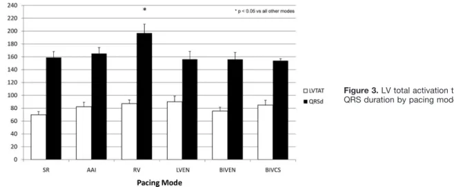
Corresponding changes in LV total endocardial activation time and QRS duration (derived from NCM and the surface ECG, respectively) are shown in Figure 3. LV total activation time (LVTAT) was not reduced in any mode versus sinus rhythm. QRS duration was significantly increased by RV pacing versus all other modes (P⬍0.05).
Site Specificity of Response to LV Stimulation Importance of Zones of Slow Conduction
For each endocardial and epicardial LV lead position, we ascertained whether the lead tip was inside or outside a zone
of slow conduction, as defined by NCM. The mean hemody-namic response was expressed as a percentage change from baseline and is shown in Figure 4, plotted against the LV lead position in relation to the zone of slow conduction.
LV stimulation within an area of slow conduction was associated with a reduction in the degree of hemodynamic response in all LV endocardial pacing configurations (P⬍0.001).
LVTAT and QRS duration were calculated according to the position of the LV lead in or outside of areas of slow conduction. The mean LVTAT when the LV lead was in an area of slow conduction was 78.6⫾22.4 ms versus 84.9⫾21.8 ms when the lead was not within these areas (P⫽0.59). The corresponding values for QRS duration were 148.6⫾25.5 and 154.9⫾32.9 ms, respectively (P⫽0.95). LV total activation time and QRS duration were not shortened when the LV lead was outside the area of slow conduction, suggesting that mechanism of hemodynamic benefit may be an improvement in mechanical efficiency, which is not underpinned by more rapid electric activation of the myocardium.
Importance of LV Lead Position by Region
For LV endocardial pacing configurations, there was consid-erable variability in the degree of hemodynamic response in different regions of the LV endocardium. The smallest acute hemodynamic response across all patients in LV-EN mode
Figure 3.LV total activation time and QRS duration by pacing mode.
Figure 4.Effect of LV lead position in relation to areas of slow conduction on acute hemodynamic response, shown as percentage change from baseline sinus rhythm.
was 12.6⫾10.4% (range, 0% to 28.2%) from baseline and the largest increase was 40.6⫾23.0% (range, 16.8% to 92.5%). In BIVEN mode, the smallest increase was 12.8⫾10.1% (range,
⫺0.12% to 29.3%) and the largest was 40.8⫾21.9% (range, 16.8% to 90.6%).
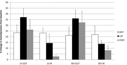
The hemodynamic response to pacing in these modes is shown in Figure 5, according to the relationship of the lead position to areas of slow conduction.
When the LV endocardial lead was outside an area of slow conduction, a lateral position of the LV lead was optimal, with posterior position being the next best. An anterior position of the LV lead gave the least hemodynamic response in this situation. When the LV lead was in an area of slow conduction, the hemodynamic response was superior when the LV lead was placed anteriorly.
Hemodynamic response according to the position of the endocardial LV lead in the long axis of the heart is shown in Figure 6. There were no significant differences in acute hemodynamic response between basal, mid, and apical LV lead positions.
Comparison of NCM With DE-CMR
Table 2 summarizes MRI and NCM patient characteristics. Ten patients had nonischemic etiology of heart failure on the basis of the history, a normal coronary angiogram, and
absence of subendocardial late enhancement on MRI. Of these, none had late gadolinium enhancement of any distri-bution on CMR. Six of 10 patients with nonischemic cardio-myopathy had lines of conduction block,17 despite the ab-sence of late enhancement on DE-CMR (Figure 7).
This suggests that DE-CMR is not capable of identifying areas of slow conduction in this patient group. However, late enhancement was demonstrated in all 5 patients with ische-mic cardiomyopathy. In this group, there was a reasonable correlation between areas of slow conduction and distribution of scar, as shown in Figure 8.
Discussion
Hemodynamic Effects of Endocardial and Epicardial Pacing
This small mechanistic study demonstrates that endocardial LV pacing has the potential to confer a superior hemodyna-mic benefit compared with conventional CRT delivered from the coronary sinus. The degree of acute hemodynamic re-sponse at the optimal site was surprisingly high in both endocardial and epicardial LV pacing configurations (79.8⫾49.0% and 59.6⫾49.5%, respectively). This reflects the fact that this was a highly selected population with marked LV dyssynchrony (with mean QRS duration of
Figure 5.Change in hemodynamics from sinus rhythm by LV pacing region (short axis) and position of the endocardial LV lead in relation to zone of slow conduction.
Figure 6.Change in hemodynamics from sinus rhythm by LV pacing region (long axis) and position of the endocardial LV lead in relation to zone of slow conduction.
164 ms) in which a nonischemic heart failure etiology predominated. Furthermore, comparison was made with base-line sinus rhythm rather than fixed rate atrial pacing, so a proportion of this percentage increase may be secondary to rate-dependent changes in LV dP/dt.
Importantly, the site of optimal benefit differs widely between patients. Pacing within areas of slow conduction was associated with a lesser degree of acute hemodynamic bene-fit. Areas of fibrosis/scarring correlating with NCM could be seen with DE-CMR in all patients with ischemic cardiomy-opathy. However, 60% of patients with nonischemic cardio-myopathy had areas of slow conduction associated with poor response that could not be predicted using DE-CMR.
These findings are in keeping with those of a recent acute hemodynamic study5that demonstrated that endocardial and epicardial pacing in the same region of the left ventricle gave rise to similar improvements in LV dP/dt max. Garrigue et al18evaluated the chronic effects of biventricular endocardial pacing and demonstrated improved systolic performance with this approach compared with conventional CRT. One key mechanism of potential benefit of endocardial over epicardial pacing may be that this approach facilitates pacing outside areas of slow conduction because a greater area of the myocardium is accessible when lead delivery is not con-strained by the coronary venous anatomy. In addition, an endocardial to epicardial mechanical activation is more phys-iological. Endocardial pacing reproduces the gradient of LV contraction in systole in an endocardial to epicardial direc-tion19 engaging the subendocardial Purkinje network. This may result in more rapid myocardial recruitment, maximizing the contractile response of the viable recruited myocytes. The difference between the angles of fiber orientation between the
endocardial and epicardial layers could also facilitate this more rapid myocardial recruitment.
Electric Versus Mechanical Mechanisms of Benefit
We have also demonstrated that the acute hemodynamic benefit from LV pacing is independent of LV endocardial activation time, which is not reduced in comparison with sinus rhythm. These findings suggest that the mechanism of acute hemodynamic benefit may be more effective mechan-ical recruitment of the LV myocardium rather than shortening of the LV electric activation time. Detailed MR and mapping studies in the canine ventricle have also shown that mechan-ical benefit occurs independent of electric resynchroniza-tion.20Clinically, the concept that mechanical resynchroniza-tion is a more important mechanism of benefit of CRT is supported by the fact that positioning of the LV lead at the site of latest mechanical activation confers the maximal clinical benefit.3,21 The magnitude of the hemodynamic re-sponse will depend on the rate of myocardial recruitment, the viability of the contracting tissue, and the proportion of the ventricle that can contribute to the stroke volume.
Variations in Hemodynamic Response to LV Pacing
We have shown that LV lead positioning within zones of slow conduction is associated with a lesser degree of acute hemodynamic response. This is consistent with the results of a previous study that we conducted that evaluated the hemodynamic response from pacing inside and outside zones of slow conduction.10 This is likely to reflect less effective capture of the myocardium and slower mechanical propaga-tion across the left ventricle. This mechanism may in part explain the significant intraindividual and interindividual variability that has been found in other studies in the optimal LV lead position in patients undergoing CRT.4,5In our study, a lateral position of the LV endocardial lead was shown to be superior to posterior or anterior lead positions when pacing outside areas of slow conduction. This is consistent with the premise that resynchronization therapy is best delivered using a laterally positioned electrode to reverse the effect of LV dyssynchrony, which gives rise to late activation of the lateral LV wall. When the endocardial LV lead was positioned within an area of slow conduction, it appeared that an anterior position may achieve greater hemodynamic benefit than a lateral position. This is in keeping with the finding in other studies of patients with ischemic cardiomyopathy that posi-tioning the LV lead in an area of posterolateral scar confers an adverse outcome.7,22,23
Electroanatomic and MRI Correlates
The relationship between myocardial scar and areas of late enhancement on DE-CMR in patients with ischemic cardio-myopathy is well known.24In our study, there was a corre-lation between areas of scar and zones of slow conduction, as previously noted.11
Myocardial fibrosis can also be visualized using this technique in patients with nonischemic cardiomyopathy in some cases.12However, in our study, areas of slow conduc-tion were demonstrated using NCM in 60% of patients with
Table 2. MRI and NCM Patient Characteristics
Patient No. Etiology Late Enhancement Conduction Block Activation Pattern 1 ICM Yes No I 2 DCM No Yes I* 3 DCM No No I 4 ICM Yes No I 5 DCM No Yes II 6 DCM No Yes II 7 DCM No NA NA 8 DCM No Yes I*
9 ICM Yes Yes I*
10 DCM No Yes II
11 ICM Yes No I
12 DCM No Yes II
13 DCM No No I
14 ICM Yes Yes II
15 DCM No No I
ICM indicates ischemic cardiomyopathy; DCM, dilated cardiomyopathy. Type I activation pattern represents smooth, homogeneous activation of the LV from septum to lateral wall. Type II describes a U-shaped activation pattern around a region of conduction block.8
*Very lateral line of conduction block resulted in type I activation pattern.
nonischemic cardiomyopathy in whom no late enhancement was seen using DE-CMR. It is feasible that cardiac MR is not capable of detecting the low degrees of diffuse fibrosis, which are thought to be associated with slow conduction in this condition.25 Late gadolinium enhancement using CMR re-mains a qualitative rather than quantitative evaluation. It provides a relative comparison of degrees of scarring or fibrosis in different regions. As a result, homogeneous diffuse fibrosis at the microscopic level may not manifest as late enhancement to the observer. An alternative explanation for the failure of CMR to detect zones of slow conduction is that fibrosis is not present in these areas but that other properties of the myocardium are affected by the disease process. This may include gap junction or ion channel remodeling, which interfere with activation wave front propagation.
Clinical Implications
Current understanding of response to CRT in patients with ischemic cardiomyopathy is that overall scar burden as well as scar density in proximity to the LV lead tip are associated with an adverse or diminished clinical or echocardiographic response to CRT.7,26,27Our findings suggest that the presence of areas of slow conduction may account for the variability in response to LV pacing in nonischemic as well as ischemic cardiomyopathy. Although these areas can be visualized in ischemic cardiomyopathy using DE-CMR, this is not the case for patients with nonischemic cardiomyopathy. This is a potential explanation for lack of response to CRT in a significant proportion of this patient population and rein-forces the need for positioning the LV lead on an individual basis. An alternative approach to addressing this problem is
Figure 7.Upper panel, Short-axis stack of DE-CMR images of a patient with non-ischemic cardiomyopathy. There is no evidence of myocardial scar or fibrosis. Middle panel, Dynamic substrate map of the same patient in anteroposterior proj-ection. The zone of slow conduction is delineated by a solid white line, which covers the lateral aspect of the anterior LV wall. Virtual unipolar electrograms from 2 points equidistant (34 mm) from the array are shown in yellow from within (left) and outside the zone of slow con-duction (right). The sample electrogram from within the region of slow conduction is fractionated and of lower amplitude, demonstrating that the reduced amplitude of electrograms in these regions is not artifact arising as a result of distance from the array. Lower panel, Images 1 to 10 show the activation map of the same patient in 25-ms steps (LAO view on the left of each image, RAO view on the right). The wave front is seen to propa-gate from the anterior septum toward the lateral LV wall. When it reaches the line of conduction block on the border of the slow conduction zone, the activation wave front passes inferior to this region, around the LV apex, and on to the poste-rior wall.
the use of multipolar leads or multisite pacing, thereby avoiding areas of slow conduction that are associated with a diminished response.
Our data suggest that the principal mechanism of acute hemodynamic benefit from CRT is an improvement in LV mechanical rather than electric activation. Endocardial LV or biventricular pacing gives rise to a superior hemodynamic response compared to conventional CRT. The clinical utility
of this approach remains limited at present due to the risk of thromboembolic complications and left-sided valvular endocarditis.
However, when transvenous delivery of the coronary sinus lead has failed, endocardial pacing represents a viable alter-native to surgical LV lead placement, in particular if the risks of general anesthetic are high. Another group of patients who may benefit from this approach are nonresponders to
conven-Figure 8.Upper panel, Late-enhancement images of patient 11 with a previous myo-cardial infarction affecting the territory of the left anterior descending coronary artery. Middle panel, Dynamic substrate map of patient 11 in the right lateral proj-ections. The zone of slow conduction is delineated by a solid white line, which covers the anterior aspect of the LV wall. Virtual unipolar electrograms (yellow) from within (left) and outside the zone of slow conduction (right) are equidistant (30 mm) from the array. A sample electrogram from within the area of slow conduction is seen to be fractionated and of low ampli-tude, whereas the electrogram from a region of normal myocardium at the same distance from the array is of greater amplitude. Lower panel, Activation map of patient 11. The progression of the depo-larization wave front can be seen crossing from a breakout point in the septum toward the lateral wall (images 1 to 4). The wave front then reaches a line of block and regresses before passing inferi-orly and posteriinferi-orly (images 5 to 8) to ac-tivate the rest of the LV.
tional CRT without suitable coronary venous anatomy for epicardial lead placement.
Study Limitations
Our study involved a small population of carefully charac-terized of patients, and further work is required to confirm these findings in a larger population. Our protocol incorpo-rated pacing endocardially in the left ventricle at multiple sites to assess the effect of different endocardial positions. The order of pacing sites was not randomly selected and therefore this could be a source of bias. We have acquired information on LV endocardial activation times and QRS duration (reflecting biventricular activation). We do not have full information on transmural LV activation, which would be too invasive to acquire in vivo. It is recognized that the accuracy of NCM reduces with increasing cavity size of the LV, which represents an important limitation in this study of patients with LV dilatation. The mean equatorial distance from the center of the array to the endocardial surface in this study was 37⫾11 mm. In the study by Schilling et al16 validating NCM against contact electrograms, perfect timing matches were obtained as far as 52 mm from the center of the array, although differences in timing of electrograms in-creased gradually at distances over 34 mm. Chamber dilata-tion may therefore be anticipated to affect accuracy of activation time measurement rather than identification of regions of slow conduction.
Conclusions
Endocardial CRT may offer a potential for a superior re-sponse in patients undergoing CRT. Our data suggest that the response to endocardial pacing is site-specific and is nega-tively affected by stimulation within area of slow conduction or fibrosis. In patients with ischemic cardiomyopathy, DE-CMR can identify these areas and may be used to guide LV lead placement. DE-CMR was not able to detect areas of slow conduction in patients with nonischemic cardiomyopathy. It is feasible that LV lead positioning within such areas would confer an adverse response to therapy, analogous to position-ing of the LV lead in an area of scar in patients with ischemic cardiomyopathy. Therefore, this may represent a mechanism of nonresponse to CRT in this patient group.
Sources of Funding
This work was supported by St Jude Medical, Stratford-upon-Avon, United Kingdom.
Disclosures
Dr Ginks received an educational grant from St Jude Medical. Dr Lambiase received an educational grant and is a member of the speaker bureau for St Jude Medical and received funding from the National Institute for Health Research. Drs Razavi and Rhode received funding from the European Commission Framework Pro-gramme 7 and the Engineering and Physical Sciences Research Council—Medical Research Council. Marcus Simon is an employee of St Jude Medical. Dr Rinaldi is an advisor to St Jude Medical and Medtronic.
References
1. Singh JP, Fan D, Heist EK, Alabiad CR, Taub C, Reddy V, Mansour M, Picard MH, Ruskin JN, Mela T. Left ventricular lead electrical delay
predicts response to cardiac resynchronization therapy.Heart Rhythm. 2006;3:1285–1292.
2. Alonso C, Leclercq C, Victor F, Mansour H, de Place C, Pavin D, Carre F, Mabo P, Daubert JC. Electrocardiographic predictive factors of long-term clinical improvement with multisite biventricular pacing in advanced heart failure.Am J Cardiol. 1999;84:1417–1421.
3. Ypenburg C, van Bommel RJ, Delgado V, Mollema SA, Bleeker GB, Boersma E, Schalij MJ, Bax JJ. Optimal left ventricular lead position predicts reverse remodeling and survival after cardiac resynchronization therapy.J Am Coll Cardiol. 2008;52:1402–1409.
4. van Campen CMC, Visser FC, de Cock CC, Vos HS, Kamp O, Visser CA. Comparison of the haemodynamics of different pacing sites in patients undergoing resynchronisation treatment: Need for individuali-sation of lead localiindividuali-sation.Heart. 2006;92:1795–1800.
5. Derval N, Steendijk P, Gula LJ, Deplagne A, Laborderie J, Sacher F, Knecht S, Wright M, Nault I, Ploux S, Ritter P, Bordachar P, Lafitte S, Re´ant P, Klein GJ, Narayan SM, Garrigue S, Hocini M, Haissaguerre M, Clementy J, Jaïs P. Optimizing hemodynamics in heart failure patients by systematic screening of left ventricular pacing sites: the lateral left ven-tricular wall and the coronary sinus are rarely the best sites.J Am Coll Cardiol. 2010;55:566 –575.
6. van Deursen C, van Geldorp IE, Rademakers LM, van Hunnik A, Kuiper M, Klersy C, Auricchio A, Prinzen FW. Left ventricular endocardial pacing improves resynchronization therapy in canine left bundle-branch hearts.Circ Arrhythm Electrophysiol. 2009;2:580 –587.
7. Bleeker GB, Kaandorp TAM, Lamb HJ, Boersma E, Steendijk P, de Roos A, van der Wall EE, Schalij MJ, Bax JJ. Effect of posterolateral scar tissue on clinical and echocardiographic improvement after cardiac resyn-chronization therapy.Circulation. 2006;113:969 –976.
8. Fung JWH, Chan JYS, Yip GWK, Chan HCK, Chan WWL, Zhang Q, Yu C-M. Effect of left ventricular endocardial activation pattern on echocar-diographic and clinical response to cardiac resynchronization therapy.
Heart. 2007;93:432– 437.
9. Jacobson JT, Afonso VX, Eisenman G, Schultz JR, Lazar S, Michele JJ, Josephson ME, Callans DJ. Characterization of the infarct substrate and ventricular tachycardia circuits with noncontact unipolar mapping in a porcine model of myocardial infarction.Heart Rhythm. 2006;3:189 –197. 10. Lambiase PD, Rinaldi A, Hauck J, Mobb M, Elliott D, Mohammad S, Gill JS, Bucknall CA. Non-contact left ventricular endocardial mapping in cardiac resynchronisation therapy.Heart. 2004;90:44 –51.
11. Codreanu A, Odille F, Aliot E, Marie P-Y, Magnin-Poull I, Andronache M, Mandry D, Djaballah W, Re´gent D, Felblinger J, de Chillou C. Electroanatomic characterization of post-infarct scars: comparison with 3-dimensional myocardial scar reconstruction based on magnetic res-onance imaging.J Am Coll Cardiol. 2008;52:839 – 842.
12. Assomull RG, Prasad SK, Lyne J, Smith G, Burman ED, Khan M, Sheppard MN, Poole-Wilson PA, Pennell DJ. Cardiovascular magnetic resonance, fibrosis, and prognosis in dilated cardiomyopathy.J Am Coll Cardiol. 2006;48:1977–1985.
13. Strickberger SA, Conti J, Daoud EG, Havranek E, Mehra MR, Pina IL, Young J. Patient selection for cardiac resynchronization therapy: from the Council on Clinical Cardiology Subcommittee on Electrocardiography and Arrhythmias and the Quality of Care and Outcomes Research Inter-disciplinary Working Group, in collaboration with the Heart Rhythm Society.Circulation. 2005;111:2146 –2150.
14. Lima JAC, Judd RM, Bazille A, Schulman SP, Atalar E, Zerhouni EA. Regional heterogeneity of human myocardial infarcts demonstrated by contrast-enhanced MRI: potential mechanisms. Circulation. 1995;92: 1117–1125.
15. Rhode KS, Hill DLG, Edwards PJ, Hipwell J, Rueckert D, Sanchez-Ortiz G, Hegde S, Rahunathan V, Razavi R. Registration and tracking to integrate x-ray and MR images in an XMR facility. IEEE Trans Med Imaging. 2003;22:1369 –1378.
16. Schilling RJ, Peters NS, Davies DW. Simultaneous endocardial mapping in the human left ventricle using a noncontact catheter: comparison of contact and reconstructed electrograms during sinus rhythm.Circulation. 1998;98:887– 898.
17. Auricchio A, Fantoni C, Regoli F, Carbucicchio C, Goette A, Geller C, Kloss M, Klein H. Characterization of left ventricular activation in patients with heart failure and left bundle-branch block. Circulation. 2004;109:1133–1139.
18. Garrigue S, Jaïs P, Espil G, Labeque J-N, Hocini M, Shah DC, Haïssa-guerre M, Clementy J. Comparison of chronic biventricular pacing between epicardial and endocardial left ventricular stimulation using
Doppler tissue imaging in patients with heart failure. Am J Cardiol. 2001;88:858 – 862.
19. Derumeaux G, Ovize M, Loufoua J, Pontier G, Andre-Fouet X, Cribier A. Assessment of nonuniformity of transmural myocardial velocities by color-coded tissue Doppler imaging: characterization of normal, ische-mic, and stunned myocardium.Circulation. 2000;101:1390 –1395. 20. Leclercq C, Faris O, Tunin R, Johnson J, Kato R, Evans F, Spinelli J,
Halperin H, McVeigh E, Kass DA. Systolic improvement and mechanical resynchronization does not require electrical synchrony in the dilated failing heart with left bundle-branch block.Circulation. 2002;106:1760 –1763. 21. Ansalone G, Giannantoni P, Ricci R, Trambaiolo P, Fedele F, Santini M.
Doppler myocardial imaging to evaluate the effectiveness of pacing sites in patients receiving biventricular pacing.J Am Coll Cardiol. 2002;39: 489 – 499.
22. White JA, Yee R, Yuan X, Krahn A, Skanes A, Parker M, Klein G, Drangova M. Delayed enhancement magnetic resonance imaging predicts response to cardiac resynchronization therapy in patients with intraven-tricular dyssynchrony.J Am Coll Cardiol. 2006;48:1953–1960. 23. Chalil S, Foley PWX, Muyhaldeen SA, Patel KCR, Yousef ZR, Smith
REA, Frenneaux MP, Leyva F. Late gadolinium
enhancement-cardiovas-cular magnetic resonance as a predictor of response to cardiac resynchro-nization therapy in patients with ischaemic cardiomyopathy.Europace. 2007;9:1031–1037.
24. Kim RJ, Fieno DS, Parrish TB, Harris K, Chen E-L, Simonetti O, Bundy J, Finn JP, Klocke FJ, Judd RM. Relationship of MRI delayed contrast enhancement to irreversible injury, infarct age, and contractile function.
Circulation. 1999;100:1992–2002.
25. Kawara T, Derksen R, de Groot JR, Coronel R, Tasseron S, Linnenbank AC, Hauer RNW, Kirkels H, Janse MJ, de Bakker JMT. Activation delay after premature stimulation in chronically diseased human myocardium relates to the architecture of interstitial fibrosis.Circulation. 2001;104: 3069 –3075.
26. Adelstein EC, Saba S. Scar burden by myocardial perfusion imaging predicts echocardiographic response to cardiac resynchronization therapy in ischemic cardiomyopathy.Am Heart J. 2007;153:105–112. 27. Ypenburg C, Schalij MJ, Bleeker GB, Steendijk P, Boersma E,
Dibbets-Schneider P, Stokkel MP, van der Wall EE, Bax JJ. Impact of viability and scar tissue on response to cardiac resynchronization therapy in ischaemic heart failure patients.Eur Heart J. 2007;28:33– 41.
CLINICAL PERSPECTIVE
The absence of clinical response in 30% to 40% of patients receiving cardiac resynchronization therapy poses a great challenge to heart failure clinicians and device implanters. It is well documented that positioning of the left ventricular (LV) lead in areas of myocardial scar in patients with ischemic cardiomyopathy is associated with a diminished response to cardiac resynchronization therapy (CRT). Regions of slow conduction exist in both nonischemic and ischemic cardiomyopathy that can be delineated using noncontact mapping, whereby the electrophysiological properties of a chamber can be characterized using a multielectrode array. Using this technique, we evaluated the effect of pacing inside and outside regions of slow conduction on acute hemodynamic response to CRT. Procedures were performed in a combined x-ray and MRI environment so that tissue characterization by delayed-enhancement cardiac magnetic resonance imaging could be correlated with electrophysiological assessment. Both endocardial and transvenous epicardial LV pacing were performed with the hypothesis that endocardial pacing may be more effective as a result of reproducing the physiological pattern of activation of the LV myocardium, as well as lack of constraint by the coronary venous anatomy. We found that zones of slow conduction could be identified using delayed-enhancement cardiac magnetic resonance in patients with an ischemic heart failure etiology but not in nonischemic cardiomyopathy. The acute effect of CRT was superior in response to endocardial compared with epicardial pacing. Stimulation within zones of slow conduction was associated with a diminished response to CRT. This is a potential explanation for lack of response to CRT and reinforces the need for positioning the LV lead on an individual basis.