EXPRESSION OF INTERLEUKIN-37 (IL37) AND THE
GENETIC VARIATIONS OF IL37 IN RELATION TO CHRONIC PERIODONTITIS
Shaoping Zhang
A thesis submitted to the faculty of the University of North Carolina at Chapel Hill in partial fulfillment of the requirements for the degree of Master of Science in the Department of
Periodontology in the School of Dentistry.
Chapel Hill 2014
Approved by:
Steven Offenbacher
James D. Beck
ii © 2014 Shaoping Zhang
iii ABSTRACT
Shaoping Zhang: Expression of Interleukin-37 (IL37) and the Genetic Variations of IL37 in Relation to Chronic Periodontitis
(Under the direction of Steven Offenbacher)
Objectives: To study the transcription of IL37 in gingival biopsies and the association between transcription of IL1B, IL6 and IL37 and genotypes at the rs3811046 and
rs3811047 IL37 loci. Materials and Methods: Gingival biopsies were collected from chronic periodontitis patients and control subjects. Pyrosequencing was applied for
genotyping. Real-time PCR was used to examine the transcription of genes from biopsies and
cell culture models. Results: Transcription of IL6 was significantly higher in periodontitis samples. Transcription of IL37 was induced in oral epithelial and THP.1 cells by LPS. The
presence of a minor allele at both loci in IL37 was significantly related to a reduced IL6
transcription in Caucasians, while the major allele was associated with a higher transcription
of IL6 in African Americans. Conclusion: Gingival epithelium and monocytic cells are the major source for IL-37. The genetic variants of IL37 related to the altered transcription of IL6
iv
TABLE OF CONTENTS
LIST OFTABLES……….………..…………..…vi
LIST OF FIGURES……….…....vii
LIST OF ABBREVATIONS……….….viii
CHAPTER I Review: Interleukin-37 (IL37), a novel anti-inflammatory IL-1 family member………...1
Introduction ………..2
Anti-inflammatory mechanisms involved in periodontal disease………….….3
Genetic variation of genes involved in inflammatory response and periodontal disease……….5
Structure, expression profile, activity and genetic variation of interleukin-37 (IL37)……….…….8
Statement of purpose, hypothesis and specific aims………...12
References……….…...15
II Expression of Interleukin-37 (IL37) and the Genetic Variations of IL37 in Relation to Chronic Periodontitis……….………..19
Introduction……….…...20
Materials and methods……….……24
Results……….…...29
Discussion………...……….……35
v
Figures………..44
vi
LIST OF TABLES
Table 1. Primers and sequencers used for IL37 genotyping…….……….26
Table 2. Demographic information and periodontal indicies………42
Table 3. Genotypes at rs3811026 locus of IL37 in the study participants………42
Table 4. Genotypes at rs3811047 locus of IL37 in the study participants………43
vii
LIST OF FIGURES
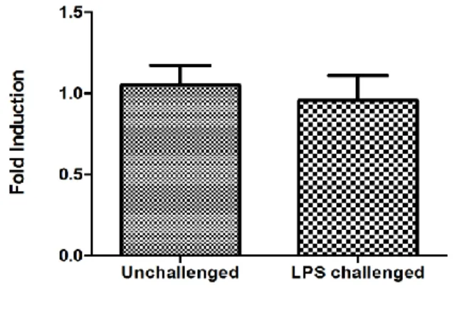
Figure 1. Transcriptional level of IL37, ILB and IL6 in gingival biopsy samples from
sites with periodontitis and periodontal health………... 44
Figure 2. Transcription of IL37, IL1B and IL6 in samples with different genotypes at
the rs3811026 locus of IL37....……….………….………46
Figure 3. Transcription of IL37, IL1B and IL6 in samples with different genotypes at
the rs3811046 locus of IL37 from Caucasian participants………48
Figure 4. Transcription of IL37, IL1B and IL6 in samples with different genotypes at
the rs3811046 locus of IL37 from African American participants………....…………50
Figure 5. Trasncriptaion of IL6 in samples with different genotypes at the rs3811047
locus of IL37 from Caucaisan and African American participants…………...52
Figure 6. Immunofluorescence staining of IL-37 in gingival biopsies………...51
Figure 7. Transcription of IL1B, IL6 and IL37 in gingival fibroblasts upon challenge
with E. Coli LPS (1μg/ml) at different time poin...………...…...55
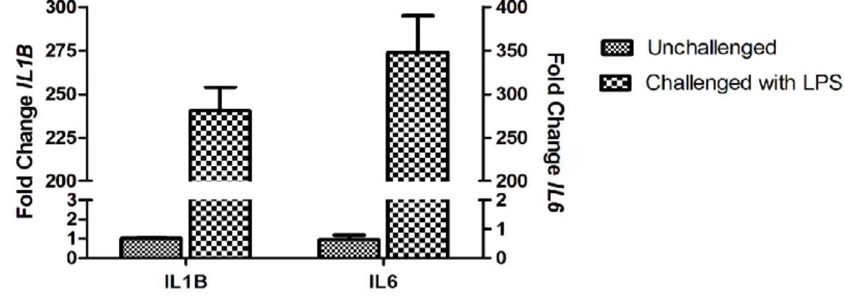
Figure 8. Transcription of IL1B, IL6 and IL37 in THP.1 cells upon challeng with E. Coli
LPS (1μg/ml) at different time points………..……..56
Figure 9. Transcription of IL6, IL1B and IL37 in an oral epithelial cell line challenged
viii
LIST OF ABBREVIATIONS
BOP Bleeding upon probing
CAL Clinical attachmen tloss
CG Cytosine-guanine dinucleotide
DSS Dextran sodium sulfate
FcγR Fc receptors for immunoglobulin G (IgG)
GCF Gingvial crevicular fluid
GWAS Genom wide association study
HGF Human gingival fibroblast
Ig Immunoglobulin
IFNG/IFN-γ Interferon-γ
IL1 Interleukin-1
IL1A Interleukin-1α
IL1B/IL-1β Interleukin-1 β
IL1F7 Interleukin-1 family member 7
IL33 Interleukin-33
IL37/IL-37 Interleukin-37
IL6/IL-6 Interleukin-6
IL8 Interleukin-8
IL10 Inlerleukin-10
LPS Lipopolysaccharide
NSAID Non steroidal anti-inflammatory drug
MMP Matrix metalloproteinase protein
PAI-I Plasminogen activator inhibitor-1
ix
PI Plaque index
PTGS2/Cox-2 Prostaglandin-endoperoxide synthase-2
RT-PCR Real-time polymerase chain reaction
SNP Single nucleotide polymorphisms
TGF-β Transformation growth factor- β
TIMP Tissue inhibitors of metalloproteinase
TLR Toll-like receptor
TNFA/TNF-α Tumor necrosis factor-α
1
CHAPTER 1: REVIEW OF INTERLEUKIN-37 (IL37), A NOVEL ANTI-INFLAMMATORY IL-1FAMILY MEMBER
Introduction
Periodontal disease has been described as an abnormal inflammatory response to the
pathogenic bacteria present in the bioflim. The uncontrolled inflammatory and immune
responses initiated by the bacteria are the major etiology of periodontal destruction1, 2
Activation of the innate immune system in periodontal disease leads to the rapid secretion of
various chemokines and cytokines, such as interleukin-1β, (IL-1β), IL-6, IL-8, TNF-α, etc.,
most of which propagate inflammation and may potentially cause destruction in
periodontium. For example, an elevated level of gingival crevicular fluid (GCF) IL-1β has
previously been established as a robust biomarker for a hyper-inflammatory phenotype and
for mediating severe inflammation, bone loss and periodontal disease progression3. Although
feedback mechanisms might be present to dampen inflammation, only several secreted
molecules have been described to possess anti-inflammatory activities, such as resolvins,
IL-10 and transforming growth factor β (TGF-β) among more than 50 known cytokines4.
Pathways to downregulate the extent and severity of inflammation in hosts may provide
insight for the therapeutic potential to reduce the destruction caused by periodontal disease.
The course of inflammation is not only determined by the proinflammatory molecules
but critically modified by resolution mechanisms that counteract the inflammation induced
by pathogens. The disequilibrium of the pro- and anti- inflammatory pathways in
2
supporting apparatus. During the prolonged battle with pathogenic microorganisms that are
present in the biofilm, the host may have developed dampening mechanisms that inhibit the
uncontrolled inflammation. It has been suggested that periodontal inflammation resulting in
the destruction of tooth-supporting tissue is partially due to the lack of adequate resolution
activities 5.
Anti-inflammatory mechanisms involved in periodontal disease
A class of small lipid molecules, which are termed resolvins, has been elucidated to
possess the capacity to promote the resolution of inflammation5, 6. Resolvins are derived from
eicosapentaenoic acid and the production of resolvins shares the similar pathway of the
metabolism of arachidonic acid. The activities of resolvins include reduction of neutrophile
trafficking, control of cytokine production and reactive oxygen species, and mitigation of the
magnitude of the inflammatory responses. Van Dyke et al. proved that the application of
resolving E1 in a rabbit model at the same time when the experimental periodontitis was
induced inhibited the onset and progression of periodontal destruction5. Interestingly,
resolving E1 also demonstrated therapeutic efficacy because the application of this molecule
in the established periodontitis in the rabbit model completely eliminated inflammation and
the pocket depth was returned to pre-ligature condition5.
Another molecule that possesses the inflammation resolving activity is transforming
growth factor-β (TGF-β). TGF-β antagonizes IL-1 activity, and it also promotes both the
synthesis of extracellular matrix proteins and proteinase inhibitors, such as tissue inhibitors
of metalloproteinases (TIMPs) and plasminogen activator inhibitor-1 (PAI-I), which inhibit
the synthesis of matrix metalloproteinase proteins (MMPs) 7. Matsuda et al. reported that
3
ligament fibroblastic cells 8. In addition, the stimulation with TGF-β progressively decreased
the expression of procollagenase and increased both the mRNA and protein levels of TIMP 9.
TGF-β has also been identified to play a pivotal role in the mediation of the
immunosuppressive activity of regulatory T (Treg) cells 10,11. In gingival crevicular fluid, the
level of TGF-β was elevated in both chronic and aggressive forms of periodontitis and such
an increase was interpreted as a mechanism to act against detrimental immune and
inflammatory responses and, therefore, may prevent further loss of periodontium 10.
Epigenetic modifications have recently been suggested as one of the most important
regulatory mechanism for cytokine production in inflammatory disease including
periodontitis 12, 13. DNA methylation and histone modifications, which are two major types of
epigenetic regulation, modulate host response at the chromatin level to environmental
stressors, including smoking, nutrient deficiency, bacterial infection, inflammation, etc 13. It
is well known that stimuli as mentioned above can regulate cytokine gene expression through
various signaling pathways. However, the enrichment of transcriptional factors in the nuclei
induced by signal transduction may not account for the only driving force for gene
expression. The state of chromatin confirmation, to which mammalian genes are packed,
determines the transcriptional readiness of genes when the orchestration of signal
transduction is not impaired and transcriptional factors are available14, 15. Therefore
epigenetic regulation also plays a fundamental role in the control of gene expression. DNA
methylation, which almost always occurs to the cytokine nucleotide in the cytokine-guanine
(CG) dinucleotides, is a more stable epigenetic mark and less likely to be reversed as
4
We have proposed that the persistence challenge from the pathogenic microflora in
the biofilm may modify the methylation level of the promoter regions of certain host
inflammatory genes that are involved in the pathogenesis of chronic periodontitis. Employing
clinical gingival biopsies, we have discovered a hypermethylation pattern within the
promoter region of prostaglandin-endoperoxide synthase-2 (PTGS2 or COX2) and tumor
necrosis factor α (TNFA) 17, 18. Using the same biological samples, we also found that the
transcriptional level of both PTGS2 and TNFA genes was dampened and failed to elevate in
comparison to the control samples that exhibited periodontal health 17. Such a
hypermethylation pattern in the promoter region and an inhibited transcriptional profile also
confirm the generally accepted paradigm that the degree of promoter methylation is inversely
related to the transcriptional levels 19. Since TNF-α and PGE2, in which PTGS2 is a key
regulatory gate for its production, are among the most critical pro-inflammatory cytokines in
periodontal disease, the dysfunction of the regulatory mechanism for those molecules leads
to disease progression. The increased methylation level change in the promoter regions of
those genes may reflect a metastable mechanism that can create a new “set-point” of the
inflammatory homeostasis in the disease state 18. Chronic periodontitis is a slow-progressing
inflammatory/infectious disease featured by episodic periodontium destruction with long
intervals of quiescence. In the chronic battle with the periodontal pathogens the host may
develop a dampening compensatory mechanism through DNA methylation that helps to
control the pronounced inflammatory response and therefore serves to prevent the
unrestricted further tissue destruction.
5
Periodontal disease, similar to most other complex inflammatory diseases, is a
polygenic disorder involving gene to gene interaction. The susceptibility to periodontal
disease, which is an inflammatory disease exhibiting a complex genetic trait, has been
explored through association studies aiming to target genetic markers and candidate
disease-modifying genes by identification of single nucleotide polymorphisms (SNPs). Many
previous studies have discovered important SNPs encoding molecules of the host defense
system in chronic periodontitis 20, 21. Kornman et al first described a composite IL1
polymorphism including -889bp (C to T transition) in IL1A and +3953bp (C to T transition)
in IL1B that was significantly related to the severity of periodontitis in non-smoking
Caucasians 22. Additionally, monocytes from individuals with homozygous IL1B +3953T
allele were shown to produce 4-fold more IL-1β, and monocytes with heterozygous IL1B
+3935T allele exhibited two-fold more IL-1β than cells without this SNP at the +3953 locus 22, 23. Several later studies relating this composite IL1 polymorphism to either susceptibility to
or severity of periodontal disease showed diverse outcomes. Papapanou et al in a
cross-sectional study found that although this IL1 genotype failed to distinguish control subjects
from patients with periodontitis, it was related to the severity of periodontal disease 24.
Walker and colleagues reported from a cross-sectional study that IL1 composite
polymorphism is not associated with the susceptibility to localized aggressive periodontitis in
an African American population 25. Armitage et al demonstrated that this composite was
almost non-existent in a Chinese population and didn’t show any association with the onset
of periodontal disease 26. McGuire and Nunn in a longitudinal study discovered that this
composite polymorphism was related to the tooth loss 27. From those studies investigating the
6
concluded that IL1 genotype might be regarded as a severity factor in certain racial groups,
especially non-smoking Caucasians population48.
Other studies examined the association of polymorphisms of several other
inflammatory genes with periodontal disease. A recent meta-analysis showed that the T
variant at -819bp locus of IL10 seemed to be a risk factor among Caucasians for chronic
periodontitis 28. Similarly, the A allele at -592bp locus of the same gene was also associated
with an increased risk for chronic periodontitis in Caucasian population. Another recent
meta-analysis concluded that -308bp A/G variations in the promoter region of TNFA is
significantly related to the susceptibility to periodontitis in Brazilian, Asian and Turkish
population 29. The same meta-analysis also found that the G/C polymorphism at -174bp IL6
may increase the susceptibility to periodontitis in Brazilians, while the G/C variation at
-572bp locus of the same gene is more prevalent in European decedents with chronic
periodontitis29. It is evident that the identified association of certain SNPs present in genes
that are critically involved in immune responses in periodontal disease is usually
population-based and usually cannot be generalized across different racial groups.
In addition to cytokine genes, polymorphisms of other genes linking cellular and
humoral immune responses to bacterial infection in periodontium have also been investigated
extensively. Fc receptors for immunoglobulin G (IgG) or FcγRs play a pivotal role in
phagocytosis of macrophages, neutrophil activation, and antibody-dependent cell-mediated
cytotoxicity of natural killer cells. The phagocytic activity of macrophages is triggered by
opsonization, a process that the FcγR on the macrophage surface binds Fc portion of
antibodies that coat the invading bacteria. The presence of polymorphisms in the FcγR gene
7
inflammatory diseases such as rheumatoid arthritis, systemic lupus erythematosus and
periodontitis 30. So far several studies have targeted the association between various
polymorphisms of different FcγR genes and periodontal disease. Yamatomo et al reported
that the polymorphism in the FcγRIIa gene resulting in a homozygous H/H at the 131 residue
was associated with chronic periodontitis in a Caucasian population 31. However, Sugita and
colleagues found that a G to T transition at nucleotide 559 within FcγRIIIa that results in an
amino acid substitution at 158 from valine to phenylalanine was significantly related to the
recurrence of chronic periodontitis in a Japanese population32 . A study by Yasuda et al
demonstrated that rather than polymorphism that resulting in the H allele in FcγRIIa, the A
allele at the nucleotide 646 -184 in the intron 4 of FcγRIIb gene was over represented in
chronic periodontitis patients as compared to healthy control in a Japanese population33.
Similar to studies of IL1 composite polymorphism, the association between a specific allele
or genotype in the FcγR genes and periodontal disease is affected by the racial groups.
Structure, expression profile, activity and genetic variation of Interleukin-37 (IL37) We have recently completed a genome-wide association study (GWAS) of 4910
Caucasians with known levels of GCF IL-1β to identify candidate genes whose changes due
to polymorphism are associated with elevated secretion of GCF IL-1β. We have identified
two novel quantitative trait loci with missense mutations within the coding region of IL37
that are both inversely and significantly associated with high GCF-IL-1β levels. IL37 was
previously designated as IL-1 family member 7 (IL1F7) 34. IL37 is mapped to the cluster of
genes on human chromosome 2, which contains other IL-1 family members except IL18 and
IL33. IL37 May be exclusively expressed in human, since the mouse homolog hasn’t been
8
and five splicing variants present (IL1F7a-e). IL1F7b has been found to be the largest and
include five of those six exons. The protein structure of IL37 comprises a 12 β-barrel strand
and shares the common pattern of other IL-1 family particularly that of IL-18 4, 35. Although
none of the variants of IL-37 contains a typical signal peptide, both b-isoform and c-isoform
encode a putative caspase-1 cleavage site. Many cell types or tissues have been reported to
express IL-37 35. The constitutive IL37 mRNA has been found in the testis, thymus, uterus
and colon35. Upon induction, peripheral blood mononuclear cells (PBMCs), dendritic cells
and colon epithelial cells can express IL-3736, 37. It has been reported that microbial products
such as lipopolysaccharide (LPS) that bind to toll-like receptors (TLRs) constitute the strong
stimulation for IL-37 expression 36. In addition, this molecule can be also detected in
monocytes, plasma cells and breast carcinoma cells at the protein level 35. Similar to IL18,
the control of IL37 expression is mainly at the post-transcriptional level by an instability
element encoded in exon 5 in its coding region. Although it is structurally similar to other
IL-1 family members, IL-37 has recently been reported to possess a unique and potent
anti-inflammatory activity, which is in sharp contrast to the pro-anti-inflammatory activity of IL-1β
and IL-18 36. Because mice don’t have known structural homolog of IL37, transgenic mice
carrying human IL37 offers an excellent tool for the studying the role of IL37 in
inflammation38. McNamee and colleagues compared the susceptibility of transgenic mice
carrying human IL37 with control mice to colitis induced by dextran sodium sulfate (DSS) in
drinking water 38. DSS-mediated colonic inflammation is significantly reduced in the
transgenic mice, and such amelioration is associated with a reduced leukocyte infiltration and
dampened expression of IL-1β, and TNF-α. Nold et al. observed a significant attenuation of
9
IL37 transgenic mice 36. In addition to weaker hypothermia, transgenic mice achieved
respiratory compensation of the metabolic acidosis, protection against a rise in blood
potassium concentration, and prevention of liver damage in the LPS induced endotoxic
model. Those physiological findings in IL37 transgenic mice were paralleling a reduction in
various cytokines, such as IL-6, IL-1β, IFN-γ in different compartments as compared with
control wild-type mice. Those protected effects observed in IL37 transgenic mice showed
dosage response because homozygotes always demonstrated more pronounced protection
from LPS induced endotoxic shock and less pro-inflammatory cytokine production than
heterozygotes 36.
The anti-inflammatory activity of IL37 can be further demonstrated in vitro cell
culture experiment. THP-1 cells, a human monocytic cell line, transfected with IL37
exhibited reduced expression of several inflammatory molecules such as 1β, TNF and
IL-8 upon challenge by LPS 36.Similar pattern of reduction in those pro-inflammatory cytokines
upon IL-1β challenge was also observed in epithelial cell line A549 transfected with IL37 36.
Further mechanistic studies have shown that the anti-inflammatory activity of IL-37 is
mediated by enhancing TGF-β signals through a direct association with Smad3, a
transcriptional factor that constitutes the major effector of signaling downstream of TGF-β 4, 36. From both in vivo and in vitro experimental models, it has been clearly indicated that
IL-37 is a natural suppressor of innate inflammatory and immune responses that reduces the
extent and severity of inflammatory insult.
Other than colitis, hepatitis, dermatitis and rheumatoid arthritis, the activity of IL-37
hasn’t been explored in other diseases 39, 38. The knowledge gap of how IL-37 is involved in
10
provided some clues of the involvement of IL-37 in the inflammatory pathogenesis of
periodontal disease. The first trait locus contains two non-synonymous, coding single
nucleotide polymorphisms (SNPs) within exon 2 [rs3811046 T/G and rs3811047 G/A] that
result in a predicted altered IL-37 protein structure [(A34V) and (A42T)], respectively. These
two SNPs demonstrate linkage disequilibrium because they are almost always present in the
same minor allele. This variant 1 [IL37v1] is relatively common with a minor allelic
frequency of 30% and is found to be significantly associated with high levels of IL-1β within
the GCF [OR=1.70, (1.53-1.89), p=6.8 X10⁻²¹]. This p value is statistically significant, as it
readily exceeds the strict genome-wide significance threshold value of p<5X10⁻⁸, which is
required by Bonferroni correction for multiple comparisons. This IL37 variant is also
associated with more severe periodontal disease [e.g. greater inflammation with higher
gingival index scores (GI, p=0.016), greater extent probing depth>4mm (PD4+mm, p=0.026)
and greater bone loss as reflected in attachment loss >4mm (AL4+mm, p=0.04)]. A second,
less common locus within IL37 (8.5% frequency of minor allele) has 5 non-synonymous,
coding SNPs [rs2708943(exon 4), rs2723183(exon 4) , rs2723187(exon 5) , rs2708947(exon
6) and rs2723192 (exon 6), OR=1.52 (1.31-1.84), p<4.2X10⁻⁷]. The association between this
trait locus with IL-1β within the GCF is approaching to the significance threshold. The
predicted protein sequence for this variant [IL37V2] is estimated to have a high probability of
inducing structural alterations in IL-37 (p=0.04). The data from this unpublished study
suggest a similar anti-inflammatory role of IL37 in periodontal disease because the predicted
alteration of protein structure due to the missense mutations present in both trait loci may
disrupt the normal activities of this cytokine in periodontium. The altered protein structure of
11
biofilm in such a way that skews the inflammatory responses to favor the destruction of
attachment and bone resportion.
According to literature and data from our GWAS study, we hypothesize that genetic IL37 variants cause a functional defect in IL-37 that is associated with an excessive pro-inflammatory innate immune response in chronic periodontitis. Epidemiological studies have shown that approximately 90% of the US population exhibits gingivitis, with 48% of
the US adult population having periodontitis40, 41. More importantly, evidence suggests that
periodontal disease carries potential risks for worsening various systemic conditions
including atherosclerosis, stroke, diabetes, and etc42, 43, 44. Genetics plays a role in the
progression of periodontal disease. Studies by Michalowicz in monozygotic and dizygotic
twins provided the first estimates of heritability and suggested that about half (48%) of the
variance in disease could be attributable to genetics 45. Identification of important genetic risk
variants that contribute to periodontal disease is the approach to study diseases with complex
trait, such as periodontal disease. Several genetic polymorphisms of several
pro-inflammatory molecules, such as IL1, FcγRs, TNFA, have been associated with periodontitis 46, 47. However, no association linking IL37 and periodontal disease has been reported so far.
Based on our GWAS study, IL37 genetic variation may confer the increased susceptibility to
chronic periodontitis due to functional alteration of this molecule.
One of the scientific merits of this study is to understand how a genetic basis of an
anti-inflammatory gene, IL37, is involved in the presentation of chronic periodontitis.
Unraveling the genetic basis of pathogenesis will lead to improved diagnosis and prognoses
to identify individuals at risk for disease to optimize prevention strategies. In addition,
12
helps to develop effective and novel therapies to target this anti-inflammatory molecule to
restore periodontal health.
Statements of purpose, hypothesis and specific Aims:
The role of IL-37 hasn’t been explored in periodontal disease. Our GWAS study
suggested an anti-inflammatory activity of IL37 in the pathogenesis of periodontal disease.
We are seeking to understand the expression pattern of IL37 in gingival biopsies and
compare the transcriptional level of IL37 in biological samples from chronic periodontitis
patients with control subjects of periodontal health. We are also aiming to study the
association of SNPs in the first IL37 trait locus identified from our GWAS data with the
transcriptional expression of inflammatory genes including IL37, IL1B and IL6. Further, we
will use in vitro cell culture models representing different cell populations present in human
gingiva to study the expression profile of IL37 upon stimulation by E. coli LPS. Two specific
aims are developed to test our hypothesis that genetic IL37 variants are associated with an altered pro-inflammatory innate immune response in chronic periodontitis.
Aim 1: to compare the transcriptional expression levels of IL37, IL1B and IL6 in biopsies collected from chronic periodontitis patients (case) and subjects with periodontal health (control); and characterize the IL37 genotypes (rs3811046 T/G and rs3811047 G/A in the first trait locus) and correlate different IL37 genotypes with the transcriptional profiles of those genes;
In this aim, we will first to identify the expression pattern of IL-37 in biopsied
gingival tissues by immuno- fluorescence technique in frozen sections and then compare the
transcriptional level of IL37 in gingival biopsies collected from periodontitis patients with
13
carrying two SNPs (rs3811046 T/G and rs3811047 G/A) by extracting DNA from biopsies
and pyro-sequencing technique. We will also associate the presence of minor allele and
different genotypes of IL37 with periodontal disease status and transcriptional profiles of
those inflammatory genes including IL1B and IL6, in addition to IL37.
Aim2: to investigate the kinetic of IL37 transcriptional expression in gingival fibroblasts, THP.1 Cells and oral keratinized cells in the response to E. Coli LPS challenge
We will analyze the transcription of IL37, IL1B and IL6 by real-time PCR in vitro in
three different cell cultures that represent the major cell populations present in human
gingiva in response to the challenge by E.Coli PLS. This information will help to map the
14 REFERENCES
1. Darveau RP, Tanner A, Page RC. The microbial challenge in periodontitis. Periodontol 2000 1997;14:12-32.
2. Madianos PN, Bobetsis YA, Kinane DF. Generation of inflammatory stimuli: how bacteria set up inflammatory responses in the gingiva. J Clin Periodontol 2005;32 Suppl 6:57-71.
3. Lang NP, Tonetti MS, Suter J, Sorrell J, Duff GW, Kornman KS. Effect of interleukin-1 gene polymorphisms on gingival inflammation assessed by bleeding on probing in a periodontal maintenance population. J Periodontal Res 2000;35(2):102-107.
4. Kim G, Kronenberg M. Cooling the fires of inflammation. Proc Natl Acad Sci U S A 2011;108(40):16493-16494.
5. Van Dyke TE. Control of inflammation and periodontitis. Periodontol 2000 2007;45:158-166.
6. Freire MO, Van Dyke TE. Natural resolution of inflammation. Periodontol 2000 2013;63(1):149-164.
7. Holla LI, Fassmann A, Benes P, Halabala T, Znojil V. 5 polymorphisms in the transforming growth factor-beta 1 gene (TGF-beta 1) in adult periodontitis. J Clin Periodontol 2002;29(4):336-341.
8. Matsuda N, Lin WL, Kumar NM, Cho MI, Genco RJ. Mitogenic, chemotactic, and synthetic responses of rat periodontal ligament fibroblastic cells to polypeptide growth factors in vitro. J Periodontol 1992;63(6):515-525.
9. Overall CM, Wrana JL, Sodek J. Independent regulation of collagenase, 72-kDa progelatinase, and metalloendoproteinase inhibitor expression in human fibroblasts by transforming growth factor-beta. J Biol Chem 1989;264(3):1860-1869.
10. Gurkan A, Emingil G, Cinarcik S, Berdeli A. Gingival crevicular fluid transforming growth factor-beta1 in several forms of periodontal disease. Arch Oral Biol 2006;51(10):906-912.
11. Wahl SM, Chen W. Transforming growth factor-beta-induced regulatory T cells referee inflammatory and autoimmune diseases. Arthritis Res Ther 2005;7(2):62-68.
12. Barros SP, Offenbacher S. Modifiable risk factors in periodontal disease: epigenetic regulation of gene expression in the inflammatory response. Periodontol 2000
2014;64(1):95-110.
15
14. Bierne H, Hamon M, Cossart P. Epigenetics and bacterial infections. Cold Spring Harb Perspect Med 2012;2(12):a010272.
15. Lod S, Johansson T, Abrahamsson KH, Larsson L. The influence of epigenetics in relation to oral health. Int J Dent Hyg 2014;12(1):48-54.
16. Bird AP, Wolffe AP. Methylation-induced repression--belts, braces, and chromatin. Cell 1999;99(5):451-454.
17. Zhang S, Barros SP, Moretti AJ, et al. Epigenetic regulation of TNFA expression in periodontal disease. J Periodontol 2013;84(11):1606-1616.
18. Zhang S, Barros SP, Niculescu MD, Moretti AJ, Preisser JS, Offenbacher S. Alteration of PTGS2 promoter methylation in chronic periodontitis. J Dent Res 2010;89(2):133-137.
19. Bird AP, Wolffe AP. Methylation-induced repression--belts, braces, and chromatin. Cell 1999;99(5):451-454.
20. Zhang J, Sun X, Xiao L, Xie C, Xuan D, Luo G. Gene polymorphisms and periodontitis. Periodontol 2000 2011;56(1):102-124.
21. Laine ML, Morre SA, Murillo LS, van Winkelhoff AJ, Pena AS. CD14 and TLR4 gene polymorphisms in adult periodontitis. J Dent Res 2005;84(11):1042-1046.
22. Kornman KS, Crane A, Wang HY, et al. The interleukin-1 genotype as a severity factor in adult periodontal disease. J Clin Periodontol 1997;24(1):72-77.
23. Pociot F, Molvig J, Wogensen L, Worsaae H, Nerup J. A TaqI polymorphism in the human interleukin-1 beta (IL-1 beta) gene correlates with IL-1 beta secretion in vitro. Eur J Clin Invest 1992;22(6):396-402.
24. Papapanou PN, Neiderud AM, Sandros J, Dahlen G. Interleukin-1 gene polymorphism and periodontal status. A case-control study. J Clin Periodontol 2001;28(5):389-396.
25. Walker SJ, Van Dyke TE, Rich S, Kornman KS, di Giovine FS, Hart TC. Genetic polymorphisms of the IL-1alpha and IL-1beta genes in African-American LJP patients and an African-American control population. J Periodontol 2000;71(5):723-728.
26. Armitage GC, Wu Y, Wang HY, Sorrell J, di Giovine FS, Duff GW. Low prevalence of a periodontitis-associated interleukin-1 composite genotype in individuals of Chinese heritage. J Periodontol 2000;71(2):164-171.
16
28. Zhong Q, Ding C, Wang M, Sun Y, Xu Y. Interleukin-10 gene polymorphisms and chronic/aggressive periodontitis susceptibility: a meta-analysis based on 14 case-control studies. Cytokine 2012;60(1):47-54.
29. Song GG, Choi SJ, Ji JD, Lee YH. Association between tumor necrosis factor-alpha promoter -308 A/G, -238 A/G, interleukin-6 -174 G/C and -572 G/C polymorphisms and periodontal disease: a meta-analysis. Mol Biol Rep 2013;40(8):5191-5203.
30. Chai L, Song YQ, Leung WK. Genetic polymorphism studies in periodontitis and Fcgamma receptors. J Periodontal Res 2012;47(3):273-285.
31. Yamamoto K, Kobayashi T, Grossi S, et al. Association of Fcgamma receptor IIa genotype with chronic periodontitis in Caucasians. J Periodontol 2004;75(4):517-522.
32. Sugita N, Yamamoto K, Kobayashi T, et al. Relevance of Fc gamma RIIIa-158V-F polymorphism to recurrence of adult periodontitis in Japanese patients. Clin Exp Immunol 1999;117(2):350-354.
33. Yasuda K, Sugita N, Kobayashi T, Yamamoto K, Yoshie H. FcgammaRIIB gene polymorphisms in Japanese periodontitis patients. Genes Immun 2003;4(8):541-546.
34. Dunn E, Sims JE, Nicklin MJ, O'Neill LA. Annotating genes with potential roles in the immune system: six new members of the IL-1 family. Trends Immunol 2001;22(10):533-536.
35. Boraschi D, Lucchesi D, Hainzl S, et al. IL-37: a new anti-inflammatory cytokine of the IL-1 family. Eur Cytokine Netw 2011;22(3):127-147.
36. Nold MF, Nold-Petry CA, Zepp JA, Palmer BE, Bufler P, Dinarello CA. IL-37 is a fundamental inhibitor of innate immunity. Nat Immunol 2010;11(11):1014-1022.
37. Imaeda H, Takahashi K, Fujimoto T, et al. Epithelial expression of interleukin-37b in inflammatory bowel disease. Clin Exp Immunol 2013;172(3):410-416.
38. McNamee EN, Masterson JC, Jedlicka P, et al. Interleukin 37 expression protects mice from colitis. Proc Natl Acad Sci U S A 2011;108(40):16711-16716.
39. Bulau AM, Fink M, Maucksch C, et al. In vivo expression of interleukin-37 reduces local and systemic inflammation in concanavalin A-induced hepatitis. ScientificWorldJournal 2011;11:2480-2490.
40. Albandar JM. Epidemiology and risk factors of periodontal diseases. Dent Clin North Am 2005;49(3):517-32, v-vi.
17
(University of Washin. Prevalence of periodontitis in adults in the United States: 2009 and 2010. J Dent Res 2012;91(10):914-920.
42. Humphrey LL, Fu R, Buckley DI, Freeman M, Helfand M. Periodontal disease and coronary heart disease incidence: a systematic review and meta-analysis. J Gen Intern Med 2008;23(12):2079-2086.
43. Mealey BL, Oates TW, American Academy of Periodontology. Diabetes mellitus and periodontal diseases. J Periodontol 2006;77(8):1289-1303.
44. Beck JD, Offenbacher S. Systemic effects of periodontitis: epidemiology of periodontal disease and cardiovascular disease. J Periodontol 2005;76(11 Suppl):2089-2100.
45. Michalowicz BS, Diehl SR, Gunsolley JC, et al. Evidence of a substantial genetic basis for risk of adult periodontitis. J Periodontol 2000;71(11):1699-1707.
46. Kornman KS, Crane A, Wang HY, et al. The interleukin-1 genotype as a severity factor in adult periodontal disease. J Clin Periodontol 1997;24(1):72-77.
47. Schulz S, Schlitt A, Lutze A, et al. The importance of genetic variants in TNFalpha for periodontal disease in a cohort of coronary patients. J Clin Periodontol 2012;39(8):699-706.
18
CHAPTER 2: EXPRESSION OF INTERLUKIN-37 (IL37) AND THE GENETIC VARIATIONS OF IL37 IN RELATION TO CHRONIC PERIODONTITIS Introduction
Periodontitis is caused by the abnormal inflammatory and immune responses initiated
by the bacteria 1, 2. Activation of the innate immune system in periodontal disease results in
the synthesis and secretion of various chemokines and cytokines, such as interleukin-1β,
(IL-1β), IL-6, IL-8, tumor necrosis factor-α (TNF-α), etc., most of which propagate inflammation
and may potentially cause destruction in periodontium. However, the periodontal destruction
caused by inflammation is usually episodic and slow-progressing in chronic periodontitis
patients. The homeostasis of inflammation, though altered in periodontal disease, must be
maintained by anti-inflammatory mechanisms in the long intervals between breakdowns of
periodontal supporting apparatus. So far several molecules have been described to possess
anti-inflammatory activities, such as resolvins, IL-10 and transforming growth factor β
(TGF-β) 3, 4, 5. Pathways to down regulate the extent and severity of inflammation in hosts
may provide insight for the novel therapeutic potential to reduce the destruction caused by
periodontal disease.
Periodontal disease, similar to most other complex inflammatory diseases, is a
polygenic disorder involving gene to gene interaction and exhibits complex genetic traits.
The susceptibility to periodontal disease has been explored by association studies aiming for
targeting genetic markers and candidate disease-modifying genes by identification of single
19
in genes encoding molecules of the host defense system in chronic periodontitis 6, 7. Kornman
et al first described a composite IL1 polymorphism including -889bp (C to T transition) in
IL1A and +3953bp (C to T transition) in IL1B that was significantly related to the severity of
periodontitis in non-smoking Caucasians 8. Additionally, monocytes from individuals with
homozygous IL1B +3953T allele were shown to produce 4-fold more IL-1β, and monocytes
with heterozygous IL1B +3935T allele exhibited two-fold more IL-1β than cells without this
SNP at the +3953 locus 8, 9. It has been concluded that IL1 genotypes might be considered as
a risk factor for the severity of periodontitis in certain racial groups, especially non-smoking
Caucasians population (Lindhe J clinical Periodontology 4th edition). Other association studies have identified certain SNPs of other genes involved in immune response including
IL10, TNFA, Fc receptor for immunoglobulin G (FcRs), etc 10, 11, 12. All those studies have
suggested that the association of certain SNPs with periodontitis is usually race specific and
cannot be generalized across all racial groups.
From a genome-wide association study (GWAS) including 4910 Caucasians with
known levels of gingival crevicular fluid (GCF) IL-1β, we have identified two novel
quantitative trait loci with missense mutations within the coding region of IL37 that are both
inversely and significantly associated with GCF-IL-1β levels. IL37, which was previously
designated as IL-1 family member 7 (IL1F7) 13, is mapped to the cluster of genes on human
chromosome 2, which contains other IL-1 family members except IL18 and IL33.
Sequencing analyses have shown that IL1F7 gene structure contains 6 exons and five
splicing variants present (IL1F7a-e) with IL1F7b to be the best characterized. Upon
induction with bacterial endotoxins such as lipopolysaccharine (LPS), peripheral blood
20
Although it is structurally similar to other IL-1 family members, IL-37 has been reported to
possess a unique anti-inflammatory activity 14. McNamee and colleagues 16 discovered that
the dextran sodium sulfate (DSS)-mediated colonic inflammation is significantly reduced in
the IL37 transgenic mice, and such amelioration is associated with a reduced leukoc yte
infiltration and dampened expression of IL-1β, and TNF-α. Nold et al. observed a significant
attenuation of the effects induced by non-lethal endotoxic shock through LPS intraperitoneal
injection in IL37 transgenic mice 14.Those physiological findings in IL37 transgenic mice
paralleled a reduction of a panel of pro-inflammatory cytokines, such as IL-6, IL-1β, IFN-γ
in different compartments as compared with control wild-type mice. 14.
The anti-inflammatory activity of IL37 was further demonstrated in vitro cell cultures.
THP-1 cells transfected with IL37 exhibited reduced expression of several inflammatory
molecules such as IL-1β, TNF-α and IL-8 upon challenge by LPS 14.Similar pattern of
reduction in those pro-inflammatory cytokines upon IL-1β challenge was also observed in
epithelial cell line A549 transfected with IL37 14.
The activity of IL-37 hasn’t been explored in diseases other than colitis, hepatitis,
dermatitis and rheumatoid arthritis 17, 16 yet. The knowledge gap of how IL-37 is involved in
periodontal disease pathogenesis remains to be filled. Our GWAS Caucasian study has
provided some clues of the involvement of IL-37 in the pathogenesis of periodontal disease.
The identified novel trait locus contains two non-synonymous, coding single nucleotide
polymorphisms (SNPs) within exon 2 [rs3811046 T/G and rs3811047 G/A] that result in a
predicted alteration of IL-37 protein structure [(A34V) and (A42T)], respectively. These two
SNPs demonstrate linkage disequilibrium because they are almost always present in the same
21
to be significantly associated with high levels of IL-1β within the GCF [OR=1.70,
(1.53-1.89), p=6.8 X10⁻²¹].
In this study, we are seeking to understand the expression pattern of IL37 in gingival
biopsies and compare the transcriptional level of IL37 in biological samples collected from
chronic periodontitis patients with control subjects with periodontal health. We are also
aiming to study the association of both SNPs in this identified IL37 trait locus (rs3811046
and rs3811047) with the transcriptional expression of inflammatory genes including IL37,
IL1B and IL6. Further, we will use in vitro cell culture models representing different cell
populations present in human gingiva to study the expression profile of IL37 upon
stimulation by E. coli LPS. By employing both in vivo and in vitro approaches, we will test
our hypothesis that genetic IL37 variants are associated with an altered pro-inflammatory
innate immune response in chronic periodontitis. Materials and Methods
Participants and tissue collection
This study was approved by the Biomedical Institutional Review Board of the
University of North Carolina at Chapel Hill (UNC). Written informed consent was obtained
from all study participants. All subjects are aged between 18-70 years of different races
including Caucasians, African Americans, Hispanics, Asians and others.
In this cross-sectional clinical study, a total of 61 subjects were enrolled. 24 of the
subjects were patients diagnosed with chronic periodontitis, exhibiting probing depth
(PD) >4mm and clinical attachment loss >3mm. Another 37 subjects served as control
subjects with periodontal health, which is defined by no site of PD greater than 3mm or
22
of Periodontics, University of North Carolina at Chapel Hill, School of Dentistry, while
control subjects were enrolled either from the General and Oral Health Clinical Center at the
School of Dentistry who volunteered for this study after screening or from the Department of
Periodontics and presented for clinical crown lengthening procedure. Major exclusion criteria
are: 1) use of either antibiotics or non-steroidal anti-inflammatory drugs (NSAIDs) within 1
month before enrollment; 2) medical treatment for systemic diseases 3 months prior to the
gingival biopsy or periodontal surgery; 3) active infectious diseases, pregnancy, cancer or
systemic diseases with oral manifestations; 4) severe unrestored caries or aggressive
periodontitis. Periodontal clinical measurements, including probing depth (PD), clinical
attachment loss (CAL), bleeding on probing (BOP), and plaque index (PI) will be obtained
prior to the biopsies. Biopsies obtained from periodontitis patients were collected from the
most severe site during the scheduled periodontal surgeries.
Upon removal, all biopsied gingival tissues were divided at least into two pieces. One
piece, used for genotyping analysis, was kept in the −80°C freezer immediately, while the
other half, used for real-time polymerase chain reaction (RT-PCR), was incubated with
RNA-later (Applied Biosystems/Ambion, Austin, TX, USA) overnight at 4°C, and then
transferred to −80°C freezer for storage. In addition, another piece of tissue from 5 chronic
periodontitis biopsies and 5 control biopsies were immersed in Tissue-Tek® O.C.T™
compound (Sakura Finetek, Netherland) and snap frozen by dry ice and then stored in −80°C
freezer for histological study.
DNA isolation and genotyping of IL37
Genomic DNA was extracted from the collected gingival biopsies using a DNeasy
23
and sequencers designed by Assay Design Software 2.0 (QIAgen) for SNP detection were
listed in table 1. For each PCR amplicon generated by forward and reserve primers was
directly genotyped in a real-time DNA synthesis approach by prosequencing technology. In
brief, 6-8 ul of PCR product was mixed with 2ul of streptavidin-coated sepharose beads and
40ul of PyroMark binding buffer (QIAgen). After captured by the PyroMark vacuum prep
filter tips (QIAgen), the beads to which biotin-labeled single strand PCR product attached
were washed sequentially with 70% ethanol, denaturing buffer, and PyroMark washing
buffer, and then released into a PyroMark Q96 HS plate (QIAgen) onto which 12ul of
PyroMark annealing buffer mixed with 3.6pmol of sequencer corresponding to each
amplicon has already been preloaded. After heated for 2 minutes at 86°C, the plate was then
loaded onto a PSQ HS 96 Pyrosequencing system (QIAgen). The sequencing was determined
by Pro Gold CDT reagents (QIAgen). The presence of variation or the copy of the minor
allele (heterozygote or homozygote) was determined by the pyrogram pattern based on the
peak height.
Table 1. Primers and sequencers used for IL37 genotyping
SNP sites Sequences AnNealing Temperature (°C)
Rs3811046 and Rs3811047
Rs2708943 and Rs2723183
Forward: 5’-TGCTAACCTCACTGCGTCTGA C-3’
Reverse: 5’-ATCACCTCACCCCGA GGC-3’
Sequencer: 5’-CCTTACTTGTGTGAACAAA-3’
Forward: 5’-AAAGGGCTGA GTCTTCCATTT-3’
Reverse: 5’- TTGTGATCCTGGTCATGAATG-3’
Sequencer:5’- TCTGGTGATATTGATCTA GG-3’
65
63
24
A human monocytic cell line (THP.1), which was originally purchased from
American Type Culture Collection (ATCC, Manassas, VA) , was maintained in RPMI 1640
culture medium (Invitrogen, Grand Island, NY) supplemented with 10% fetal bovine serum
(FBS) (Mediatech, Manassas, VA), 5X10⁻⁵ M 2-mercaptoethanol (Sigma-Aldrich, St. Louise,
MO), and 1% penicillin/streptomycin (Invitrogen). Cells with a density of 1X10⁶ were
seeded into a 6-well plate just before use.
A primary human gingival fibroblast cell culture (HGF), which was originally
purchased from ATCC, was maintained in DMEM culture medium supplemented with 10%
fetal bovine serum (FBS, Mediatech) and 1% penicillin/streptomycin (Invitrogen). 0.4X10⁵
cells were seeded into a 6-well plate 48 hours prior to challenge.
A primary human gingival epithelial progenitor cell culture (HGEPp, CELLnTEC
Advanced Cell Systems, Bern, Swizerland) was maintained in CnT basal medium
(CnT-BM.1) supplemented with CnT-24.S (CELLnTEC Advanced Cell Systems). 0.5X10⁶ cells
were seeded into a 24-well plate 24 hours prior to challenge.
E.coli Lipopolysaccharide (LPS) (Sigma-Aldrich) was diluted with each individual
cell culture medium and added to each cell culture to achieve either 100ng/μl or 1μg/μl
according to experimental protocols. After challenging for different intervals, cells were
collected and RNA was isolated.
RNA isolation and quantitative Real-time RT-PCR
Total RNA was isolated from gingival biopsy samples and cells with a RNeasy Mini
Kit (QIAgen, Valencia, CA). cDNA from 300-600ng isolated RNA was synthesized with a
SuperScript VILO cDNA synthesis kit (Invitrogen). Quantitative Real-time RT-PCR of the
gene-25
expression assay mix (Hs01555410_m1, Hs00985639_m1, and Hs00367201_m1,
respectively, Applied Biosystems/Ambion, Austin, TX) in a 7000 Sequence Detection
System (Applied Biosystems/Ambion). A TaqMan® GAPDH control reagents kit was used
as an internal control (Applied Biosystems/Ambion).
Immunofluorescence
The O.C.T embedded and frozen preserved biopsy samples were stored at -80°C
freezer until use. After cryostat sectioning, the frozen gingival biopsies from periodontal
health and chronic periodontitis were fixed with 70% ethanol for 30 seconds and then
immersed in acetone for another 15 minutes. After fixation, the tissue slides were blocked for
1 hour at room temperature, and then incubated overnight a t 4°C with monoclonal antibody
specific for IL-37 (6.7μg/ml, Cat #1975-IL, R&D Systems, Minneapolis, MN) or vehicle.
The blocking PBS buffer contains 8% bovine serum albumin, and 0.1% Triton-X-100, and
the antibody was diluted with the same blocking buffer. After vigorous washing with 1X
PBS containing 0.1% Triton-X-100 for, the slides were incubated for another hour at room
temperature with a donkey anti-goat IgG secondary antibody labelled with northenlights 557
Fluorochrome (NL007, R & D Systems). Sections were then washed with PBS, mounted
with ProLong Gold antifade reagent with DAPI (Invitrogen) and coverslipped.
Immunofluorescence signals were observed using confocal microscopy (Carl Zeiss LSM 710
Confocal Microscope, Thornwood, NY).
Statistics:
Pearson’s chi-square or exact Pearson chi square test was applied to test the null
hypothesis that the distribution of genotypes at both rs3811046 and rs3811047 loci was not
26
populational major alleles and heterozygous alleles/homozygous for populational minor
alleles. The same statistical analysis was also performed to test the gender difference between
periodontitis patient group and control group of subjects with periodontal health.
Two sample student-t test was used to detect the difference of periodontal indices and
age between diseased and control groups. To compare the transcriptional expression of
cytokines between diseased and control subjects, we applied non-parametric
Wilcoxon-Mann-Whitney rank test. Using the same statistical analysis, we compared the transcription
of those cytokines between genotypes at both loci in Caucasian subjects and
African-American subjects. The level of significance was set at p=0.05.
Results
Demographic information of the clinical subjects:
The demographic information, periodontal indices and racial status of the study
participants are listed in Table 2. The mean age of patients with chronic periodontitis was significantly older than subjects with periodontal health (Table 2, p=0.014). The proportion
of males in the chronic periodontitis group was significantly higher than the control group
(56% vs. 30%, respectively, p=0.04, Table 2). The distribution of different racial groups was
not significantly different between the two groups of participants. As expected, sites with
chronic periodontitis exhibited significantly higher measurements for both probing depth and
attachment loss when compared to the periodontally healthy sites (p<0.0001 for bo th, Table
2).
Genotypes of IL37 in the clinical subjects:
21 out of 37 (56.8%) Caucasian subjects and all the participants from Hispanic, Asian
27
genotype (or 1.1), which is in contrast to only 2 out of 17 (11.8%) African Americans who
were homozygous for “T” allele at this locus. The prevalence for 1.1 genotype at this locus
was significantly higher in non-African American subjects than African American
participants (p=0.0005, Table 3). However, the prevalence of 1.1 genotype and other
genotypes TG/GG (or 1.2/2.2) in periodontitis group was not significantly different from that
of periodontal health group (p=0.3, data not shown).
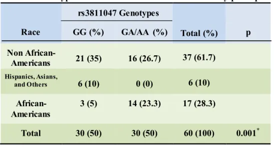
Similarly, 27 out of 44 (61.4%) non-African American subjects presented
homozygous “G” genotype (GG or 1.1) at the rs3811047 locus, while only 3 out of 17
(17.6%) African Americans were homozygous for “G” allele. The distribution of 1.1
genotype was significantly different among different racial groups with higher percentage of
GA/AA genotype (or 1.2/2.2) at this locus in African American subjects than in non-African
American group (Table 3, p=0.001). When disease status was considered, again the
prevalence of 1.1 genotype in periodontitis patients was not statistically different from the
subjects with periodontal health (p=0.4, data not shown).
Changes of transcriptional levels of cytokines IL37, IL1B and IL6 in gingival biopsies with periodontitis
The transcriptional level of IL37 was not significantly different from gingival
biopsies between periodontitis and periodontal health group (p=0.97, Figure 1A). The
transcriptional level of IL1B from the gingival biopsies with periodontitis was higher than
that in the periodontal healthy group, though the difference was also not statistically
significant (p=0.11, Figure 1B). However, the messenger level of IL6 was significantly
elevated in samples collected from chronic periodontitis patients in comparison to biopsies
28
Transcriptional levels of cytokines IL37, IL1B and IL6 in gingival biopsies in relation to genotypes of IL37 at rs3811046 and rs3811047 loci
Because the presence of genetic variations in the loci of IL37 may affect its
transcriptional activity, messenger stability, and the transcription activity of other
proinflammatory cytokine genes, we observed the messenger levels of IL1B and IL6, in
addition to IL37, in relation to the genotypes of IL37 at both rs3811046 and rs3811047 loci.
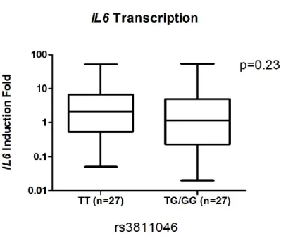
The transcriptional levels of IL37, IL1B and IL6 were not significantly different between the
homozygous major T allele (TT or 1.1) and TG/GG genotypes (1.2/2.2) at rs3811046 locus
(p=0.15, 0.97, and 0.23, respectively, Figure 2A, 2B and 2C).
Because the G (2) minor allele was more prevalent only in African American subjects,
which is different from other racial groups, we next compared the transcriptional levels of
those cytokine genes between different genotypes stratified by race. Since there were very
few non-Caucasian and non-African American subjects, we only focused on Caucasian and
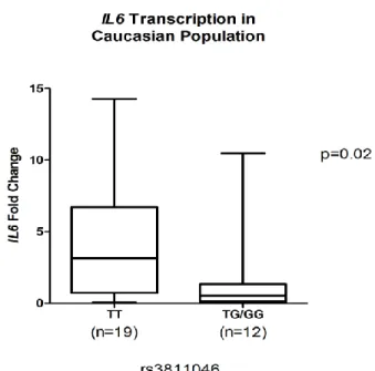
African American participants. In Caucasian subjects, the transcriptional level of IL37 in
gingival samples positive for TT (1.1) genotype was not significantly different from samples
with TG/GG (1.2/2.2) genotypes at the rs3811046 locus (p=0.12, Figure 3A). Nor was the
transcriptional level of IL1B from samples with 1.1 genotype significantly different from
biopsies with 1.2/2.2 genotypes (p=0.11, Figure 3B). However, a trend of higher level of
IL37 and IL1B was present in the 1.1 genotype. The messenger level of IL6 in samples
present GG (1.1) genotype was significantly higher than TG/TT (1.2/2.2) samples in
Caucasian participants (p=0.02, Figure 3C).
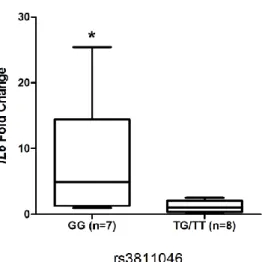
In African American participants who exhibited a G (or 2) dominating allele, we
29
allele genotype (GG or 2.2) with samples present for other allelic variations (GT/TT or
2.1/1.1). We found that the transcriptional expression of both IL37 and IL1B from samples
with 2.2 genotypes was not significantly different from samples present 1.1/1.2 genotypes
(p=0.09 and p=0.45, respectively, figure 4A and 4B). However, the transcriptional level of
IL6 in samples with 2.2 was significantly higher than other genotypes in African American
subjects (p=0.014, Figure 4C).
Due to the similar consideration that in African Americans the A allele is more
dominating than G allele, while the G allele is more prevalent in non-African American
population, we separated the Caucasian participants from the African-Americans for
comparisons. Because rs3811047 presented strong linkage disequilibrium with rs3811046,
the results for rs3811047 mirrors what we obtained from rs3811046. Briefly, the
transcriptional levels of IL37, IL1B and IL6 were not significantly different between the GG
genotype (GG or 1.1) and GA/AA (1.2/2.2) genotypes at rs3811047 locus in overall
participants (p=0.11, 0.71, and 0.38, respectively, data not shown). The expression of IL37
and IL1B among Caucasians were not significantly different between 1.1 genotype and
1.2/2.2 genotypes (p=0.1 and p=0.14, respectively, data not shown), while the expression of
IL6 was significantly higher in samples with genotype GG (1.1) than from samples with
GA/AA (1.2/2.2) genotypes (p=0.03, Figure 5A).
In African American subjects, the transcriptional levels of IL37 and IL1B in samples
present AA (2.2) genotypes were not significantly different from samples with AG/GG
(1.2/1.1) (p=0.2 and p=0.9, respectively, data not shown). Again, the messenger level of IL6
was significantly elevated in samples with 2.2 genotype at this locus than 1.2/1.1 variations
30
Immunofluorescence staining of IL-37 in frozen sections of gingival biopsy samples: IL-37 staining was prevalently present across the epithelial layer of the gingival
biopsy samples (Figure 6A-C). The staining was primarily intracellular. Scattered staining of
IL-37 can be also identified in the sub-epithelial layer to a milder extent. The staining pattern
of IL-37 in frozen sections collected from the periodontitis site (Figure 6A and 6B) was not
distinctly different from the sample collected from a control subject (Figure 6C), though the
staining seemed stronger in the diseased sample.
Transcriptional levels of IL1B, IL6 and IL37 in gingival fibroblasts challenged with E.coli LPS:
Upon challenge by E.coli LPS (1 µg/ml) for 24 hours, the messenger level of IL1B in
oral gingival fibroblasts was elevated about 287-fold as compared to the control cells
challenged with cell culture medium (Figure 7A). Similarly, the transcriptional level of IL6
was induced about 40-fold in LPS challenged cells in comparison to mock challenged cells at
24 hours (Figure 7A). However, the transcriptional level of IL37 in fibroblasts was
unchanged in response to LPS stimulation (Figure 7B). This may suggest that gingival
fibroblast present in periodontium is not the major source for IL37 production.
THP.1 cells, which are a monocytic cell line, exhibited elevated IL1B and IL6
transcriptional expression by about 240-fold and 274-fold, respectively, in response to LPS
challenge in comparison to mock challenge at 24 hours (figure 8A). Treatment with LPS
increased IL37 in a time-dependent manner as compared to control cells. After 4 hours of
treatment messenger level of IL37 was induced about 1.7-fold, while the transcription was
31
the magnitude of IL37 transcriptional induction in THP.1 cells is not as dramatic as other two
cytokines.
The gingival epithelial cells were challenged with a lower dose of E.coli LPS at the
100ng/ml and a higher dose at 1µg/ml at 6 hours, 24 hours and 48 hours. The increase of
IL1B and IL6 transcriptional levels in gingival epithelial cells demonstrated both dose- and
time-dependent manner (figure 9A, 9B): the higher concentration of LPS in the cell culture
resulted in more elevation of transcription for both molecules than the lower concentration,
and the messenger levels of both cytokines increased as cells were treated longer by LPS.
The IL37 transcriptional expression pattern mimicked the kinetics of IL1B and IL6
production by LPS treatment (Figure 9C). It was noticeable that the fold change of IL37 upon
challenge by LPS in gingival epithelial cells was higher than that in THP.1 cells.
Discussion
In this study, we used human gingival biopsy samples collected from periodontitis
patients and subjects with periodontal health to analyze the genetic variations at rs3811046
and rs3811047 loci of IL37 and related the genotypes at both loci to disease status and race.
We also compared the transcriptional levels of several cytokine genes including IL37, IL1B
and IL6 from samples with periodontitis to control biopsies and genotypical variations of
IL37 stratified by race. We further observed the expression pattern of IL-37 in gingiva by
immunofluorescence approach. At last, using different cell cultures that represent different
cell populations in periodontium, we explored the readiness of IL37 transcriptional activation
in different cell types upon challenge by E.coli LPS.
IL37 was first identified by several independent groups in 2000 18,19, 20. It has been