Mesenchymal stem cell
extraction from human
umbilical cord tissue:
processing to understand and
minimise variability in cell
yield
This item was submitted to Loughborough University's Institutional Repository by the/an author.
Additional Information:
• A Doctoral Thesis. Submitted in partial fullment of the requirements for the award of Doctor of Philosophy of Loughborough University.
Metadata Record: https://dspace.lboro.ac.uk/2134/13643 Publisher: cAndreea Diana Iftimia-Mander
author and is made available in the Institutional Repository
(https://dspace.lboro.ac.uk/) under the following Creative Commons Licence conditions.
For the full text of this licence, please go to:
Centre for Biological Engineering
Wolfson School of Mechanical and Manufacturing Engineering
Loughborough University
Mesenchymal Stem Cell Extraction from Human
Umbilical Cord Tissue: Processing to Understand
and Minimise Variability in Cell Yield
Thesis submitted for the degree of Doctor of Philosophy
Andreea Diana Iftimia-Mander
November 2013
I, Andreea Diana Iftimia-Mander, confirm that the work presented in this thesis
is my own. Where information has been derived from other sources, I confirm
that this has been indicated in the thesis.
Andreea Diana Iftimia-Mander
Abstract
Human tissue banks are a potential source of cellular material for the emerging cell-based therapy industry; umbilical cord tissue (UCT) private banking is increasing in such facilities as a source of mesenchymal stem cells for future therapeutic use. However, early handling of UCT is relatively uncontrolled due to the clinical demands of the birth environment and subsequent transport logistics. It is therefore necessary to develop extraction methods that are robust to real world operating conditions, rather than idealised operation. This will be critical for all processes using primary tissue or cell sources.
The research work undertaken in this PhD project was initiated by the collaboration with one of the leading private cord blood banks in the UK and later driven by the prospect of expanding the cell therapy and business potential of the bank. The investigation described in this thesis has focused on:
Developing an extraction method for human mesencymal stem cells (hMSCs) from UCT. Understanding and minimizing the noticed variability in cell yield extracted from UCT by mapping the operating environment and assessing the risk factors before empirically determining their effect on the process.
Establishing the necessary process controls in the production of high quality hMSCs, through a series of wet experiments, targeted at narrowing down the sources of variability down to sub-process level.
Finding a novel method for assessing the cell content and viability of cords prior to processing. Therefore, helping the tissue processing facility to predict the risk of sub-optimal cell yield from a given cord tissue section and processing method, given different operating ranges.
Determining the tissue storage requirements and isolation method with acceptable risk of adequate cell recovery.
Characterization of cells extracted from UCT via different extraction methods and comparison to primary cells extracted from other tissue sources.
Investigation of cryopreservation method for UCT.
The result of this work provides a solid example of the type of data and analysis that will be required to inform a Quality-by-Design type approach for cell product development and manufacture. It will help tissue processing facilities and banks to predict the probability of cell yields from tissue sections given different operating ranges, and to aid and inform the experimental approach of others.
Acknowledgements
Having had the opportunity to study for a PhD amongst mentors and scientists, who are leaders in the exciting field of regenerative medicine, has been a great honour and privilege. This journey has not been something I could have undertaken without the support of a number of individuals and institutions.
Firstly, I would like to thank the Doctoral Training Centre and the Centre for Biological Engineering at Loughborough University for providing the facilities for my studies. I would also like to thank Professor David Williams and Dr Rob Thomas for their advice and support during my three years at Loughborough University. A special thank you goes to Dr Rob Thomas for his support, useful advice and criticism during the three years spent at Loughborough and during the writing and the review phase of this thesis.
Secondly, I would like to thank Future Health Technologies for the extra funding and direct involvement in my research project. I hope my findings will have truly made a difference in their procedures and that there are more answers than questions now then at the beginning of this journey.
I would also like to thank my fellow PhD and post-doctoral students at Loughborough University and Doctoral Training Centre, the university staff, the university administration, and the laboratory staff for their support.
In particular I would like to thank Petra, Karina, Maz, Mark, Dave, and Will for their discussions, friendship, laughter and support. You guys made my journey easier and worthwhile, and most of all made me feel like home in a country that it is not my own. Whatever we achieve in life is nothing if we do not have people with whom to share the highs and lows.
Finally I would like to thank my dear friends Joan and Phillippe Louise for adopting me into their family, for their continuous support and friendship from the very first day I stepped foot in England. A big thank you to my parents who always encouraged me to aim as high as possible and for raising me with the belief that anything in life can be achieved if you believe and work hard enough for it. Most of all I would like to thank my dear husband Christian, for believing in me, for his constant encouragement, for making me laugh every day, for his advice and support, for loving me and above all for being my sanctuary and my rock.
Table of Contents
Chapter one ... 9
1. INTRODUCTION ... 10
1.1 Regenerative medicine ... 10
1.2 Stem cells ... 11
1.2.1 Embryonic stem cells (ESCs) ... 12
1.2.2 Foetal stem cells (FSCs)... 13
1.2.3 Adult stem cells (ASCs) ... 13
1.3 The human umbilical cord (hUC) ... 15
1.4 Human umbilical cord mesenchymal stem cells ... 18
1.4.1 Isolation methods for umbilical cord tissue (UCT) hMSCs ... 18
1.4.1.1 Isolation by enzymatic digestion ... 20
1.4.1.2 Isolation by explants culture ... 20
1.4.2 Cryopreservation methods for UCT and hMSCs isolated from UCT ... 21
1.4.3 Differentiation potential of UCT hMSCs ... 22
1.4.4 Immunophenotyping of UCT hMSCs ... 24
1.5 Process control and optimization ... 25
1.5.1 Variability in hMSCs extraction from hUCT ... 25
1.5.2 Quality by design (QbD) ... 26
1.5.2.1 QbD implementation ... 27
1.5.3 Six Sigma ... 30
1.6 Thesis objectives ... 32
Chapter two ... 34
2. METHODS AND INITIAL DEVELOPMENT ... 35
2.1 Isolation methods for human umbilical cord tissue mesenchymal stem cells ... 35
2.1.1 Extraction of hMSCs from umbilical cord tissue via enzymatic digestion. Basic method development ... 35
2.1.1.1 Protocol for enzymatic digestion of frozen cord tissue ... 40
2.1.1.2 Protocol for enzymatic digestion of fresh cord tissue ... 44
2.1.1.3 Protocol for enzymatic digestion of fresh and frozen cord tissue with the cord bank’s method... 45
2.1.2 Isolation of hMSCs from whole lengths of fresh umbilical cords ... 46
2.1.2.1 Umbilical cord collection and transportation ... 46
2.1.2.3 Extraction of hMSCs from umbilical cord tissue via explant culture ... 51
2.2 Metabolic activity assay for UCT ... 53
2.3 Cell passaging, seeding, expansion, cryopreservation and defrosting procedures for human mesenchymal stem cells extracted from umbilical cord tissue (UCT), dental pulp tissue (DPT) and adipose tissue (AT) ... 56
2.3.1 hMSCs passaging procedure ... 57
2.3.2 Cell density and viability assessment ... 59
2.3.3 hMSC seeding and expansion procedure ... 60
2.3.4 hMSC cryopreservation and defrosting procedure ... 61
2.4 Characterization of cell functionality by the use of differentiation assays ... 63
2.4.1 Adipogenic differentiation assay ... 64
2.4.2 Osteogenic differentiation assay ... 66
2.4.3 Chondrogenic differentiation assay ... 67
2.4.4 Hepatogenic differentiation assay ... 68
2.5 Flow cytometry and histology characterization assays for hMSCs extracted from UCT, DPT and AT ... 69
2.5.1 Flow cytometry characterization ... 70
2.5.1.1 Staining Protocol for surface markers: ... 71
2.5.1.2 Staining Protocol for internal markers: ... 72
2.5.1.3 Labeling protocol for unconjugated monoclonal antibodies (Albumin, AFP, HNF-4 alpha and STRO-1) with Alexa Fluor 488® labeling kit: ... 73
2.5.2 Histology staining protocols ... 74
2.5.2.1 Staining Protocol for Adipogenesis ... 75
2.5.2.2 Staining protocol for Osteogenesis ... 76
2.5.2.3 Staining protocol for Chondrogenesis ... 77
2.5.2.4 Staining protocol for hepatic differentiation ... 77
2.6 Cryopreservation of umbilical cord tissue (UCT) ... 78
2.6.1 Preliminary investigation of cryopreservation method ... 78
2.6.2 Further investigation of cryopreservation method ... 82
Chapter three ... 94
3. PROCESS ANALYSIS AND IDENTIFICATION OF PROCESS PERFORMANCE ... 95
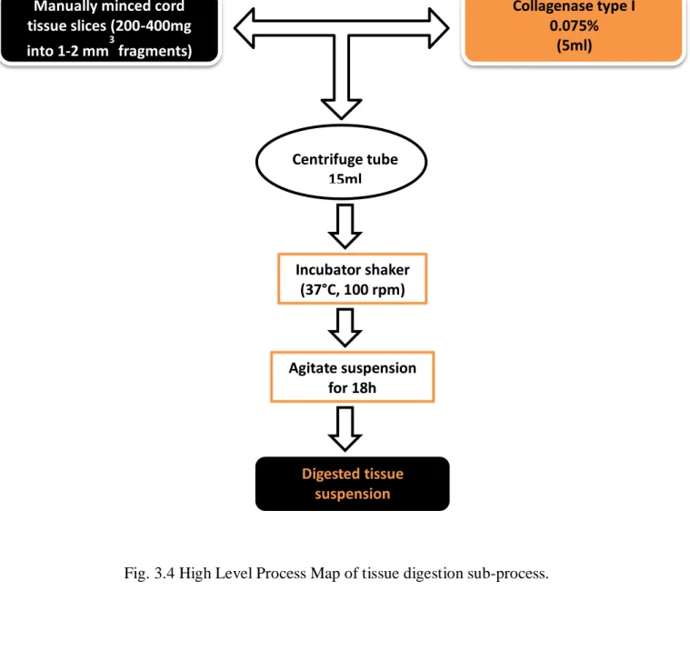
3.1 Process mapping... 95
3.1.1 How to generate a High Level Process Map (HLPM) ... 96
3.2 Process mapping and sub-process definition ... 98
3.3 A systematic approach to process improvement ... 110
3.4 Analysis of major points for variability and control from historical process data ... 111
Chapter four ... 115
ISOLATION OF hMSCs FROM hUCT UNDERSTANDING AND MINIMISING VARIABILITY IN CELL YIELD FOR PROCESS OPTIMIZATION ... 115
4. Development of isolation methods ... 116
4.1 Preliminary development of isolation methods for human mesenchymal stem cells (hMSCs) from umbilical cord tissue (UCT). ... 117
4.2 Metabolic activity analysis and correlation to cell yield for fresh and frozen umbilical cord tissue (UCT) sections ... 130
4.3 Isolation of hMSCs from whole lengths of fresh umbilical cords ... 133
4.3.1 Effect of isolation method on cell yield ... 137
4.3.2 Effect of cord storage time (age) on cell yield ... 139
4.3.3 Cord to cord variability and the influence of sampling location on cell yield ... 143
4.4.4 Operational significance ... 146
Chapter five ... 151
5. CELL FUNCTIONALITY CHARACTERIZATION... 152
5.1 A comparison study between hMSCs extracted from umbilical cord tissue (UCT), adipose tissue (AT) and dental pulp tissue (DPT) ... 153
5.1.1 Adipogenic lineage differentiation of hMSCs ... 155
5.1.2 Osteogenic lineage differentiation of hMSCs ... 157
5.1.3 Chondrogenic lineage differentiation of hMSCs ... 159
5.1.4 Hepatic lineage differentiation of hMSCs ... 161
5.1.5 Immunophenotyping of hMSCs extracted from UCT, AT and DPT ... 163
5.2 Characterization of hMSCs extracted from UCT via enzymatic digestion and explant culture. A comparison study ... 176
Chapter six ... 182
6. CRYOPRESERVATION OF UMBILICAL CORD TISSUE (UCT) METHOD INVESTIGATION ... 183
6.1 Preliminary investigation of cryopreservation method ... 183
6.2 Further investigation of cryopreservation method ... 186
7. CONCLUSIONS AND FUTURE WORK ... 196
7.1 Conclusions... 196
7.2 Future work ... 201
REFERENCES ... 203
Chapter one
1. INTRODUCTION
1.1Regenerative medicine
“Regenerative medicine replaces or regenerates human cells, tissue or organs, to restore or establish normal function1.”
With an increasingly aging population, a lack of donors for transplantation and the limited existing therapies to treat degenerative diseases such as cancer, Parkinson’s, or Alzheimer, regenerative medicine represents a field of great interest and enthusiasm in the present and future outlook of science2. Cell therapy, tissue engineering and gene therapy are some of the most novel and promising regenerative medicine approaches for major paradigm shifts in healthcare1.
Tissue engineering is a fast growing, multidisciplinary area of research that combines engineering, physical sciences, biology, and medicine with the aim to restore or replace tissue’s and organ’s functions by creating tissue equivalents of blood vessels, heart muscle, nerves, cartilage, bone, and other organs3.
Construction of engineered tissues and organs in vitro for transplantation, or regeneration of the tissue in vivo could be achieved by the use of stem cells4. Biological advances in the last decade have made it possible to study the path of differentiation and the effect of different parameters in stem cell proliferation for regenerative medicine applications5.
The therapeutic use of stem and progenitor cells in treating a variety of human diseases requires the development of validated, clinical-grade cell therapies6. In order to achieve successful results of transplantation and treatment of significant diseases as Parkinson’s disease, Alzheimer’s disease, leukaemia, diabetes, stroke, muscular dystrophy, hepatic, renal failure, optimum methods need to be developed on how to extract stem cells from various primary tissue sources, the way in which they are expanded, how and for how long they are cryopreserved7-9.
1.2Stem cells
“The central focus of regenerative medicine is human cells. These may be somatic, adult stem or embryo-derived cells1”.
Stem cells are cells found in all multi cellular organisms. They are undifferentiated cells characterized by the ability to renew themselves through mitotic cell division and differentiate into a diverse range of specialized cell types, generating multiple cell lineages10,11. Therefore, they have the potential to contribute to tissue homeostasis by replenishment of cells or regeneration of tissue after injury12-14.
Stem cells possess three main characteristics that differentiate them from somatic cells15:
• self-renewal, or the ability to generate at least one daughter cell after mitosis with identical characteristics to the mother cell;
• multi-lineage differentiation of a single cell into one of the three germ layer cells that form an organism;
• in vivo functional reconstitution of a given tissue; There are different ways of classifying stem cells:
Depending on their differential potential, stem cells can be classified as9:
• Totipotent (a.k.a omnipotent) stem cells can differentiate into embryonic and extra-embryonic cell types. Such cells can construct a complete, viable, organism. These cells are produced from the fusion of an egg and sperm cell. Cells produced by the first few divisions of the fertilized egg are also totipotent.
• Pluripotent stem cells are the descendants of totipotent cells and can differentiate into nearly all cells, i.e. cells derived from any of the three germ layers (ectoderm, mesoderm and endoderm) but cannot form a whole organism.
• Multipotent stem cells can differentiate into a number of cells, but only those of a closely related family of cells (e.g. skin stem cell, would give rise to the various types of skin cells4.
• Oligopotent stem cells can differentiate into only a few cells, such as lymphoid or myeloid stem cells.
• Unipotent stem cells can produce only one cell type, their own, but have the property of self-renewal which distinguishes them from non-stem cells (e.g. pancreatic, endothelial and fibroblastic stromal progenitor stem cells, blast cells).
According to their origin, stem cells can also be classified into three categories : • Embryonic stem cells (ESCs).
• Foetal stem cells (FSCs). • Adult stem cells (ASCs).
1.2.1 Embryonic stem cells (ESCs)
ESC lines are derived from the inner cell mass of the blastocyst that originates five days after the fertilization of a female egg with a spermatozoid. ESCs are pluripotent and give rise during development to all derivatives of the three primary germ layers: ectoderm, endoderm and mesoderm. In other words, they can develop into each of the more than 200 cell types of the adult body when given sufficient and necessary stimulation for a specific cell type. They do not contribute to the extra-embryonic membranes or the placenta. Embryonic cells were first obtained from mouse, but it was not until 1998 when J. Thomson derived the first line o f human embryonic stem cells16. These cells can be propagated in vitro while maintaining pluripotency for extended periods of time. However, embryonic stem cell lines are an artefact of cell culture techniques, since they do not remain as such in vivo. The cells that constitute the blastocyst differentiate quickly into different lineages to form tissues and organs for the development of an organism. Embryonic stem cells can also be propagated in vitro for months while retaining self-renewal potential and pluripotent characteristics17. Current research focuses on differentiating ESCs into a variety of cell types for eventual use as cell replacement therapies18. Some of the cell types that have or are currently being developed
include cardiomyocytes, neurons, hepatocytes, bone marrow cells, islet cells
and endothelial cells. However, the derivation of such cell types from ESCs is not without obstacles and hence current research is focused on overcoming these barriers133, 134.
1.2.2 Foetal stem cells (FSCs)
FSCs are primitive cell types found in the organs of foetuses. The classification of foetal stem cells remains unclear and this type of stem cell is currently often grouped into an adult stem cell. However, a more clear distinction between the two cell types appears necessary19.
Although the literature is lacking more information about this type of stem cells, several differences between FSCs and ASCs have been distinguished and described recently. First, FSCs appear to have a greater expansion capacity in vitro and faster doubling times than ASCs, which may be due to their having longer telomeres than ASCs20,21. Second, FSC’s appear to lack some of the immune suppression properties observed in ASCs; also appear to synthesize HLA-G, which is absent in ASCs22. Third, FSCs appear to lack class II human leukocyte antigens (HLA), in contrast to ASCs23. Fourth, FSCs express a slightly different cytokine profile than ASCs23. In conclusion, primitive stem cells have a greater ability to expand in culture, are less lineage committed than ASC, and have a different physiology that is most likely due to their immature condition.
1.2.3 Adult stem cells (ASCs)
ASCs, also known as somatic stem cells, are undifferentiated cells found in many organs and tissues in the body including brain, bone marrow, peripheral blood, blood vessels, skeletal muscle, skin, teeth, heart, gut, liver, ovarian epithelium, and testis. They are thought to reside in a specific area of each tissue, called a "stem cell niche". Although they possess stem cell characteristics of self-renewal and differentiation, they have limited proliferation potential in vitro24. Their self-renewal potential is maintained in the body, since these cells are involved in the maintenance and repair of tissues and organs throughout the life span of the individual25. Most adult stem cells are multipotent and are generally referred to by their tissue origin (e.g. mesenchymal stem cell, adipose-derived stem cell, endothelial stem cell)26,27. Pluripotent adult stem cells are rare and generally small in number but can be found in a number of tissues including umbilical cord and cord blood. A great deal of adult stem
cell research has focused on clarifying their capacity to divide or self-renew indefinitely and their differentiation potential28.
Mesenchymal stem cells (MSCs) are a type of multipotent adult stem cell; a common progenitor, not just of skeletal tissues, but of ‘‘mesenchymal’’ tissues, meaning virtually all non-hematopoietic derivatives of the mesoderm; and although found in the bone marrow, it is not unique to the bone marrow135. MSCs have also been isolated from placenta, adipose tissue, lung, blood, Wharton's jelly from the umbilical cord,136 and teeth (perivascular niche of dental pulp and periodontal ligament)137. Although MSCs have become more recently attractive for clinical therapy due to their ability to differentiate, provide trophic support, and modulate innate immune response135, the origin of the concept of a ‘‘mesenchymal’’ stem cell goes back to the pioneering experiments of Tavassoli and Crosby in the 1960s138.
While the terms Mesenchymal Stem Cell and Marrow Stromal Cell have been used interchangeably, neither term is sufficient to describe the differentiation potential of this type of cells. Mesenchyme refers to the embryonic connective tissue that is derived from the mesoderm and that differentiates into hematopoietic and connective tissue, whereas MSCs do not differentiate into hematopoietic cells28. Stromal cells are connective tissue cells that form the supportive structure in which the functional cells of the tissue reside. While this is an accurate description for one function of MSCs, the term fails to convey the relatively recently discovered roles of MSCs in the repair of tissue28.
The International Society for Cellular Therapy (ISCT) differentiates between MSCs and mesenchymal stem cells based upon in vivo characterization; for example, mesenchymal stem cells undergo self-renewal and multipotential differentiation following engraftment. Currently, a compromise marker set that would allow for a prospective identification of mesenchymal stem cells from the in vitro MSC population has not yet been portrayed. There is no single surface marker, but rather a panel of surface markers that define hMSC’s, derived from fresh tissues or cryopreserved samples. Due to different hMSC tissue sources, differences exist among these cells29. The matter is further complicated because these stem cells have poorly characterized growth conditions, such as low glucose DMEM containing fairly high concentrations of foetal bovine serum (FBS, 10–20%) and because not all lots of FBS are equal in terms of their ability to maintain MSCs growth30. Therefore in 2006, the Mesenchymal and Tissue Stem Cell Committee of the International Society for Cellular Therapy summoned a group of researchers to discuss, analyse and define the
immune-phenotype of MSCs. There were proposed 3 minimal criteria to define human MSCs (hMSCs) 30:
1. the ability to adhere to plastic;
2. presence of ≥ 95% expression of hMSC-specific antigen markers (CD13, CD44, CD90, CD73, CD105) and ≤ 2% positive for hematopoietic/endothelial marker expression (CD14, CD11b, CD79, CD 34, CD45 and HLA-DR);
3. the differentiation of the hMSCs into osteoblasts, adipocytes, and chondroblasts in vitro.
While there might be a lot more answers in regard to mesenchymal stem cells from other, more explored, sources, like bone marrow; the exploration of human umbilical cord tissue as a source of MSCs still raises a lot of questions, presenting researchers with the opportunity of new challenges and discoveries.
1.3The human umbilical cord (hUC)
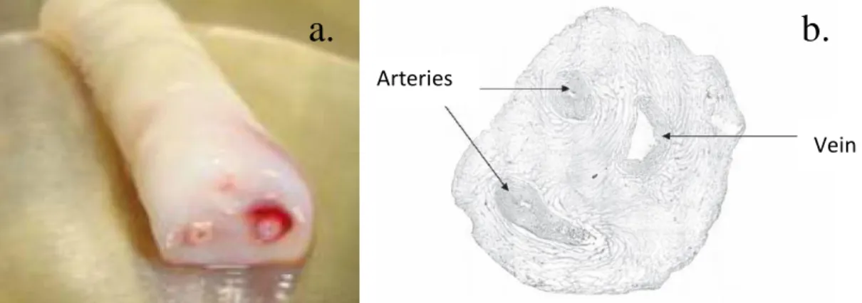
The hUC, embryologically formed at day 26 of gestation32, represents the link between mother and foetus during pregnancy. It is composed of a special embryonic mucous connective tissue, called Wharton’s jelly, lying between the covering amniotic epithelium and the umbilical vessels (Fig.1.1).
Fig. 1.1 (a) Umbilical Cord Sample; (b) Cross-section of an umbilical cord displaying the two arteries (left) and vein (right), which has a larger lumen. Note that the lower artery is
sectioned tangentially41-43.
a.
b.
Arteries
The main role of this jelly-like material is to prevent the compression, torsion, and bending of the enclosed vessels, which provide bidirectional blood flow between foetal and maternal circulation33, 34. The human UC weighs approximately 40 g, its length reaches to approximately 60–65cm, and it has a mean diameter of 1.5 cm at term35,36. It is covered by a single/multiple layer(s) of squamous-cubic epithelial cells called umbilical epithelium, which is generally thought to be derived from amniotic epithelium37, 38. Those epithelial cells display ultra-structural and functional characteristics to those seen in keratinocytes39 and were also shown to possess stem cell characteristics40. The inner tissue architecture is composed of a set of two arteries and one vein and a surrounding matrix of mucous connective tissue comprised of specialized fibroblast-like cells and occasional mast cells embedded in an amorphous ground substance rich in proteoglycans, mainly hyaluronic acid. Neither capillaries nor lymphatics are found in the UC. Vessels are normally organized as left spiral (counter clockwise) turns (Fig. 1.2). In clinical practice, determining the “umbilical coiling index” (number of complete coils divided by the length of the cord; average 0.24 coils per centimetre) may identify the foetus at risk of foetal demise40.
Fig. 1.2 Different regions for the isolation of hMSCs in the umbilical cord41. Cord Arteries (Blue)
Wharton’s Jelly
Cord Vein (Red)
Cord blood Stem Cells
Umbilical cord MSCs
Vascular Endothelial Cells
The part of the cord, mentioned as Wharton’s jelly, appears to serve the function of adventitia (connective tissue), which the UC vessels lack, binding and encasing the umbilical vessels. It has been suggested that the stromal cells of Wharton’s jelly may participate in the regulation of UC blood flow and that, at least in some cases; the reduction in foetal growth could be the consequence of stromal diminution leading to hypoplasia of umbilical vessels44,45.
At least six distinctive zones are now recognized based on the structural and functional studies, from outer to inner (Fig. 1.3): (1) umbilical cord blood; (2) umbilical vein subendothelium, (3) perivascular stroma, (4) intervascular stroma (named classically as Wharton’s jelly), (5) sub amnion, and (6) amnion32
. Fine structural, immunohistochemical46,48 and in vitro functional studies49,50 proved that there are significant differences in the number and nature of cells among subamniotic, intervascular, and perivascular regions, which leads to the hypothesis that those regions might be originating from different pre-existing formations. For instance, myofibroblastic cells of the intervascular stroma might have derived from adjacent vascular smooth muscle cells or, alternatively, from pre-existing fibroblasts32.
Fig. 1.3 Compartments within the umbilical cord. Separate regions, which have been shown to contain mesenchymal stromal cells. Wharton’s Jelly is the connective tissue surrounding umbilical vessels and includes the perivascular, intervascular, and sub amnion regions (zones
3-5)29.
3. Perivascular
4. Interivascular
6. Amnion 5. Sub amnion
1. Umbilical cord blood
1.4Human umbilical cord mesenchymal stem cells
1.4.1 Isolation methods for umbilical cord tissue (UCT) hMSCs
UCT hMSCs have been reportedly isolated from the different areas that form the umbilical cord (Fig. 1.2 and 1.3).
More precisely they have been described to be isolated from: Umbilical cord blood5, 50-59.
Umbilical vein sub-endothelium19, 49, 60-64. Wharton’s jelly29, 32, 41, 65-69
.
Within the Wharton’s jelly, MSCs have been isolated from three relatively indistinct regions: the perivascular zone, the intervascular zone, and the sub amnion. It is still undecided whether MSCs isolated from the different compartments of the umbilical cord represent different populations48, 139,140. The nomenclature used in the literature for these various compartments has been misleading and not standardized, with terms such as ‘cord lining’, ‘subamnion’, ‘intervascular’, ‘perivascular’ and ‘hUVEC’ being used. Stem cell populations with varied stemness properties have been reported for each of these compartments140, but the various individual derivation protocolspublished in the literature for stem cells from the UCT areambiguous and do not pay regard to the differences in stem cell populations between compartments. At the same time it is notknown whether the stem cell populations between compartments are one and the same as there is no clear demarcation histologically between some of these compartments. Given the reports that stem cell populations in different compartments have varied stemness characteristics the derivation protocols involving entire cord pieces containing all the compartments will result in mixed heterogeneous stem cell populations making a meaningful assessment of investigations difficult. It is therefore urgently necessary to standardizea derivation protocol for MSCs of the UCT that yields definedor minimally heterogeneous cell populations140.
Bongso A and Fong CY have discussed in a recent review diverse methods for isolation of MSCs from UCT141. They grouped these methods into six representative isolation methods
conclusion of their study was that even though MSCs have been reported from the various compartments of the human UCT, the compartment with stem cell populations of the most optimal therapeutic value remains debatable. Robust comparisons between the stem cell populations of these various compartments in order to identify the most optimum source and subpopulation is urgently necessary for standardization and comparison of results between groups and to ensure reliability in terms of stemness properties, product quality, safety and efficiency for attaining regulatory approval for future clinical trials. Currently, stem cells from the Wharton’s Jelly compartment appear to be the most defined with several unique characteristics141.
The fact that there is no standardized method for extraction of MSCs from hUCT comes to show that there is a gap in knowledge and that there is a real need for developing practical methods that apply to real processing environment, such as autologous and allogeneic tissue banking. There are pros and cons to each of the methods described in literature but the real challenge is represented by the fact that these methods are usually compared under idealized conditions. However, due to the nature of tissue collection in a birthing environment the early period of tissue processing is relatively uncontrolled; the priority is maternal and neonate safety. Further, tissue often needs to be transported from maternity units to distant processing sites, especially in the case of private banks. Such factors make imposing tight process controls on early handling challenging. In addition, innate biological variation in the tissue will affect the cell yield. Therefore cell isolation methods should be assessed and engineered for robustness to innate biological tissue variation or arising variation due to tissue collection procedures. This is particularly important for tissue stored for autologous use (private banking), where a processing facility will not be able to select tissue based on favourable characteristics.
Current procedures presented in literature for enzymatic digestion and explant methods of extraction for MSCs from UCT have been detailed below.
1.4.1.1Isolation by enzymatic digestion
Protocols applied by different investigators seem to have as a general trend the use of collagenase-containing solutions, which have strong collagenase activity as well as caseinase, clostripain, and tryptic activities. Type I collagenase, or collagenase type A, is extensively used for the isolation of mesenchymal-like cells from the cord tissue. Furthermore, recent literature describes the use of a combination of collagenase with hyaluronidase, which seems to facilitate the degradation of matrix ground substance and shortens the time required for the isolation process49, 65, 69, 70. The use of type II collagenase, which is stronger for its clostripain activity, or collagenase type B, which is more efficient in solubilizing the UC microfibrils than other types of collagenases, is also worth taking into consideration48, 71, and 72.
The duration of collagenase treatment is critically important, especially if collagenase/ hyaluronidase cocktails are used, since there is always a risk of degradation of cellular external lamina, a phenomenon preventing cells from adhering to the culture substrate after isolation and even causing severe cellular damage32. The time required for tissue digestion ranges from 30 minutes49 to 16 hours68 depending on the quantity/concentration of enzyme and duration of treatment with digesting reagents.
It has been noted that filtration of the digested material through 70–100µm pore sized cell strainers facilitates the removal of any unwanted tissue debris73.
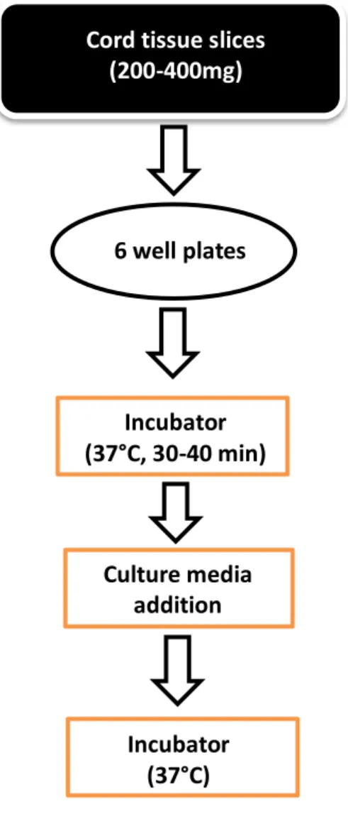
1.4.1.2Isolation by explants culture
Enzymatic digestion can be avoided if an explant culture is employed. Unfortunately a limited number of studies that involve the explant method have been conducted so far. The principle of the method is generally described as fine chopping of the Wharton’s jelly sections of the cord tissue, after excision of the blood vessels, with a scalpel, plating of the fine fragments in sterile culture plates or Petri dishes, and culturing of these with low-glucose DMEM, supplemented with foetal bovine serum (10-20% v/v), L-glutamine and antibiotics/antimycotics66, 74-78.
1.4.2 Cryopreservation methods for UCT and hMSCs isolated from UCT
Cryopreservation of cord tissue and/or cells extracted from the tissue represents an important stage to overcome in the view of the therapeutic use of stem cells. The banking model of our industrial partners is to extract MSCs from both cryopreserved and fresh cord tissue. Therefore, the choice of an appropriate cryopreservation protocol is essential for maintenance of cryopreserved tissue. UCT cryopreservation should be able maintain the cellular metabolism in a dormancy state for an indefinite period of time.
Freezing conditions of both isolated UC MSCs and UCT could have a serious impact on the viability rates after thawing and they have to be evaluated. Most researchers prefer to use defined culture media supplemented with high amounts of foetal bovine serum and 7%– 10% (v/v) dimethyl sulfoxide (DMSO)48 or glycerol69 and freeze the cells gradually (eg. 10C/min) and keep them between -135°C and -196°C. After rapid thawing at 37°C, viability rates of over 50% were achieved49. The use of higher levels of foetal bovine serum especially during the first week after thawing the cells has proven to substantially increase their growth69. Certainly, more controlled studies are needed to maximize the freeze-thaw efficiency, especially when their routine use is concerned in clinical trials and even more when it comes to cord tissue itself.
The success of regenerative medicine and its components depends on the ability to physically distribute the products to patients in need and to produce these products ‘‘off-the-shelf’’9
. For this reason, the ability to cryogenically preserve not only cells, but also tissue fragments, is an important part of a complete technology platform.
In a recent study Da-Croce L, et al., have tested two protocols of cryopreservation on hUCT: slow cooling and vitrification. The samples were frozen for a period of time ranging from 5 to 78 days. The efficiency of cryopreservation was evaluated by testing cell viability, histological analysis, cell culture, cytogenetic analysis and comparison with the results of the fresh samples. The results showed that the slow cooling protocol was more efficient than the vitrification, for cryopreservation of umbilical cord tissue, because it has caused fewer changes in the structure of tissue (edema and degeneration of the epithelium) and, despite the significant decrease cell viability compared to fresh samples, the ability of cell proliferation
in vitro was preserved in most samples. In conclusion, this study showed that it is possible to cryopreserve small fragments of tissue from the umbilical cord and, to obtain viable cells capable of proliferation in vitro after thawing, contributing to the creation of a frozen tissue bank142.
Also, in another recent study, Mahmood S, et al., have studied the utility of cryopreserved UCT by characterizing MSCs isolated from cryopreserved and fresh cord tissue. They found no significant functional differences between MSCs from frozen cord tissue as compared to fresh cord tissue143.
Cryopreserving cord tissue could allow for isolation of MSCs at the point of care in the near future. This may be advantageous as MSC isolation protocols continue to be optimized dependent on intended use. More studies with large numbers of samples, testing various cryoprotectants, and assessing other parameters such as, viability and ability of preserving stemness, should be conducted142, 143.
1.4.3 Differentiation potential of UCT hMSCs
One of the main goals of regenerative medicine is to achieve the potential to use stem cells in cell-based therapies. Since UC is one of the most easily reached stem cell sources both ethically and technically, both in vitro and in vivo studies are definitely desirable. There have been several reports of successfully differentiated lineages using a variety of cell culture techniques and reagents32:
In vitro differentiation: Adipocytes48, 71, 75, 79. Chondrocytes48, 68, 70, 79. Osteocytes48, 49,71,75,79. Cardiomyocytes62, 68. Skeletal myocytes75. Neuronal/glial precursors48, 71 ,80. Dopaminergic neurons65, 69. 81
In vivodifferentiation:
Dopaminergic neurons65, 69.
Photoreceptor rescues73.
Endothelial cells81.
Skeletal myocytes75.
As the umbilical cord stromal cells originate from the extra embryonic mesoderm, adipogenic, chondrogenic, osteogenic, cardiomyogenic, and skeletal myogenic inductions have been the most studied cell lineages32.
Despite morphological and immunophenotypical similarities, human mesenchymal stem cells (hMSCs) from diverse origins vary in regard to their differentiation potential28. Bone marrow hMSCs, for example, can differentiate along all known mesodermal differentiation pathways82. In contrast, umbilical cord blood (UCB) hMSCs and umbilical cord (UC) hMSCs display a reduced sensitivity to undergoing adipogenic differentiation5, 83, although they can differentiate into adipocytes51. Independently of their origin, the adipogenic potential of hMSCs is inversely related to the length of in vitro culture, and sharply declines when hMSCs become senescent84. Contrary, prolonged culturing increases their osteogenic differentiation85. In vitro expansion of hMSCs should therefore be performed with limited passaging, to avoid changes in their differentiation ability. Gradual shortening of the telomeres during a cell’s life continues, until the presence of critically short telomeres triggers a senescence pathway, which results in proliferation arrest28. Because of that, a normal human cell can only divide 50 to 100 times in in vitro conditions; hMSCs are no exception51, 86. UC blood hMSCs, however, have slightly longer telomeres than other hMSCs, and thus can be cultured for longer periods before they senesce87. Proliferation arrest in hMSCs results in their senescence, which is described by the appearance of large senescent cells with flat shape, circumscribed nuclei and increased lysosome compartment. These morphological changes are not restricted to the senescent stage only, but represent continuous alterations in the course of hMSCs long-term culture85.
1.4.4 Immunophenotyping of UCT hMSCs
The scientific literature is abundant in information about marker profiles that, theoretically, characterize stromal cells from the umbilical cord. Investigators suggest different paths towards achieving a correct characterization of mesenchymal stem-like cells from the umbilical cord, thus, making the process of accurate evaluation even more confusing and harder to reach32. Furthermore the existence of various populations of mesenchymal-like stem cells in the different areas that form the umbilical cord76 sets hurdles in establishing a standard marker profile for these cells88.
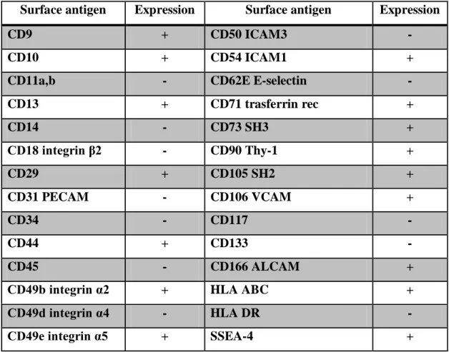
At the present time, characterisation of hMSCs is generally accomplished by flow-cytometry analysis of surface markers. Stro-1 has been identified as a marker for cells that can differentiate into multiple mesenchymal lineages89. However, other scientists’ findings90 suggested that Stro-1 is not essential for the differentiation potential of hMSCs. Moreover, it has been demonstrated that a CD9/CD90/CD166 triple positive subpopulation of hMSCs showed multipotency for chondrogenic, osteogenic and adipogenic differentiation providing a basis for identification of hMSCs91. It has been indicated that positive expression of CD166 is indicative of multipotency in hMSCs. However, the level of expression has been shown to decrease with increasing cell density in culture and regained during inoculation of successive passages92. Expression levels of CD90 and CD105 are maintained over sequential passages and they can be important for validating cultures of hMSCs intended for therapy93. A good indication of hMSC identity can be reached by expression of CD90, CD105 and CD166 and lack of expression of CD34 and CD45 as a minimum set of surface markers. A more extensive list has been compiled from results obtained by different groups (Table 1.1)32.
Table 1.1 Expression of surface markers for human mesenchymal stem cells (hMSCs).
Surface antigen Expression Surface antigen Expression
CD9 + CD50 ICAM3 -
CD10 + CD54 ICAM1 +
CD11a,b - CD62E E-selectin -
CD13 + CD71 trasferrin rec + CD14 - CD73 SH3 + CD18 integrin β2 - CD90 Thy-1 + CD29 + CD105 SH2 + CD31 PECAM - CD106 VCAM + CD34 - CD117 - CD44 + CD133 - CD45 - CD166 ALCAM +
CD49b integrin α2 + HLA ABC +
CD49d integrin α4 - HLA DR -
CD49e integrin α5 + SSEA-4 +
1.5Process control and optimization
1.5.1 Variability in hMSCs extraction from hUCT
There are some sources of variability in all primary cell isolation processes that need to be taken into consideration when designing such a process. There are ways of minimizing variations between lots produced, by controlling process parameters, and by screening the raw materials that will be in contact with the cells and cell source94. There are other non-controllable parameters such as the source of the cells, which represents a real challenge for regenerative medicine applications. Each cell extraction method is produced with cells from a different patient/donor, with intrinsic characteristics that result in variations of cell growth patterns and differentiation95. It is therefore necessary to develop extraction methods that are
robust to real world operating conditions, rather than idealized operation. Biological variation in patients, or biological material introduced into samples due to isolation and handling will have a major effect on the safety and efficacy of clinical applications. It is necessary to map the operating environment and assess risk factors before empirically determining the effect on the process. This will be particularly critical for processes using primary tissue or cell sources where the biological variation at input is likely to be high. Regulated therapeutic products will require characterized and risk assessed manufacturing processes96. This fits the philosophy of process control industry tools such as quality by design (QbD) 97 and Six Sigma98, 99; represent approaches to understanding process operating space and risks of associated variables.
1.5.2 Quality by design (QbD)
“Quality by design means designing and developing manufacturing processes during the product development stage to consistently ensure a predefined quality at the end of the manufacturing process100.”
QbD was born out of the need for the pharmaceutical industry and the US Food and Drug Administration (FDA) to move pharmaceutical development toward a new, more scientific, risk-based, complete and proactive methodology. This new approach requires a ‘built in’ quality for the product and manufacturing process. This is achieved through a deep understanding of process components, starting at the product development stage. The design and advance of the final product involves identification of critical quality attributes (CQA’s) and a clear outline of product performance. Understanding the impact of raw material characteristics and process parameters on the CQAs in the process development of a product is crucial in finding and controlling sources of variability. Once these sources are identified, measurements for control can be implemented in the manufacturing process and methods can be designed to deliver the desired product attributes97.
QbD ensures a systematic approach to product development that allows companies to achieve consistent product quality. This is seen in Figure 1.4, which shows the different phases during the life cycle of a pharmaceutical process: define, design, characterize, validate, and monitor and control. The final link between “monitor and control” and “define”
represents process changes that are initiated based on process improvement opportunities identified during process monitoring or introduced otherwise to improve process performance or robustness97.
Fig. 1.4 Different phases during the life cycle of a pharmaceutical process97.
1.5.2.1QbD implementation
A popular concept used for implementation of QbD is ‘design space’, which has been defined as “the multidimensional combination and interaction of input variables (e.g., material attributes) and process parameters that have been demonstrated to provide assurance of quality. Working within the design space is not considered as a change. Movement out of the design space is considered to be a change and would normally initiate a regulatory post-approval-change process. Design space is proposed by the applicant and is subject to regulatory assessment and approval101.”
Though design space has primarily been used in the context of pharmaceutical processes, it can also be applied to represent the clinical and product-quality aspects of a product97. Define Design Characterize Validate Monitor& Control
Because QbD is a novel concept, there is very limited literature on its application. It has mainly been used in small molecule manufacture102, 103 and in biopharmaceuticals in the production of monoclonal antibodies104.
Another important tool for gathering process knowledge and increasing process understanding is multivariate data analysis (MVDA). The use of MVDA as a tool to establish process parameters and their interactions, therefore to define the design space for a particular manufacturing process has been reported in case studies involving cell-culture processes105, 106 and by other biotech companies107. In these circumstances MVDA was used to identify parameter interactions that adversely affect cell culture process performance and to support some of the key activities required for successful manufacturing of biopharmaceutical products, including scale-up, process comparability, process characterization and fault diagnosis.
In all cases, it was concluded that it is possible to design control systems that rely on measurement of product CQAs and enable real-time decisions. Once the design space for a particular manufacturing process has been defined, it can be continually reassessed and changed, as appropriate108.
Process analytical technology (PAT) is a complementary concept to that of design space, it is “a system for designing, analysing, and controlling manufacturing through timely measurements (i.e., during processing) of critical quality and performance attributes of raw and in-process materials and processes, with the goal of ensuring final product quality109” and it’s goal is “to enhance understanding and control the manufacturing process, which is consistent with our current drug quality system: quality cannot be tested into products; it should be built-in or should be by design109”.
Appropriate use of PAT tools and principles ensures that the process operates within the approved process design space, and enables process understanding, therefore facilitates process control and optimization. “A process is generally considered well understood when (1) all critical sources of variability are identified and explained; (2) variability is managed by the process; and, (3) product quality attributes can be accurately and reliably predicted over the design space established for materials used, process parameters, manufacturing, environmental, and other conditions. The ability to predict reflects a high degree of process understanding. A focus on process understanding can reduce the burden for validating
systems by providing more options for justifying and qualifying systems intended to monitor and control biological, physical, and/or chemical attributes of materials and processes. Structured product and process development on a small scale, using experimental design and on- or in-line process analysers to collect data in real time, can provide increased insight and understanding for process development, optimization, scale-up, technology transfer, and control109”.
The application of PAT has been reported in the use of a commercially available online HPLC system for real-time pooling of process chromatography columns 110. This case study shows the practicality of online-HPLC for analysis and its capacity to enable real-time decisions for column pooling based on product-quality attributes. Thus, the quality systems approach allows continuous improvement of the manufacturing process97, 100, and 111.
There are many tools available that enable process understanding for scientific, risk-managed pharmaceutical development, manufacture, and quality assurance. These tools, when used within a system, can provide effective and efficient means for acquiring information to facilitate process understanding, continuous improvement, and development of risk-mitigation strategies. In the PAT framework, these tools can be categorized according to the following109:
Multivariate tools for design, data acquisition and analysis. Process analyzers.
Process control tools.
Continuous improvement and knowledge management tools.
An appropriate combination of some, or all, of these tools may be applicable to a single-unit operation, or to an entire manufacturing process and its quality assurance109.
The application of QbD principles to pharmaceutical manufacturing has received more and more interest recently102, 103. The biotech and traditional small-molecule pharmaceutical industry has been working actively on applying the concepts of Quality by Design to the development and manufacture of drug products108. Case studies mentioned previously serve as useful tools in establishing common ground on how to develop and define a design space. They provide examples of how to carry out three key steps in process characterization108:
2) Developing studies based on a design-of-experiments approach to study those parameters and their interactions;
3) Executing those studies and analysing them to determine which parameters are critical and how the design space should be defined.
1.5.3 Six Sigma
Six Sigma represents a framework for quality improvement and business excellence that has been adopted by high-profile companies such as Motorola and General Electric99. It has been defined as “a disciplined method of using extremely rigorous data gathering and statistical analysis to pinpoint sources of errors and ways of eliminating them112”. Also Minitab, popular software used to perform statistical analysis, describes Six Sigma as “an information-driven methodology for reducing waste, increasing customer satisfaction and improving processes with a focus on financially measurable results99”.
There are several features that distinguish Six Sigma from other quality improvement techniques. First is the use of DMAIC framework, where techniques such as QFD (quality function deployment), FMEA (failure mode and effects analysis), DOE (design of experiments) and SPC (statistical process control) are integrated into a logical flow99. DMAICis used for projects aimed at improving an existing business process. The DMAIC project methodology has five phases113:
1. Define the problem, the voice of the customer, and the project goals, specifically. 2. Measure key aspects of the current process and collect relevant data.
3. Analyse the data to investigate and verify cause-and-effect relationships. Determine what the relationships are, and attempt to ensure that all factors have been considered. Seek out root cause of the defect under investigation.
4. Improve or optimize the current process based upon data analysis using techniques such as design of experiments, mistake proofing, and standard work to create a new, future state process. Set up pilot runs to establish process capability.
5. Control the future state process to ensure that any deviations from target are corrected before they result in defects. Implement control systems such as statistical process control, production boards, visual workplaces, and continuously monitor the process.
Some organizations add a Recognize step at the beginning, which is to recognize the right problem to work on, thus yielding an RDMAIC methodology113.
The idea of information-based improvement has been extended to design activities, in the form of DMADV or DFSS (design for Six Sigma) 114, 115. DFSS (typically in the form of IDOV or Identify–Design–Optimize– Validate) aims to design products, services and processes that are ‘Six Sigma capable’, emphasizing the early application of Six Sigma tools and the fact that as far as defect elimination goes, prevention is better than cure99.
DFSS features five phases as well113:
1. Define design goals that are consistent with customer demands and the enterprise strategy.
2. Measure and identify characteristics that are critical to quality (CTQs), product capabilities, production process capability, and risks.
3. Analyse to develop and design alternatives, create a high-level design and evaluate design capability to select the best design.
4. Design details, optimize the design, and plan for design verification. This phase may require simulations.
5. Verify the design, set up pilot runs, implement the production process and hand it over to the process owner(s).
Within the individual phases of a DMAIC or DMADV/DFSS project, Six Sigma utilizes many established quality-management tools that are also used outside Six Sigma, such as design of experiments (DOE), analysis of variance (ANOVA), control charts, general linear model, histograms, process capability, process mapping, to name a few113, 116.
It can be concluded, therefore, that statistical thinking and statistical methodologies constitute the backbone of Six Sigma. The results achieved through implementation of Six Sigma are a far better from the days when quality had to depend on testing and inspection (T&I). More and more the emphasis of quality improvement has been moving upstream through the years: from T&I on the product to statistical process control (SPC) on the process, then Six Sigma on the system, and finally DFSS as a pre-emptive move for achieving the desired performance. Certainly Six Sigma and DFSS represent a far more fundamental approach to problem solving and problem anticipation, respectively, in any given situation99.
1.6Thesis objectives
Cell-based therapeutic products will require sources of input for human cell material to underpin clinical supply. Different therapies will require different constituent cell types; human tissue banks are a potential source of cellular material for the nascent cell-based therapy industry; supply strategies are likely to include both large single cell-line banks for allogeneic application, as well as large banks of individual donor units of primary tissue for either autologous or allogeneic applications. In the latter category, a significant international industry, both public and private, now exists to bank human tissue for potential future therapeutic use.
Umbilical cord tissue is increasingly privately banked in such facilities as a source of mesenchymal stem cells for future therapeutic use. However, early handling of cord tissue is relatively uncontrolled due to the clinical demands of the birth environment and subsequent transport logistics. It is therefore necessary to develop extraction methods that are robust to relevant operating conditions, rather than idealized operation.
The research work described in this thesis was driven by the opportunity to expand the therapeutic and business potential of one of the leading private cord blood banks in the UK. The primary objectives of this collaboration were to understand and minimize variability in cell yield extracted from human umbilical cord tissue (hUCT) and to help the tissue processing facility to predict the probability of cell yields from 200-400 mg tissue sections given different operating ranges, and inform the experimental approach of others.
In order to achieve this goal it was understood that tight control and characterization of the process was critical. Therefore a systematic approach and work program was developed; this allowed the necessary process controls in the production of high quality hMSCs from hUCT, to be established, and also for a statistically capable production process, to be achieved. The systematic side of this approach was rooted in industrial systems such as Six Sigma and QbD, described previously.
The aim was to direct the final product of the research work towards a Product 1 type (Fig. 1.5), that has a large and diverse clinical market.
Fig. 1.5 Diagram represents an overview of potential products that could be obtained by expansion of hMSCs from the hUCT.
Products 1-3 will likely satisfy basic hMSCs criteria at isolation, providing a basis for banking, however as clinical science progresses and banked samples will need to become practical, different cell banks will see differing results from their cryopreserved ‘hMSCs’ because they will have stored/isolated different cell populations, with different potentials.
P1 is representative for a MSC population that has high clinical utility and safety, at low passage number and therefore the best proliferation and differentiation potential. P2 is representative for a senescent MSC population that has low clinical utility and safety, at a high passage number and therefore a considerably reduced proliferation and differentiation potential. P3 is representative for a cell population that has the least clinical utility and safety, this population possesses basic proliferation or differentiation potential characteristic for MSCs at isolation, but will fail to maintain these after expansion.
Therefore, selection of cells from a processing method based on P1’s potential is preferable for long term success.
0 Passage/Time E x p an sio n Product 1 (P1) Product 2 (P2) Product 3 (P3)
Chapter two
METHODS
AND
2. METHODS AND INITIAL DEVELOPMENT
This thesis is focused on method development for extraction of cells from human umbilical cord tissue. This has presented some structural challenges in separating the methods chapter from the novel development work. This methods chapter contains the core common methods used throughout the length of the research work. Where these methods have been further developed as the focus of a chapter this was clearly stated as appropriate in the relevant chapter. Some preliminary work was conducted in order to establish some ‘base-line’ processes and this preliminary development was also detailed within this methods chapter.
Umbilical cords used for the research purposes of this project were sourced by the private cord blood bank, which was our industrial partner and from a public UK hospital. All umbilical cords used for this study were collected with parental consent. Procedures for collection and transportation of umbilical cords have been described further in this chapter.
2.1Isolation methods for human umbilical cord tissue mesenchymal stem cells
2.1.1 Extraction of hMSCs from umbilical cord tissue via enzymatic digestion. Basic method development
Several methods of enzymatic digestion were initially tested; on 200-400 mg cord slices from multiple cords, both fresh and frozen, with the purpose to screen different methods for an assessment of method success with a view to downstream standardization of the isolation and expansion of mesenchymal stem cells from the UCT (Table 2.1).
Table 2.1. Enzymatic digestion methods for the extraction of hMSCs from UCT.
No. Method of digestion
No. of cords used State of the cord 1
2 hours enzyme digestion of cord tissue with 3 different enzymatic solutions:
9 (200 – 400 mg) slices of cord tissue were digested in 3 ml of enzymatic solution/each; solutions obtained after digestion were
filtered through 40 µm cell strainers and centrifuged at 1500 rcf for 10 min/each;
3 Frozen
2
4 hours enzyme digestion of cord tissue with 3 different enzymatic solutions:
9 (200 – 400 mg) slices of cord tissue were digested in 3 ml of enzymatic solution/each; solutions obtained after digestion were
filtered through 70 µm cell strainers and centrifuged at 1500 rcf for 10 min/each;
3 Frozen
3
18 hours enzyme digestion of cord tissue with 3 different enzymatic solutions:
9 (200 – 400 mg) slices of cord tissue were digested in 3 ml of enzymatic solution/each; solutions obtained after digestion were
filtered through 70 µm cell strainers and centrifuged at 1500 rcf for 10 min/each;
No. Method of digestion No. of cords used State of the cord 4
2, 4 and 18 hours enzyme digestion of cord tissue with 3 different enzymatic solutions:
18 (200 – 400 mg) slices of cord tissue were digested in 3 ml of enzymatic solution/each; solutions obtained after digestion were
filtered through 70 µm cell strainers and centrifuged at 1500 rcf for 10 min/each;
3
Fresh (2 days old)
5
2, 4 and 18 hours enzyme digestion of cord tissue with 3 different enzymatic solutions:
18 (200 – 400 mg) slices of cord tissue were digested in 5 ml of enzymatic solution/each; solutions obtained after digestion were
filtered through 100 µm cell strainers and centrifuged at 500 rcf for 10 min/each;
6 Frozen
6
2, 4 and 18 hours enzyme digestion of cord tissue with 3 different enzymatic solutions:
9 (200 – 400 mg) slices of cord tissue were digested in 5 ml of enzymatic solution/each; solutions obtained after digestion were
filtered through 100 µm cell strainers;
no centrifugation for slices digested with 2 of the enzymatic solutions and 1000 rcf
centrifugation speed for slices digested with the 3rd type of enzymatic solution;
No. Method of digestion No. of cords used State of the cord 7
2, 4 and 18 hours enzyme digestion of cord tissue with 3 different enzymatic solutions:
9 (200 – 400 mg) slices of cord tissue were digested in 5 ml of enzymatic solution/each; solutions obtained after digestion were
filtered through 100 µm cell strainers;
no centrifugation for slices digested with 2 of the enzymatic solutions and 1000 rcf
centrifugation speed for slices digested with the 3rd type of enzymatic solution;
1 Fresh
8
18 hours enzyme digestion of cord tissue with cord bank’s method and reagents:
(200 – 400 mg) slices of cord tissue were digested in 5 ml of enzymatic solution/each; solutions obtained after digestion were
filtered through 100 µm cell strainers and diluted with 3ml of culture media, before being plated in T25 flasks;
no centrifugation.
Reagents and materials
Media A_Dulbecco's Modified Eagle Medium Low-Glucose with GlutaMAXTM
(D-MEM LG - 1X , Life Technologies, UK) + 10% prescreened foetal bovine serum v/v (FBS - reserved batch of pre-screened FBS for MSCs, Fisher Scientific, UK, country of origin USA) + 1% antibiotics/antimycotics (100x) v/v (10,000 units/mL of penicillin, 10,000 µg/mL of streptomycin, and 25 µg/mL of Fungizone®, Life Technologies, UK);
Media B_Stemline expansion media (Sigma-Aldrich, UK) + 10% prescreened
foetal bovine serum v/v (FBS, reserved batch of pre-screened FBS for MSCs, Fisher Scientific, UK, country of origin USA) + 1% antibiotics/antimycotics antimycotics (100x) v/v (10,000 units/mL of penicillin, 10,000 µg/mL of streptomycin, and 25 µg/mL of Fungizone®, Life Technologies, UK) + 2mM GlutaMAXTM-I supplement (Life Technologies, UK)_media combination used by cord bank;
CellGro media (Mediatech, UK);
DPBS, Dublecco’s phosphate-buffered saline solution (without Ca2+ and Mg2+ _ Life Technologies, UK);
Collagenase A (Type I, from Clostridium histolyticum, ≥125 CDU/mg solid, cell culture tested, Sigma-Aldrich, UK);
Collagenase Type I (Collagenase NB 4 Standard Grade from Clostridium histolyticum, 100 CDU/mg solid, cell culture tested, AMS Biotechnology Ltd)_used by cord bank;
0.25% Trypsin – EDTA (1x, Life Technologies, UK);
TrypLE™ Express, trypsin substitute (Life Technologies, UK);
Hyaluronidase (Type II_from sheep testes, ≥300 units/mg, Sigma-Aldrich, UK); Trypan blue (0.4%, liquid, sterile-filtered, cell culture tested , Sigma-Aldrich,
UK);
Sterile DMSO (Sigma-Aldrich, UK);
Disposable, sterile scalpels (No. 22 blade, Scientific Laboratory Supplies, UK); Disposable, sterile plastic forceps (Cole & Parmer, UK);