Experimental and
ab initio
study of the structural and optical
properties of ZnO coatings: Performance of the DFT
+
U
approach
Agustín Apaolaza
1, Diego Richard
1,2, Matías R. Tejerina
2,3,∗1Facultad de Ciencias Exactas, Universidad Nacional de La Plata (UNLP), calle 115 y 47 (1900) La Plata,
Argentina
2Centro de Tecnología de Recursos Minerales y Cerámica (CETMIC), CIC-CONICET, Camino Centenario y
506, CC 49 (B1897ZCA) M.
3Facultad de Ingeniería, Universidad Nacional de La Plata (UNLP), Av 1 y 47 (1900) La Plata, Argentina
Received 8 May 2020; Received in revised form 5 October 2020; Accepted 31 October 2020
Abstract
In this work, ZnO coatings were produced by the spray-pyrolysis technique and characterized by scanning electron microscopy, X-ray diffraction and optical transmittance spectroscopy. The experimental results were compared to predictions obtained from electronic-structure calculations based on the Density Functional The-ory plus U (DFT+U) approach. To this purpose, the 2H, 4H and 6H polytypes of ZnO were theoretically analysed, and DFT+U was assessed for the calculation of structural, electronic and optical properties of the hexagonal ZnO structures. We found that DFT+U is an effective and accurate method that combined with ex-perimental measurements, allows a deeper insight about the coatings of the wurtzite (2H) phase synthesized in the laboratory. This comprehensive study of the pure ZnO is the first step towards the study of more complex ZnO-based coatings.
Keywords: ZnO, structure, optical properties, electronic-structure calculations
I. Introduction
Zinc oxide (ZnO) is an important semiconductor with broad applications in technological devices such as so-lar cells, optical filters, and gas sensors, among many others [1–3]. Processed as thin films and coatings, ZnO is widely studied for applications in optoelectronics [4– 7]. To enhance material performance in such applica-tions, the study of its elastic and optical properties is of paramount importance. In this sense, in the last decade many experimental investigations have addressed the
different structural and optical properties of pure and
doped ZnO systems [3–18]. As a general remark, these investigations frequently report how the material band gap energy (Eg) and its elastic moduli depend on diff
er-ent parameters related to the processing method. On the other hand, the theoretical description of the ZnO properties using computational methods based on Density Functional Theory (DFT) presents some
chal-∗Corresponding author: tel:+54 0221 484 0167 (121), e-mail:[email protected]
lenges. In particular, it is well known that the
predic-tion ofEg using the most common approximations for
the exchange and correlation energy underestimates the experimental value (the “band gap problem” [19,20]). Considering this, many DFT approaches have been
pro-posed to improve the predictions ofEg in ZnO, and
re-lated electronic properties [20–26]. Among them, the semiempirical methods that include a Hubbard
parame-terU in orbitals of the constituent atoms of the system
(DFT+U approach) have demonstrated to be accurate
in predicting these electric properties of ZnO and less computationally expensive than other methods [20,24–
29]. In this case, the choice of theUparameter requires
detailed studies of its incidence in other properties of
ZnO besidesEg [24,25,27,30]. Based on its good
per-formance in the pure compound, the DFT+Uapproach
has also been proposed to model the case of ZnO doped with transition metals and lanthanide atoms [28,29,31– 36]. So, nowadays, investigations that combine
experi-mental determinations and DFT+Upredictions are quite
common, because this combination allows a deeper un-derstanding of the materials through the relation of the
observed properties and the modelling of their origins [34,36–40].
In this work, we analyse the performance of the DFT
and DFT+U methods to simultaneously describe the
structural, electronic and optical properties of the 2H, 4H and 6H polytypes of ZnO. We aim to assess the
DFT+U method as a tool for predicting and
describ-ing the different properties of wurtzite ZnO phase
coat-ings prepared by spray-pyrolysis (SP) in the labora-tory. To the best of our knowledge, the
implementa-tion of DFT+Umethod to analyse the 4H and 6H
poly-types is reported for the first time. In this paper, we firstly present a characterization of the SP manufac-tured ZnO coatings. To this end, scanning electron
mi-croscopy (SEM), X-ray diffraction (XRD), and optical
transmittance spectroscopy were employed. Then, we
have performed a wide range of DFT and DFT+U
pre-dictions for the properties in polytypes of bulk ZnO and we compare them with the results obtained for the man-ufactured coatings and also with the literature. Hence, we investigated the electronic and optical properties by performing measurements and calculations of the
trans-mittance spectra and Eg. Regarding the structure, we
covered different scales of the ZnO system, going from
the crystallographic structure (lattice parameters, elastic constants) to the atomic local environments. In this re-spect, we focused on the electric field gradient (EFG) at each atomic site of the structure because it is highly sen-sitive to small changes in the electronic charge density close to that site. Then, the EFG is a very good indicator of the prediction accuracy at the nanoscopic scale.
All these structural and electronic properties are of interest in the fabrication processes of ZnO-based thin films and coatings. Moreover, this comprehensive anal-ysis of the ZnO properties, by combining experimental andab initiotechniques, is important as a key starting
point for future research in more complex ZnO-based materials, such as doped ZnO coatings.
II. Materials and methods 2.1. Experimental procedure
The ZnO coatings were produced by spray pyroly-sis (SP) starting from a precursor solution containing zinc acetate dihydrate (ZAD), acetylacetone (AcA) as a stabilizer and ethanol as a solvent [41]. The precur-sor solution was prepared by dissolving 5 g of ZAD in a mixture composed of 50 ml of ethanol and 5 ml of AcA. Then, the sol was stirred for 30 min at room tempera-ture. For the SP coatings, microscope soda-lime glass slides were used as substrates, which were previously washed with a detergent solution, ethanol, acetone and dried at 80 °C. The precursor solution was manually at-omized through a conventional airbrush, using 3.0 bar pressurized air. The application onto the glass substrate, heated at 450 °C, was carried out in three steps to gener-ate zones of different thickness (called Z1, Z2 and Z3).
At first, 19.0 ml of the solution was applied over the
whole substrate surface, then the substrate was partially masked leaving two thirds free, and the other 19.0 ml of solution was applied over the uncovered region. After this, the procedure was repeated, we covered half of the remaining surface and applied 13.4 ml of the solution to the exposed surface (one-third of the total surface). As a result, the region Z1 was sprayed with 6.3 ml, the region Z2 with 15.8 ml and Z3 with 29.2 ml of the solution, so
different thicknesses were obtained over the same
sub-strate. This could be confirmed with naked-eye inspec-tion (Fig. 1).
Figure 1. Image of ZnO coatings onto the substrate with three zones: Z1, Z2, and Z3
The resulting coatings were characterized by SEM (JEOL JCM-6000 instrument), XRD (BRUKER D2 Phaser instrument), and optical transmittance spec-troscopy (Cary 5000, Agilent Technologies). These measurements allowed an analysis of the coating thick-ness and morphology at the micrometric scale, its crys-tal structure and the estimation ofEg.
2.2. Ab initio calculations
We performed all our calculations with the open-source Quantum Espresso (QE) package, which is based on pseudopotentials and plane waves [42]. Generalized gradient approximation (GGA) exchange-correlation functional has been used, with the Perdew-Burke-Ernzerhof (PBE) parametrization [43].
As the starting ZnO structures, we considered those
of the wurtzite (2H) type reported by Wyckoff [44],
which is available at the crystallography open database (COD ID 9008877) [45], and the 4H and 6H
poly-types reported by Zagorac et al. [22]. The 2H
struc-ture contains two formula units per unit cell, and the Zn atoms are tetrahedrally coordinated with O atoms (Fig. 2a). The 4H and 6H correspond to stacking vari-ants of the wurtzite structure (Figs. 2b and 2c). They were recently reported as theoretical stable structures of ZnO that may allow a fine-tune of electronic properties [21,22,46]. The three structures were fully optimized at
DFT and DFT+U levels. For the last, the PBE
func-tional was complemented by adding ad hoc Hubbard
potentials on the d orbitals of Zn atoms and the p
or-bitals of O atoms. Considering previous investigations,
we usedU parameters of 12 and 6.5 eV for Zn and O,
respectively [25,27,29].
Calculations were performed using a Monkhorst-Pack grid of 6×6×6k-points and a kinetic energy
cut-off of 100 Ry. The optimized structures were obtained
lat-Figure 2. Visualization of the experimentally observed wurtzite (2H) type (a) and the theoretically calculated 4H polytype (b) and 6H polytype (c) modifications of ZnO (the grey and red spheres stand for the Zn and O atoms, respectively)
tice parameteraand thec/aratio [47], and allowing the
atoms to relax until the forces on them were 0.025 eV/Å
or less.
For the three optimized structures the electronic and optical properties were analysed by calculating the den-sity of electronic states (DOS) and the dielectric func-tionε(ω) and its optical related properties: the
absorp-tion coefficientα(λ), the real part of the refractive index n(λ), and the transmittance spectraT(λ) [28,29,42].
On the other hand, by considering that among the three studied ZnO polytypes, the 2H bulk phase is the one reported experimentally, additional properties were predicted for this structure to make a comparison with previously published results. In this sense, the Zn and O local environments were analysed by considering vari-ations in the Zn–O distances and studying the EFG at the Zn and O sites. For this purpose, we used the Gauge-Included Projector Augmented Waves (GIPAW) approach [42,48,49]. Finally, from a set of strains
ap-plied to the DFT and DFT+U equilibrium structures,
the elastic constantsCi j and the bulk modulusBwere
calculated [47,50].
III. Results
3.1. Experimental results
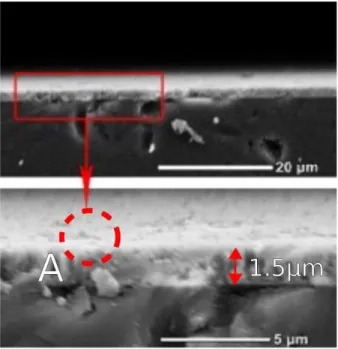
SEM images obtained for the sample Z3 after polish-ing one face of this sample up to optical quality (1/4µm)
are presented in Fig. 3. As it can be seen, the local
mea-surement of the sample thickness gives about 1.5µm at
the end-face. Also, pores or inhomogeneities were ob-served at the micrometric scale, distributed over the film
surface and volume (see the dark regions of about 1µm2
shown by enclosed curve A in Fig. 3).
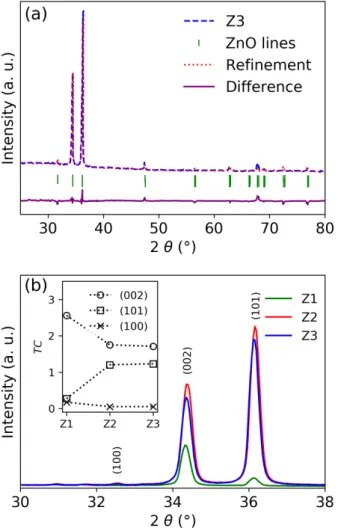
XRD pattern of the sample Z3 is shown in Fig. 4a and allows determination of the crystal structure and the coatings preferred orientation. All the sharp
diffraction peaks were assigned to the hexagonal phase
(wurtzite). We performed a Rietveld refinement con-sidering a preferred directional growth along (002) and
(101) planes, and determined the lattice parametersa=
b =3.2577(6) Å,c = 5.21456(9) Å (c/a= 1.6007(3)),
and the positional parameteru= 0.3799(1), which
de-fine the z coordinates of the O atoms in the structure.
By using these values the distances from Zn to the
apical and basal O atoms were calculated: d(Zn−Oa) =
1.981(3) Å andd(Zn−Ob)=1.982(1) Å, respectively.
XRD patterns of the coatings Z1, Z2 and Z3 for the
selected 2θrange from 30° to 38° are shown in Fig. 4b.
In this region, we observed the 002 and 101 diffraction
peaks, and also the very weak peak corresponding to (100) plane. As it can be seen, it is clear that the Z1 coating is oriented along (002) plane, which means a preferred growth direction along thec-axis for this
sam-ple. On the other hand, the samples Z2 and Z3 have a
101 diffraction peak with higher intensity than the 002
one, which is related to a preference for the (101) plane as the sample thickness increases, as also observed by other authors [51].
The preferential crystallite orientation can be quan-titatively analysed with the texture coefficient (T C),
which compares the relative peak intensities with those of the reference data card (JCPDS 00-036-1451). The
Figure 4. XRD pattern with Rietveld refinement for sample
Z3 (a) and detail of the (100), (002) and (101) diffraction
peaks (b) for samples Z1, Z2 and Z3 and
texture coefficientsTC(inset)
T Cfactor was estimated using the following relation:
T C(hkl)= Ihkl I0hkl 1 N PIhkl I0hkl (1)
whereT C(hkl)corresponds to thehklplane,Iis the
mea-sured peak intensity,I0the corresponding one of the
ref-erence card, andN is the number of considered
reflec-tion faces in the XRD pattern. The deviareflec-tion ofT Cfrom
unity implies the film growth in a preferred orientation.
TheT Cvalues for the (002), (101), and (100) planes are
shown in inset of Fig. 4b. As it can be seen, the Z1 has the highestT C(200)value, being 2.7, and it is about 1.5
for the Z3 and Z2 coatings. The opposite behaviour was observed forT C(101), being about 0.2 for the Z1 and
in-creasing up to 1.2 for both Z2 and Z3 coatings, whereas
T C(001)is about 0.1–0.2 in all three samples.
Regarding the optical properties, Fig. 5 shows the transmittance spectra of the Z1, Z2 and Z3 coatings. As it can be seen, the thicker coatings present more in-terference fringes. The maximum transmittance (about 90%) is obtained in the infrared (IR) region, whereas
at the visible range the transmittance is between 40 and 80%. The inhomogeneities observed by SEM are rep-resented in these measurements by the shrinkage of the two evolving curves: one that interpolates maximums and the other that interpolates minimum values of inter-ference fringes (dashed lines around Z3 transmittance in Fig. 5), i.e. an important reduction of the oscillation amplitude is observed as wavelength decreases [52].
From the interference fringes in the λ range of 700–
1000 nm, we determined the coating thicknesst. To this
purpose, we compared the experimental spectra with
simulations of T(λ) obtained using the widely known
Filmetrics reflectance calculator based on the complex-matrix form of the Fresnel equations [53]. By
consider-ing coatconsider-ings with a refractive indexnbetween 1.8 and
2.0 [5,7,8], and a SiO2substrate withns =1.45, we
ob-tainedt=1.3(1), 2.1(1), and 2.3(1)µm for samples Z1,
Z2, and Z3, respectively. For Z3, the difference of thet
value with the local thickness of about 1.5µm observed
by SEM (Fig. 3) can be attributed to the variation of thickness along the sample surface.
Figure 5. Transmittance spectra and Tauc’s plot (inset)
On the other hand, to determine the optical band gap
Eg, we used the Tauc’s relationαhν = A(hν−Eg)1/2,
where Ais a constant,hνis the photon energy, and the
absorption coefficientαis calculated from the
transmit-tanceT(λ) using the Beer-Lambert lawα = ln(1/T)/t.
The Tauc’s plots (αhν)2vs.hν, presented in inset of Fig.
5, were used to determine Eg by extrapolating the
lin-ear portion to zero. It was found that Eg = 3.24(1) eV
for all three samples, a value that is in good agreement with the results reported for ZnO films by other authors [8,10,12,13].
3.2. Calculation results
DFT and DFT+U optimized structures
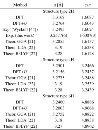
Table 1 summarizes the basal lattice parameteraand
thec/aratio for 2H, 4H and 6H polytypes obtained
us-ing the DFT and DFT+Uapproaches. For comparison,
other theoretical data available in the literature for all three polytypes were also included [21,22] together with
Table 1. Lattice parameters for ZnO polytypes obtained by
DFT and DFT+U(for three polytypes, theoretical values
predicted by other authors are also included as well as the experimental values for ZnO bulk and coating)
Method a[Å] c/a
Structure type 2H
DFT 3.3169 1.6087
DFT+U 3.2764 1.6043
Exp. (Wyckoff[44]) 3.2495 1.6024 Exp. (this work) 3.2577(6) 1.6007(3)
Theor. GGA [21] 3.2855 1.6137 Theor. LDA [22] 3.19 1.6238 Theor. B3LYP [22] 3.28 1.6128 Structure type 4H DFT 3.2501 3.2466 DFT+U 3.2156 3.2437 Theor. GGA [21] 3.2775 3.2488 Theor. LDA [22] 3.19 3.2539 Theor. B3LYP [22] 3.28 3.2439 Structure type 6H DFT 3.2460 4.8886 DFT+U 3.2003 4.9668 Theor. GGA [21] 3.2752 4.8822 Theor. LDA [22] 3.18 4.8838 Theor. B3LYP [22] 3.27 4.8962
the experimental results reported by Wyckoff [44] and
data obtained in this work for the 2H phase.
Our calculations show that the different polytypes
have a similar basal lattice parameter. The predicteda
andc/avalues are in good agreement with the available
experimental and theoretical data. As it can be seen in Table 1, the optimization of the 2H structure leads to a slight overestimation of the lattice parameters (up to about 2% of the experimental values). A closer look at these results shows that the inclusion of the Hubbard term produces lattice parameters closer to the experi-mental ones. This improvement in the prediction of the
2H unit cell with DFT+U was also observed before by
other authors [23,24,26].
DOS, bandgap and optical properties
The results of the density of electronic states (DOS) corresponding to the different ZnO structures and
calcu-lation approaches are shown in Fig. 6. As it can be seen,
for the same calculation method (DFT or DFT+U) no
significant changes of the DOS are observed from one cell to another. For the 2H type, the DOS corresponding to the starting structure (dashed lines in Fig. 6) is also presented to show that the structural optimizations do not produce significant changes in the electronic prop-erties. In general, the DOS presents the valence band
with O-2pand Zn-4dcharacter, separated from the
Zn-2sconduction band by Eg. In the case of DFT
calcu-lations (Fig. 6a), we obtainedEg values of about 1 eV,
which underestimates the experimental value and
re-Figure 6. DOS calculated for the different structures
according to the DFT (a) and DFT+Umethods (b) (the scale
of energy refers to the Fermi level and the extent ofEgis
indicated between the vertical dotted lines)
flects the well-known band gap problem. The addition
of theUparameter modifies the DOS and increases the
predictedEgto about 3.4 eV (Fig. 6b), which is in better
agreement with the measured band gaps.
Considering the improvement obtained with DFT+U,
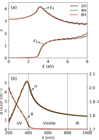
we used this approach to calculate the dielectric
func-tion ε(ω) and related optical properties. The real and
imaginary part ofε(ω) (ε1 andε2, respectively) for all
three structures are given in Fig. 7a. As it can be seen again, no significant changes in these dielectric func-tions are observed from one polytype to another. These results show the typical behaviour for a
semiconduc-tor:ε1 is a positive function, which approaches the
di-electric constant ε∞ in the limit ω → 0
(experimen-tally,ε∞ = 3.7 [16,54]). On the other hand, the firstε2
slight shoulder at about 3.4 eV corresponds to the inter-band absorption edge in the range of UV energies, ex-pected according to the transparency of the ideal ZnO [29,30,54]. By using ε1(ω) and ε2(ω), we calculated
α(λ) andn(λ) [28] (Fig. 7b). Our predictions for all these
optical properties are in good agreement with previous calculations by other DFT methods [20,26,31,33,55], as well as with the experimental measurements obtained
Figure 7. Dielectric functionε=ε1+jε2(a) and absorption
coefficientαand the real part of the refractive indexn(b) for
different fully optimized ZnO structures with DFT+U
Figure 8. DFT+Usimulated transmittance spectraTin the
range of 200–1000 nm for the 2H type ZnO, before and after structural optimization, and comparison with experimental
measurement for the Z1 sample
for thin films and bulk ZnO possessing the wurtzite phase [8,54].
Finally, we used the calculated absorption coeffi
-cient to simulate the transmittance spectra T(λ) =
exp(−α(λ)t) for the 2H structure, using a thicknesst =
1.3µm. The obtained results are presented in Fig. 8 and
we compared them with the experimental spectrum
cor-responding to the sample Z1. The predictedT(λ) does
not significantly depend on the considered lattice pa-rameters and internal coordinates, as it can be seen from the similar spectra obtained using the 2H starting
struc-ture of Wyckoff [44] and the DFT+U fully optimized
in this work. On the other hand, differences from the
experimental spectrum can be attributed to finite thick-ness, surface roughthick-ness, and sample inhomogeneity and
porosity, which affect the optical transmittance,
espe-cially in the visible range [13,52].
Local structures and EFGs in wurtzite (2H) ZnO
Considering that among the three polytypes only the wurtzite phase of ZnO is known experimentally, and it was the one observed in our ZnO coatings, further DFT
and DFT+U studies were performed for this structure.
As presented in Fig. 2a, the Zn atoms are tetrahedrally coordinated with O atoms, so the atomic environments can be characterized by the distances from the Zn atom to the apical and the basal oxygen atoms (d(Zn−Oa)and d(Zn−Ob), respectively). These distances before and after
the DFT and DFT+U structural optimization are
pre-sented in Table 2. As it can be seen, the internal atomic relaxation from the starting structure of Wyckoff[44] to
the final ones produces a change of both distances, in a way that the values approach each other, and also to the
d values obtained from our XRD measurements. This
result suggests that the four O atoms around Zn are lo-cated at the corners of a practically regular tetrahedron. We also analysed the local structures by calculat-ing the EFG because it is a quantity very sensitive to changes in the local environments. The last two columns in Table 2 present the predictions for the largest com-ponent of the diagonalized EFG tensor in the
princi-pal axis system,Vzz, at the Zn and O atomic sites. The
tensor is complemented by the asymmetry parameter
η, which for the considered wurtzite structure is null
at both atomic sites. The obtained results for the EFG show that changes of the Zn–O distances from one cell to another strongly affect the predicted values ofVzz. In
particular, we found that the structural relaxations must be taken into account to correctly reproduce the
exper-Table 2. Local structures and EFGs for the 2H ZnO unit cells (the experimental values in the last row come from [a] this work,
and [b] Ref. [56] and the sign ofVzzis unknown)
2H structure d(Zn−O
a)[Å] d(Zn−O
b)[Å] Vzz(Zn)[10
21V/m2] V
zz(O)[1021V/m2]
Starting structure of Wyckoff[44] DFT 1.796 2.042 −8.13 +2.47
DFT+U 1.796 2.042 −9.25 +2.53
DFT fully optimized 2.031 2.018 +0.61 −0.26
DFT+Ufully optimized 2.000 1.993 +0.60 −0.25
Table 3. Results corresponding to the elastic properties of the 2H structure of ZnO
C11[GPa] C12[GPa] C13[GPa] C33[GPa] C44[GPa] B[GPa]
DFT 204 89 69 207 42 119 DFT+U 244 110 86 243 53 144 GGA [58] 235 67 61 288 50 129 B3LYP [59] 218 124 106 223 44 148 Exp. [60] 209(1.5) 120(1) 104.4(2.0) 216(4) 44.3 143(5) Exp. [61] 206(4) 116(5) 118(10) 211(4) 44(1) 147(7)
imental values [56]. Therefore, as it can be seen from Table 2, the structural relaxations (involved in the fully optimized structures) are necessary to correctly describe the local structures observed in our XRD measurements and the EFG experimental values. This means that the starting structure considered for our calculations has an
incorrect value ofupositional parameter and therefore
wrong Zn–O distances, as other authors also mentioned [57]. Nevertheless, we included these results to show
that DFT and DFT+Ucan find the correct atomic
posi-tions after structural relaxaposi-tions, and also the correctVzz
values. Considering the high sensitivity of the EFG, the comparison of these predicted and measured quantities is very suitable to assess the accuracy of the calculation approaches.
Elastic properties of the wurtzite (2H) ZnO
The elastic constantsCi jand the bulk modulusB
cal-culated for the wurtzite structure are presented in Ta-ble 3. The Voigt’s notation is used according to which
i,j = 1,2, . . . ,6 (1 = xx, 2 = yy, 3 = zz, 4 = yz,
5= xz, 6= xy). In our case, theCi jcoefficients are the
five independent components of the fourth-rank tensor corresponding to the hexagonal solid [47]. Table 3 also includes theoretical data calculated by other authors us-ing GGA [58] and the hybrid functional B3LYP [59], as well as the available experimental values obtained for ZnO polycrystalline films [60,61]. It can be noted that our predictions are in a general agreement with those values. A comparison of our DFT values with the
ex-perimental data shows that the calculatedCi j
systemat-ically underestimates the experimental value, which is expected according to the systematic overestimation of the equilibrium lattice parameters obtained with DFT
(Table 1). The inclusion of the U parameter produces
an increase in the predictedCi j values, givingCi j
val-ues dispersed around the experimental ones, with diff
er-ences up to about 30% (forC13). Finally, the bulk
mod-ulus B, calculated through a linear combination of the
elastic constants [59], follows the same general agree-ment already described.
IV. Discussion
Even though there are some studies of ZnO-based
systems that combine experiments and DFT+U
calcu-lations, to the best of our knowledge, there has been no comprehensive analysis of the performance of the
DFT+U approach that simultaneously describes
struc-tural, optical and elastic properties of ZnO as it has
been done in the present investigation, where theab
ini-tiopredictions are proposed to complement
experimen-tal studies on ZnO coatings. Therefore, we produced and characterized ZnO coatings and also we also per-formed first-principles calculations for three polytypes of hexagonal ZnO. This combination of measurements and predictions allowed us to simultaneously analyse different properties of the synthesized coatings and also
to assess the DFT+Umethod for predicting those
prop-erties in the different polytypes. For the experimental
existing 2H phase, our systematic comparison of the
DFT+Upredictions with DFT and experimental results
demonstrated that DFT+Uis effective to predict and
de-scribe not only bandgap energy, as it is already known, but also the structural and elastic properties of ZnO. In this sense, its strength lies in its vast range of applica-bility: from the atomic local environments (through the Zn–O distances and EFGs) to the crystallographic scale and the elastic constants. Then, the presented combina-tion of calculacombina-tions and experiments enhances the anal-ysis of the materials. For example, we showed that after considering the structural relaxations the atomic
envi-ronments have strong differences compared to those of
the starting structure, and such relaxations are required to correctly describe the properties observed through XRD and EFG measurements. In contrast, these local environment differences are not reflected by the optical
transmittance spectra. So, we can propose interplay
be-tween theab initiomodels and a variety of
experimen-tal results to validate the calculation methods and assess the sensitivity of the involved experimental techniques.
Considering this, we found that the DFT+U predicted
properties are generally in a good agreement with our own experimental measurements performed on the ZnO coatings produced with the SP technique. The few dif-ferences between predictions and measurements could be expected according to the method and due to the simplicity of the proposed modelling approach and the
complex nature of the real samples. Then, DFT+U is
useful to obtain information about the optical and elas-tic properties of ZnO, which is of pracelas-tical importance in the design of thin films and coatings.
The present results allow us to extend this method-ology to analyse further aspects related to the electro-optical and piezo-electro-optical applications of ZnO coatings. For example, how the film deposition and growing con-ditions affect the residual stress and Ci j coefficients
ap-plied to more complex ZnO systems to predict optical and elastic properties when defects, impurities, and in-terfaces are present [3,4,6,14,22,26].
V. Conclusions
ZnO coatings were produced by the spray-pyrolysis technique and characterized by scanning electron
mi-croscopy, X-ray diffraction and optical transmittance
spectroscopy. The experimental results were compared to predictions obtained from the electronic-structure
calculations based on the DFT+U approach. We
con-sider that this study is a successful first step towards the study of more complex ZnO-based coatings, where the
ab initiocalculations support the experiments to achieve
a deeper insight into the material properties, which are central to the development of optically active nanocom-ponents like diodes, lasers, solar cells and band optical filters.
Acknowledgments: We wish to acknowledge the
Ar-gentinian funding institutions for supporting this work: Agencia Nacional de Promoción de Ciencia y Tec-nología under contract PICT 2015-0452 and Universi-dad Nacional de La Plata (UNLP) under grant “Jóvenes Investigadores”. Part of the results presented in this work has been obtained using the facilities of the CCT-Rosario Computational Center, member of the High Performance Computing National System (SNCAD, MinCyT-Argentina). We want to thank Dra. Susana Conconi (CETMIC) for assistance with XRD analysis. D. R. and M. R. T. are members of Consejo Nacional de Investigaciones Científicas y Técnicas, CONICET, Ar-gentina.
References
1. P. Rong, S. Ren, Q. Yu, “Fabrications and applications of ZnO nanomaterials in flexible functional devices - A Re-view”,Crit. Rev. Anal. Chem.,49[4] (2019) 336–349. 2. J. Theerthagiri, S. Salla, R.A. Senthil, P. Nithyadharseni,
A. Madankumar, P. Arunachalam, T. Maiyalagan, H.-S. Kim, “A review on ZnO nanostructured materials: Energy, environmental and biological applications”, Nanotechnol-ogy,30[39] (2019) 392001.
3. B. Ghanbari Shohany, A. Khorsand Zak, “Doped ZnO nanostructures with selected elements - Structural, mor-phology and optical properties: A review”,Ceram. Int.,46 [5] (2020) 5507–5520.
4. K.D.A. Kumar, R. Thomas, S. Valanarasu, V. Ganesh, M. Shkir, S. AlFaify, J. Thirumalai, “Analysis of Pr co-doped Al:ZnO thin films using feasible nebulizer spray technique for optoelectronic technology”, Appl. Phys. A,125 [10] (2019) 712.
5. G. Malik, S. Mourya, J. Jaiswal, R. Chandra, “Effect of an-nealing parameters on optoelectronic properties of highly ordered ZnO thin films”,Mater. Sci. Semicond. Process.,
100(2019) 200–213.
6. F. Baig, M.W. Ashraf, A. Asif, M. Imran, “A compara-tive analysis for effects of solvents on optical properties of Mg doped ZnO thin films for optoelectronic applications”,
Optik,208(2020) 164534.
7. M.-J. Zhao, Z.-T. Sun, C.-H. Hsu, P.-H. Huang, X.-Y. Zhang, W.-Y. Wu, P. Gao, Y. Qiu, S.-Y. Lien, W.-Z. Zhu, “Zinc oxide films with high transparency and crystallinity prepared by a low temperature spatial atomic layer depo-sition process”,Nanomaterials,10[3] (2020) 459. 8. A. Ashour, M.A. Kaid, N.Z. El-Sayed, A.A. Ibrahim,
“Physical properties of ZnO thin films deposited by spray pyrolysis technique”, Appl. Surf. Sci., 252 [22] (2006) 7844–7848.
9. R. Navamathavan, K.-K. Kim, D.-K. Hwang, S.-J. Park, J.-H. Hahn, T.G. Lee, G.-S. Kim, “A nanoindentation study of the mechanical properties of ZnO thin films on (0 0 0 1) sapphire”,Appl. Surf. Sci.,253[2] (2006) 464–467. 10. J.H. Cai, G. Ni, G. He, Z.Y. Wu, “Red luminescence in
ZnO films prepared by a glycol-based Pechini method”,
Phys. Lett. A,372[22] (2008) 4104–4108.
11. P.-F. Yang, H.-C. Wen, S.-R. Jian, Y.-S. Lai, S. Wu, R.-S. Chen, “Characteristics of ZnO thin films prepared by radio frequency magnetron sputtering”,Microelectron. Reliab.,
48[3] (2008) 389–394.
12. T.P. Rao, M.C.S. Kumar, S.A. Angayarkanni, M. Ashok, “Effect of stress on optical band gap of ZnO thin films with substrate temperature by spray pyrolysis”,J. Alloys Compd.,485[1-2] (2009) 413–417.
13. T. Prasada Rao, M.C. Santhoshkumar, “Highly oriented (100) ZnO thin films by spray pyrolysis”,Appl. Surf. Sci.,
255[16] (2009) 7212–7215.
14. M.-C. Jun, S.-U. Park, J.-H. Koh, “Comparative studies of Al-doped ZnO and Ga-doped ZnO transparent conducting oxide thin films”,Nanoscale Res. Lett.,7[1] (2012) 639. 15. Y. Cherifi, A. Chaouchi, Y. Lorgoilloux, M. Rguiti, A.
Kadri, C. Courtois, “Electrical, dielectric and photocat-alytic properties of Fe-doped ZnO nanomaterials synthe-sized by sol gel method”,Process. Appl. Ceram.,10[3] (2016) 125–135.
16. M.B. Bouzourâa, Y. Battie, S. Dalmasso, M.A. Zaïbi, M. Oueslati, A. En Naciri, “Temperature dependent optical properties of ZnO thin film using ellipsometry and pho-toluminescence”, Superlattices Microstruct., 117 (2018) 457–468.
17. D. Richard, M. Romero, R. Faccio, “Experimental and the-oretical study on the structural, electrical and optical prop-erties of tantalum-doped ZnO nanoparticles prepared via sol-gel acetate route”,Ceram. Int.,44[1] (2018) 703–711. 18. E.A. Villegas, R. Parra, L. Ramajo, “Integral nanoinden-tation evaluation of TiO2, SnO2, and ZnO thin films de-posited via spray-pyrolysis on glass substrates”,J Mater. Sci. Mater. Electron.,30[2] (2019) 1360–1365.
19. S. Lany, A. Zunger, “Assessment of correction methods for the band-gap problem and for finite-size effects in super-cell defect calculations: Case studies for ZnO and GaAs”,
Phys. Rev. B,78[23] (2008) 235104.
20. K. Bashyal, C.K. Pyles, S. Afroosheh, A. Lamichhane, A.T. Zayak, “Empirical optimization of DFT+U and HSE for the band structure of ZnO”,J. Phys. Condens. Matter,
30[6] (2018) 065501.
21. Z. Huang, T.-Y. Lü, H.-Q. Wang, J.-C. Zheng, “Thermo-electric properties of the 3C, 2H, 4H, and 6H polytypes of the wide-band-gap semiconductors SiC, GaN, and ZnO”,
AIP Advances,5(2015) 097204.
22. D. Zagorac, J.C. Schoen, J. Zagorac, M. Jansen, “Theo-retical investigations of novel zinc oxide polytypes and in-depth study of their electronic properties”, RSC Adv.,
5(2015) 259290.
23. G.-Y. Huang, C.-Y. Wang, J.-T. Wang, “Detailed check of the LDA+U and GGA+U corrected method for defect cal-culations in wurtzite ZnO”,Comput. Phys. Commun.,183 [8] (2012) 1749–1752.
24. M.K. Yaakob, N.H. Hussin, M.F.M. Taib, T.I.T. Kudin, O.H. Hassan, A.M.M. Ali, M.Z.A. Yahya, “First princi-ples LDA+U calculations for ZnO materials”,Integr. Fer-roelectr.,155[1] (2014) 15–22.
25. E.S. Goh, J.W. Mah, T.L. Yoon, “Effects of Hubbard term correction on the structural parameters and elec-tronic properties of wurtzite ZnO”,Comp. Mater. Sci.,138 (2017) 111–116.
26. K. Harun, N.A. Salleh, B. Deghfel, M.K. Yaakob, A.A. Mohamad, “DFT+U calculations for electronic, structural, and optical properties of ZnO wurtzite structure: A re-view”,Results Phys.,16(2020) 102829.
27. A. Calzolari, M.B. Nardelli, “Dielectric properties and Raman spectra of ZnO from a first principles finite-differences/finite-fields approach”, Sci. Rep., 3 (2013) 2999.
28. Q.-B. Wang, C. Zhou, J. Wu, T. Lü, “A GGA+U study of the optical properties of vanadium doped ZnO with and without single intrinsic vacancy”,Opt. Commun.,297 (2013) 79–84.
29. A. Calzolari, A. Ruini, A. Catellani, “Transparent conduc-tive oxides as near-IR plasmonic materials: The case of Al-doped ZnO derivatives”,ACS Photonics,1[8] (2014) 703–709.
30. S.Zh. Karazhanov, P. Ravindran, U. Grossner, A. Kjek-shus, H. Fjellvåg, B.G. Svensson, “Strong Coulomb cor-relation effects in ZnO”, Solid State Commun., 139 [8] (2006) 391–396.
31. P. Palacios, I. Aguilera, P. Wahnón, “Electronic structure and optical properties in ZnO:M(Co, Cd): Effect of band-gap variation”,Thin Solid Films,518 [16] (2010) 4568– 4571.
32. Y.-S. Lee, Y.-C. Peng, J.-H. Lu, Y.-R. Zhu, H.-C. Wu, “Electronic and optical properties of Ga-doped ZnO”,Thin Solid Films,570(2014) 464–470.
33. H.-C. Wu, H.-H. Chen, Y.-R. Zhu, “Effects of Al-impurity type on formation energy, crystal structure, electronic structure, and optical properties of ZnO by using Density Functional Theory and the Hubbard-U method”, Materi-als,9[8] (2016) 647.
34. S. Horzum, E. Torun, T. Serin, F.M. Peeters, “Structural, electronic and optical properties of Cu-doped ZnO: ex-perimental and theoretical investigation”,Philos. Mag.,96 [17] (2016) 1743–1756.
35. M.V. Gallegos, C.R. Luna, M.A. Peluso, L.C. Damonte, J.E. Sambeth, P.V. Jasen, “Effect of Mn in ZnO using DFT calculations: Magnetic and electronic changes”,J. Alloys Compd.,795(2019) 254–260.
36. R. Amari, B. Deghfel, A. Mahroug, A.A. Mohamad, A. Boukhari, N. Selmi, “Effects of Mn doping on the struc-tural, morphological, electronic and optical properties of ZnO thin films by sol-gel spin coating method: An exper-imental and DFT+U study”,Physica B Condens. Matter,
577(2020) 411766.
37. L. Honglin, L. Yingbo, L. Jinzhu, Y. Ke, “Experimental and first-principles studies of structural and optical proper-ties of rare earth (RE=La, Er, Nd) doped ZnO”,J. Alloys Compd.,617(2014) 102–107.
38. J.V.N. Sarma, A. Rahman, R. Jayaganthan, R. Chowd-hury, D. Haranath, “Al-doped ZnO nanostructured thin films: Density Functional Theory and experiment”,Int. J. Nanosci.,14[04] (2015) 1550015.
39. M. Bououdina, S. Azzaza, R. Ghomri, M.N. Shaikh, J.H. Dai, Y. Song, W. Song, W. Cai, M. Ghers, “Structural and magnetic properties and DFT analysis of ZnO:(Al,Er) nanoparticles”,RSC Adv.,7[52] (2017) 32931–32941. 40. J.C.A. Queiroz, J.B. Azevedo Filho, J.Q. Medeiros Neto, I.
Oliveira Nascimento, I.A. Souza, M.G. Oliveira Queiroz, E.B. Melo, T.H. Carvalho Costa, “Structural and optical properties of Al-doped ZnO thin films produced by mag-netron sputtering”,Process. Appl. Ceram.,14 [2] (2020) 119–127.
41. G. Suarez, F.C. Alvira, R. Parra, M.R. Tejerina, “Char-acterization of thin coatings based on ZnO for photonic applications”,Optoelectron. Adv. Mater.,13[9-10] (2019) 535–538.
42. P. Giannozzi, O. Andreussi, T. Brumme, O. Bunau, M. Buongiorno Nardelli, M. Calandra, R. Car, C. Cavazzoni, D. Ceresoli, M. Cococcioni, N. Colonna, I. Carnimeo1, A. Dal Corso, S. de Gironcoli, P. Delugas, R.A. DiStasio Jr., A. Ferretti, A. Floris, G. Fratesi, G. Fugallo, R. Gebauer, U. Gerstmann, F. Giustino, T. Gorni, J. Jia, M. Kawamura, H.-Y. Ko, A. Kokalj, E. Küçükbenli, M. Lazzeri, M. Mar-sili, N. Marzari, F. Mauri, N.L. Nguyen, H.-V. Nguyen, A. Otero-de-la-Roza, L. Paulatto, S. Poncé, D. Rocca, R. Sabatini, B. Santra, M. Schlipf, A.P. Seitsonen, A. Smo-gunov, I. Timrov, T. Thonhauser, P. Umari, N. Vast, X. Wu, S. Baroni, “Advanced capabilities for materials modelling with Quantum ESPRESSO”,J. Phys. Condens. Matter,29 [46] (2017) 465901.
43. J.P. Perdew, K. Burke, M. Ernzerhof, “Generalized gradi-ent approximation made simple”,Phys. Rev. Lett.,77[18] (1996) 3865–3868.
44. R.W.G. Wyckoff,Crystal structures, second edition.
Inter-science Publishers, New York, 1963.
45. S. Gražulis, A. Daškeviˇc, A. Merkys, D. Chateigner, L. Lutterotti, M. Quirós, N.R. Serebryanaya, P. Moeck, R.T. Downs, A. Le Bail, “Crystallography Open Database (COD): an open-access collection of crystal structures and platform for world-wide collaboration”,Nucleic Acids Res.,40[Database issue] (2012) D420–D427.
46. A. Menad, M.E. Benmalti, A. Zaoui, M. Ferhat, “Impact of polytypism on the ground state properties of zinc oxide: A first-principles study”,Results Phys.,18(2020) 103316. 47. A. Dal Corso, “Elastic constants of beryllium: a first-principles investigation”,J. Phys. Condens. Matter,28[7] (2016) 075401.
48. C.J. Pickard, F. Mauri, “All-electron magnetic response with pseudopotentials: NMR chemical shifts”,Phys. Rev. B,63[24] (2001) 245101.
49. M. Profeta, F. Mauri, C.J. Pickard, “Accurate first princi-ples prediction of17O NMR parameters in SiO
2: Assign-ment of the zeolite ferrierite spectrum”,J. Am. Chem. Soc.,
125[2] (2003) 541–548.
50. A. Dal Corso, The thermo_pw software, https://dalcorso. github.io/thermo_pw.
51. L. Znaidi, “Sol-gel-deposited ZnO thin films: A review”,
Mater. Sci. Eng. B,174[1-3] (2010) 18–30.
52. R. Swanepoel, “Determination of surface roughness and optical constants of inhomogeneous amorphous silicon films”,J. Phys. E,17[10] (1984) 896–903.
53. Filmetrics Reflectance Calculator, https://www.filmetrics. com/reflectance-calculator.
54. H. Yoshikawa, S. Adachi, “Optical constants of ZnO”,Jpn. J. Appl. Phys.,36[10R] (1997) 6237–6243.
55. E.A. Alkahtani, A.E. Merad, M.R. Boufatah, A. Benos-man, “DFT investigation of structural, electronic and opti-cal properties of pure and Er-doped ZnO: Modified Becke-Johnson exchange potential”,Optik,128(2017) 274–280. 56. G. Denninger, D. Reiser, “Determination of electric-field gradients in semiconductors with high precision and high sensitivity”,Phys. Rev. B,55[8] (1997) 5073–5078. 57. M. Nyberg, M.A. Nygren, L.G.M. Pettersson, D.H. Gay,
A.L. Rohl, “Hydrogen dissociation on reconstructed ZnO surfaces”,J. Phys. Chem.,100[21] (1996) 9054–9063. 58. M. Kalay, H.H. Kart, S. Özdemir Kart, T. Ça˘gın,
“Elas-tic properties and pressure induced transitions of ZnO polymorphs from first-principle calculations”, J. Alloys Compd.,484[1-2] (2009) 431–438.
59. N.L. Marana, S.M. Casassa, J.R. Sambrano, “Piezoelec-tric, elastic, infrared and Raman behavior of ZnO wurtzite under pressure from periodic DFT calculations”, Chem. Phys.,485-486(2017) 98–107.
60. A.G. Every, A.K. McCurdy, Second and Higher Or-der Elastic Constants - Elastische Konstanten zweiter und höherer Ordnung(Landolt-Börnstein series, vol 29),
Springer, Berlin,1993.
61. G. Carlotti, D. Fioretto, G. Socino, E. Verona, “Brillouin scattering determination of the whole set of elastic con-stants of a single transparent film of hexagonal symmetry”,

![Table 3. Results corresponding to the elastic properties of the 2H structure of ZnO C 11 [GPa] C 12 [GPa] C 13 [GPa] C 33 [GPa] C 44 [GPa] B [GPa]](https://thumb-us.123doks.com/thumbv2/123dok_us/10690380.2957244/7.892.165.721.135.270/table-results-corresponding-elastic-properties-structure-zno-gpa.webp)
