N A N O E X P R E S S
Open Access
Biodegradable nanoparticles of methoxy poly
(ethylene glycol)-
b
-poly(D,L-lactide)/methoxy poly
(ethylene glycol)-
b
-poly(
E
-caprolactone) blends
for drug delivery
Yodthong Baimark
*and Yaowalak Srisuwan
Abstract
The effects of blend weight ratio and polyester block length of methoxy poly(ethylene glycol)-b-poly(D,L-lactide) (MPEG-b-PDLL)/methoxy poly(ethylene glycol)-b-poly(E-caprolactone) (MPEG-b-PCL) blends on nanoparticle characteristics and drug release behaviors were evaluated. The blend nanoparticles were prepared by nanoprecipitation method for controlled release of a poorly water-soluble model drug, indomethacin. The drug-loaded nanoparticles were nearly spherical in shape. The particle size and drug loading efficiency slightly decreased with increasing MPEG-b-PCL blend weight ratio. Two distinct thermal decomposition steps from thermogravimetric analysis suggested different blend weight ratios. Thermal transition changes from differential scanning calorimetry revealed miscible blending between MPEG-b-PDLL and MPEG-b-PCL in an amorphous phase. Anin vitrodrug release study demonstrated that the drug release behaviors depended upon the PDLL block length and the blend weight ratios but not on PCL block length.
Keywords:Biodegradable polymers, Polymer blends, Diblock copolymers, Nanoparticles, Nanoprecipitation method, Controlled release
Background
In the past few decades, biodegradable and biocompat-ible nanoparticles of methoxy poly(ethylene glycol)-b -poly(D,L-lactide) (MPEG-b-PDLL) and methoxy poly (ethylene glycol)-b-poly(E-caprolactone) (MPEG-b-PCL) amphiphilic diblock copolymers have shown potential as controlled-release drug delivery carriers because of the small size of their nanoparticles, which improves circula-tion time in the body and decreases the administracircula-tion frequency when compared to microparticles which are rapidly cleared by the reticuloendothelial tissue [1,2]. Moreover, for MPEG-b-PDLL and MPEG-b-PCL nano-particles suspended in water, PDLL and PCL hydropho-bic cores are surrounded by hydrophilic MPEG blocks on the nanoparticle surface to solubilize hydrophobic
drugs; this increases blood circulation time and
decreases uptake by the liver of the nanoparticles [3-6]. The need for surfactants when preparing nanoparticles of amphiphilic diblock copolymer by the nanoprecipita-tion method can be removed. The protective effect of the hydrophilic MPEG block is adequate for preventing nanoparticle aggregation. The poly(vinyl alcohol), Span series, Tween series, poly(ethylene oxide) (PEO), and poloxamer (PEO-poly(propylene oxide) block copoly-mer) have been used as surfactants to stabilize emulsion droplets [7]. These surfactants remain at the particle sur-face and are difficult to remove which affect the bio-degradability and drug release profile of the drug-loaded particles. Also, these remaining surfactants can influence the human body, for example, causing an allergy-like reaction.
In previous studies, much attention was paid to the drug-loaded nanoparticles of these amphiphilic diblock copolymers with different types of chemical composi-tions and lengths of polymer blocks used [8-11]. Drug release profiles of the nanoparticles depended upon * Correspondence:[email protected]
Department of Chemistry and Center of Excellence for Innovation in Chemistry, Faculty of Science, Mahasarakham University, Mahasarakham 44150, Thailand
these factors. The physical blending of polymers is an al-ternative method that has been widely used to adjust the properties of biodegradable polyesters [12-15]. Thus unique properties of polymer blends, quite different from the origin polymers, were obtained. The drug re-lease rate from poly(L-lactide)/PCL blend nanoparticles prepared from the nanoprecipitation method using poloxamer 188 as the surfactant has been adjusted by varying the blend weight ratios [16].
The characteristics and drug release behaviors of the MPEG-b-PCL nanoparticles containing drug prepared by the nanoprecipitation method can be controlled by adjusting the processing parameters, such as the or-ganic/water phase volume ratio, polymer concentration, drug/polymer weight ratio, and stirring speed [17]. How-ever, the effect of MPEG-b-PDLL/MPEG-b-PCL
blend-ing on characteristics and drug release of the
nanoparticles has not been reported.
In this study, surfactant-free MPEG-b-PDLL/MPEG-b -PCL blend nanoparticles containing a model drug were prepared by the nanoprecipitation method. Indometh-acin was selected as the model drug because of its poor water solubility. The effects of different polyester block lengths and blend weight ratios on the nanoparticle characteristics, drug loading efficiency, and drug release profiles were studied and discussed.
Methods
Materials
MPEG-b-PDLL and MPEG-b-PCL diblock copolymers were synthesized by ring-opening polymerization in bulk under a nitrogen atmosphere as described in the previ-ous works [18,19]. MPEG with a molecular weight of 5,000 g/mol (Fluka Chemical Corporation, St. Louis, Milwaukee, WI, USA) and stannous octoate (95%, Sigma-Aldrich Corporation, St. Louis, MO, USA) were used as the initiating system. Stannous octoate concen-tration was kept constant at 0.02 mol%. MPEG-b-PDLL
and MPEG-b-PCL with theoretical PDLL/PCL block
lengths of 30,000 (30 K) and 60,000 (60 K) g/mol were
called MPEG-b-PDLL30K, MPEG-b-PDLL60K,
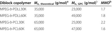
MPEG-b-PCL30K, and MPEG-b-PCL60K, respectively. The resulting number-average molecular weights (Mn) and
molecular weight distribution (MWD) of diblock copoly-mers are summarized in Table 1. Indomethacin model drug (99%, Sigma-Aldrich Corporation, St. Louis, MO, USA) was used without further purification. All reagents used were analytical grade.
Preparation of drug-loaded blend nanoparticles
Drug-loaded blend nanoparticles were prepared via a nanoprecipitation method. Briefly, 4 mg of indomethacin and 60 mg of copolymer blend were co-dissolved in 6 mL of acetone. The organic solution was added dropwise
into 60 mL of distilled water under magnetic stirring. The nanoparticles were immediately formed after solv-ent extraction. The organic solvsolv-ent was then evaporated at room temperature for 6 h in a fume hood. The drug-loaded blend nanoparticles with MPEG-b
-PDLL/MPEG-b-PCL blend weight ratios of 100/0, 75/25, 50/50, 25/75, and 0/100 (w/w) were investigated. The resultant nano-particle suspensions were centrifuged at 15,000 rpm 4°C for 2 h. The supernatant was carefully discarded and the precipitated nanoparticles were then resuspended in a phosphate buffer solution media (0.1 M, pH 7.4). The dried drug-loaded nanoparticles were obtained by freeze-drying the precipitated nanoparticles for 48 h.
Characterization of drug-loaded blend nanoparticles
Morphology of the drug-loaded nanoparticles was deter-mined by transmission electron microscopy (TEM) using a JEOL JEM 1230 TEM (JEOL Ltd., Tokyo, Japan). For TEM analysis, a drop of the nanoparticle suspension was placed on a formvar film coated on a copper grid. The specimen on the copper grid was not stained. The aver-age particle sizes of the drug-loaded nanoparticles were measured from the nanoparticle suspension by light scat-tering analysis using a Coulter LS230 particle size analyzer (Beckman Coulter Inc., Brea, CA, USA) at 25°C. The thermal decomposition behavior of the dried nano-particles was characterized by thermogravimetry (TG) analysis using a TA-Instrument SDT Q600 TG analyzer (TA Instruments, New Castle, DE, USA). For TG, 5–10 mg of nanoparticles was heated from 50°C to 600°C at the rate of 20°C/min under a nitrogen atmosphere. Thermal transition properties of the dried nanoparticles were determined by mean of non-isothermal differential scanning calorimetry (DSC) using a Perkin-Elmer Pyris Diamond DSC (Perkin-Elmer Inc., Waltham, MA, USA). For DSC, 5–10 mg of nanoparticles was heated at the rate of 10°C/min under helium flow. The degree of
crys-tallinity of the blend nanoparticles due to PCL
crystallization was calculated according to the following equation [20],
Crystallinity of blend nanoparticles %ð Þ
[image:2.595.305.540.112.180.2]¼½ΔHm=135:44 100 ð1Þ Table 1 Molecular weight characteristics of diblock copolymers
Diblock copolymer Mn, theoretical(g/mol)a Mn, GPC(g/mol)b MWDb
MPEG-b-PDLL30K 35,000 23,000 1.7
MPEG-b-PDLL60K 35,000 49,000 1.8
MPEG-b-PCL30K 65,000 25,000 2.2
MPEG-b-PCL60K 65,000 47,000 1.6
a
Calculated based on monomer/MPEG ratio and MPEG molecular weight; b
whereΔHmis the melting enthalpy of the nanoparticles,
and 135.44 J/g is the melting enthalpy for 100% crystal-line PCL [21].
The theoretical drug loading content (DLCtheoretical),
actual drug loading content (DLCactual), and drug
load-ing efficiency (DLE) were calculated from Equations 2, 3, and 4, respectively. The DLCactual is an average value
from three measurements. For the DLCactual
measure-ment, the dried drug-loaded nanoparticles were dis-solved in dichloromethane. The weight of the actual entrapped drug was then determined by ultraviolet (UV)–vis spectrophotometry using a Perkin-Elmer Lambda 25 UV–vis spectrophotometer (Perkin-Elmer Inc., Waltham, MA, USA) at 319 nm and compared to a standard curve of indomethacin.
DLCtheoreticalð Þ ¼%
Weight of feed drug Weight of feed drug and copolymer
100
ð2Þ
DLCactualð Þ ¼%
Weight of actual drug entrapped in nanoparticles Weight of drugloaded nanoparticles
100
ð3Þ
DLEð Þ ¼% DLCactual DLCtheoretical
100 ð4Þ
In vitrodrug release
In vitro release of indomethacin from the blend nano-particles was performed by dialysis bag diffusion tech-nique. The drug-loaded nanoparticle suspension (10 mL) was placed in a dialysis bag, tied, and immersed into 100 mL of phosphate buffered saline (0.1 M, pH 7.4). The entire system was kept at 37°C under shaking at 100 rpm.
At predetermined time intervals, 5 mL aliquots of the release medium were withdrawn from the release medium, and the same volume of fresh buffer solution was added to continue the release test. The concentra-tion of indomethacin released was monitored using an UV–vis spectrophotometer at 319 nm. According to a predetermined indomethacin concentration-UV absorb-ance standard curve, indomethacin concentration of the release medium was obtained. Percentage of indometh-acin release was calculated based on the ratio of drug re-lease at each rere-lease time and initial drug content in the nanoparticles. The average percentage release was calcu-lated from the three experiments.
Results and discussion
Morphology and size
Morphology of the blend nanoparticles was investigated by TEM, an example of which is shown in Figure 1 for
the MPEG-b-PDLL30K/MPEG-b-PCL30K blend
nano-particles. It can be seen that they were nearly spherical in shape for all blend weight ratios. The blend nanopar-ticles of MPEG-b-PDLL30K/MPEG-b-PCL60K,
MPEG-b-PDLL60K/MPEG-b-PCL30K, and MPEG-b-PDLL60K/ MPEG-b-PCL60K showed similar results. The morph-ology results suggested that the hydrophilic MPEG blocks act as the nanoparticle shell to stabilize the nano-particle formation [18,19]. In these nanonano-particles, the hydrophobic core of the PDLL/PCL blocks is sur-rounded by the water-soluble polar groups of the MPEG blocks that extend into the aqueous medium to prevent nanoparticle aggregation. The polyester block length and blend weight ratio did not affect the morphology of drug-loaded blend nanoparticles. The TEM images also indicate that submicron-sized particles of copolymer blends can be prepared by the nanoprecipitation method. The particle size results, from light scattering analysis, are illustrated in Figure 2. They were less than 140 nm with a narrow size distribution. The average particle size slightly decreased as the MPEG-b-PCL blend weight ratio increased, which may be due to crystallization of the MPEG-b-PCL component during particle solidifica-tion that induced nanoparticle shrinkage.
Thermal decomposition
Thermal decomposition of the blend nanoparticles was determined by TG. The TG thermograms in Figure 3 (top) showed that the thermal resistance of the drug-loaded nanoparticles of MPEG-b-PCL are better than MPEG-b-PDLL. The blend nanoparticles, comprised of two decomposition steps, had maintained distinct ther-mal stability due to MPEG-b-PDLL decomposition, fol-lowed by degradation of the MPEG-b-PCL component. It was found that the TG profiles of the blend nanoparti-cles strongly depended upon the blend weight ratio. The amount of weight loss in the first step increased signifi-cantly when the MPEG-b-PDLL blend weight ratio was increased, as would be expected, since the blend nano-particles with various blend weight ratios were success-fully prepared.
Thermal decomposition can be clearly observed from differential TG (DTG) thermograms. Figure 3 (bottom)
show DTG thermograms of the MPEG-b-PDLL30K/
MPEG-b-PCL30K blend nanoparticles as examples. The two-step decomposition process of the blend nanoparti-cles is clearly illustrated. The peak temperature of the DTG thermogram is the temperature of maximum de-composition rate (Td, max). The lower and higher Td, max
MPEG-b-PDLL and MPEG-b-PCL decompositions, re-spectively. The Td, maxof MPEG-b-PDLL and MPEG-b
-PCL decomposition peaks are in the range of 343°C to 354°C and 400°C to 418°C, respectively, which also indi-cates higher thermal stability of the MPEG-b-PCL matrix. It should be noted that the peak areas of the DTG thermograms strongly depend on the blend weight
ratio. It can be clearly seen that the peak area of the lower temperature peak due to MPEG-b-PDLL decom-position decreased steadily as the MPEG-b-PDLL blend weight ratio decreased.
The TG and DTG thermograms of the blend nanopar-ticles of MPEG-b-PDLL30K/MPEG-b-PCL60K,
MPEG-b-PDLL60K/MPEG-b-PCL30K, and MPEG-b-PDLL60K/ MPEG-b-PCL60K changed with the blend weight ratio
similar to the MPEG-b-PDLL30K/MPEG-b-PCL30K
blend nanoparticles. The thermogravimetry results sup-ported the fact that MPEG-b-PDLL/MPEG-b-PCL blend nanoparticles with different blend weight ratios were obtained.
Thermal transition
[image:4.595.61.538.89.423.2]The thermal transition of the blend nanoparticles was studied by DSC. The DSC thermograms of the MPEG-b -PDLL/MPEG-b-PCL blend nanoparticles are shown as an example in Figure 4. The melting peak of indometh-acin is around 165°C and was not observed for all nano-particles. This may be explained by the indomethacin molecules being incorporated into the MPEG-b-PDLL
Figure 1TEM micrographs of drug-loaded nanoparticles with different MPEG-b-PDLL30K/MPEG-b-PCL30K blend weight ratios.All bars = 50 nm.
[image:4.595.58.292.572.707.2]or MPEG-b-PCL in the nanoparticle matrix. The indo-methacin crystallizability was then suppressed. Thus the drug was homogeneously dispersed in an amorphous form inside the nanoparticle matrix.
The drug-loaded MPEG-b-PDLL nanoparticles were amorphous (Figure 4a). Meanwhile, the MPEG-b-PCL and blend nanoparticles exhibited a single melting peak due to MPEG-b-PCL crystalline (Figures 4b,c,d,e). The
DSC results, including melting temperature (Tm),
melt-ing enthalpy (ΔHm) and crystallinity of the nanoparticles
are given in Figure 5. TheTm,ΔHmand crystallinity
sig-nificantly decreased with the MPEG-b-PCL blend weight
ratio. This may be explained by the MPEG-b-PCL
crystallization being inhibited by blending with
MPEG-b-PDLL. The results suggest that the MPEG-b-PDLL
and MPEG-b-PCL can be miscible blended in the
amorphous nanoparticle matrix to reduce the Tm, ΔHm
and crystallinity of the MPEG-b-PCL.
Drug loading efficiency
From the feed drug and copolymer blend, the theoretical drug loading contents (DLCtheoretical) of all blend
nano-particle formulations were calculated from Equation 1, giving the result of 6.25%. The DLE of the blend nano-particles were calculated from Equation 3 based on the DLCtheoretical/actual drug loading content (DLCactual)
ratios that are shown in Figure 6. The DLE was directly related to the DLCactual. The DLCactual and DLE of
[image:5.595.58.291.90.387.2]MPEG-b-PDLL nanoparticles were higher than that of MPEG-b-PCL. The higher chain flexibility of MPEG-b -PCL is due to its lower glass transition temperature which induced faster outward drug diffusion during
Figure 4DSC thermograms of drug-loaded nanoparticles with different MPEG-b-PDLL30K/MPEG-b-PCL30K blend weight ratios.
[image:5.595.307.539.391.701.2]Figure 5DSC results. (a)Tm,(b)melting enthalpy, and (c)crystallinity of drug-loaded blend nanoparticles.
[image:5.595.58.293.565.693.2]nanoparticle solidification. Therefore, the residue drug entrapped in the MPEG-b-PCL nanoparticles was less than the MPEG-b-PDLL nanoparticles. The DLCactual
and DLE of the blend nanoparticles were between MPEG-b-PDLL and MPEG-b-PCL nanoparticles. In vitrodrug release
The in vitro drug releases from the blend nanoparticles performed in phosphate buffered saline pH 7.4 at 37°C for 7 days are illustrated in Figure 7. The drug release profiles
exhibited biphasic features containing a rapid initial burst release followed by a sustaining release. The rapid initial burst release of the drug from the blend nanoparticles was probably due to the releasing of drug that was entrapped or adsorbed near to the nanoparticle surface. The initial burst release from the blend nanoparticles was found within the first 24 h of release time. After that the slow re-lease may be due to a drug diffusion mechanism.
[image:6.595.61.540.89.270.2]It can be seen that the drug release contents from the nanoparticles are in the order MPEG-b-PCL30K
Figure 6DLE of drug-loaded blend nanoparticles.
[image:6.595.57.540.425.712.2]MPEG-b-PCL60K> MPEG-b-PDLL30K>MPEG-b -PDLL60K. The results may be explained due to the crys-tallinity of the MPEG-b-PCL component which could lead to formation of a channel structure in the nanopar-ticle matrix and results in the drug being easily released from the nanoparticles [10,16]. Zhang et al. [10] reported that the drug release content from the
MPEG-b-P(CL-co-DLL) nanoparticles increased with the CL content. For the blend nanoparticles, with increasing MPEG-b-PCL blend weight ratio, the drug release increased. The higher crystallinity of the blend nanopar-ticles is due to the larger MPEG-b-PCL blend weight ratio, inducing faster drug release from the nanoparti-cles. The drug release results indicate that the polyester block type and blend weight ratio were important factors for controlling the drug release content from the blend nanoparticles.
Conclusions
In the present study, drug-loaded MPEG-b-PDLL/
MPEG-b-PCL blend nanoparticles with various polyester block lengths and blend weight ratios were prepared by nanoprecipitation method. They were nearly spherical in shape. The average particle size slightly decreased as the MPEG-b-PCL blend weight ratio increased. The thermal decomposition behaviors confirm the difference in MPEG-b-PDLL/MPEG-b-PCL blend weight ratios of the blend nanoparticles. Melting temperature and melting enthalpy depression of MPEG-b-PCL indicate the misci-bility between MPEG-b-PDLL and MPEG-b-PCL com-ponents in the drug-loaded blend nanoparticle matrix.
The nanoparticles of MPEG-b-PDLL and MPEG-b -PCL showed the slowest and fastest drug releases, re-spectively. The drug release behaviors of the blend nano-particles were between the MPEG-b-PCL and MPEG-b -PDLL nanoparticles. The drug release from the blend nanoparticles can be tailored by adjusting the PCL block length and blend weight ratio. These blend nanoparticles without any surfactants added are considered to be promising biodegradable drug carriers for sustained re-lease of poorly water-soluble drugs.
Competing interests
The authors declare that they have no competing interests.
Authors’contributions
YB developed the concept and designed the experiments. The block copolymers and drug-loaded blend nanoparticles were prepared and characterized by YB. YB and YS performed thein vitrodrug release tests. YB wrote the manuscript. Both authors read and approved the final manuscript.
Acknowledgments
The authors would like to acknowledge financial support from the Research, Development and Engineering (RD&E) fund through The National Nanotechnology Center (NANOTEC), The National Science and Technology Development Agency (NSTDA), Thailand (Project No. NN-B-22-EN4-30-52-11) to Mahasarakham University. The authors also thank the Center of Excellence
for Innovation in Chemistry (PERCH-CIC), and the Commission on Higher Education, Ministry of Education, Thailand.
Received: 20 April 2012 Accepted: 18 May 2012 Published: 30 May 2012
References
1. Stolnik S, Illum L, Davis SS:Long circulating microparticulate drug carriers.
Adv Drug Deliv Rev1995,16:195. doi:10.1016/0169-409X(95)00025-3.
2. Stolnik S, Heald CR, Neal J, Garnett MC, Davis SS, Illum L, Purkiss SC, Barlow RJ, Gellert PR:Polylactide-poly(ethylene Glycol) miscellar-like particles as potential drug carriers: production, colloidal properties and biological performance.J Drug Target2001,9:361. doi:10.3109/10611860108998772. 3. Tanodekaew S, Pannu R, Heatley F, Attwood D, Booth C:Association and
surface properties of diblock copolymers of ethylene oxide and DL-lactide in aqueous solution.Macromol Chem Phys1997,198:927. doi:10.1002/macp.1997.021980402.
4. Lemoine D, Francois C, Kedzierewicz F, Preat V, Holman M, Maincent P:
Stability study of nanoparticles of poly(-caprolactone), poly(d, l-lactide) and poly(d, l-lactide-co-glycolide).Biomaterials1996,17:2191. doi:10.1016/ s0142-9612(96)00049-X.
5. Kim SY, Lee YM, Baik DJ, Kang JS:Toxic characteristics of methoxy poly (ethylene glycol)/poly(e-caprolactone) nanospheres;in vitroandin vivo studies in the normal mice.Biomaterials2003,24:55. doi:10.1016/S0142-9612(02)00248-X.
6. Otsuka H, Nagasaki Y, Kataoka K:Self-assembly of poly(ethylene glycol)-based block copolymers for biomedical applications.Curr Opin Colloid Interface Sci2001,6:3. doi:10.1016/S1359-0294(00)00082-0.
7. Jeon H-J, Jeong Y-I, Jang M-K, Park Y-H, Nah J-W:Effect of solvent on the preparation of surfactant-free poly(dl-lactide-co-glycolide) nanoparticles and norfloxacin release characteristics.Int J Pharm2000,207:99. doi: S0378-5173(00)00537-8.
8. Kim SY, Shin IG, Lee YM, Cho CS, Sung YK:Methoxy poly(ethylene glycol) and epsilon-caprolactone amphiphilic block copolymeric micelle containing indomethacin. II. Micelle formation and drug release behaviours.J Control Release1998,51:13. doi:10.1016/S0168-3659(97)00124-7.
9. Shuai X, Ai H, Nasongkla N, Kim S, Gao J:Micellar carriers based on block copolymers of poly(e-caprolactone) and poly(ethylene glycol) for doxorubicin delivery.J Control Release2004,98:415. doi:10.1016/j. jconrel.2004.06.003.
10. Zhang Y, Wang C, Yang W, Shi B, Fu S:Tri-component of diblock polymers of poly(ethylene glycol)-poly(e-caprolactone-co-lactide): synthesis, characterization and loading campothecin.Colloid Polym Sci2005,
283:1246. doi:10.1007/s00396-005-1306-5.
11. Zhang Y, Li X, Zhou Y, Wang X, Fan Y, Huang Y, Liu Y:Preparation and evaluation of poly(ethylene glycol)–poly(lactide) micelles as nanocarriers for oral delivery of cyclosporine A.Nanoscale Res Lett2010,5:917. doi:10.1007/s11671-010-9583-4.
12. Lopez-Rodriguez N, Lopez-Arraiza A, Meaurio E, Sarasua JR:Crystallization, morphology, and mechanical behavior of polylactide/poly
(e-caprolactone) blends.Polym Eng Sci2006,46:1299. doi:10.1002/ pen.20609.
13. Dong Y, Feng SS:Nanoparticles of poly(d, l-lactide)/methoxy poly (ethylene glycol)-poly(d, l-lactide) blends for controlled release of paclitaxel.J Biomed Mater Res2006,78A:12. doi:10.1002/jbm.a.30684. 14. Yao M, Deng H, Mai F, Wang K, Zhang Q, Chen F, Fu Q:Modification of
poly(lactic acid)/poly(propylene carbonate) blends through melt compounding with maleic anhydride.Express Polym Lett2011,5:937. doi:10.3144/expresspolymlett.2011.92.
15. Modi S, Koelling K, Vodovotz Y:Miscibility of poly(3-hydroxybutyrate-co -3-hydroxyvalerate) with high molecular weight poly(lactic acid) blends determined by thermal analysis.J Appl Polym Sci2012,124:3074. doi:10.1002/app. 35343.
16. Ge H, Hu Y, Yang S, Jiang X, Yang C:Preparation, characterization, and drug release behaviors of drug-loadede-caprolactone/L-lactide copolymer nanoparticles.J Appl Polym Sci2000,75:874. doi:10.1002/(SICI) 1097-4628(20000214)75.
18. Baimark Y, Srisa-ard M, Threeprom J, Srisuwan Y:Preparation of nanoparticle colloids of methoxy poly(ethylene glycol)-b-poly(d, l-lactide): effects of surfactant and organic solvent.Colloid Polym Sci2007,285:1521. doi:10.1007/ s00396-007-1731-8.
19. Baimark Y:Surfactant-free nanospheres of methoxy poly (ethylene glycol)-b-poly (e-caprolactone) for controlled release of ibuprofen.J Appl Sci2009,9:2287. doi:10.3923/jas.2009.2287.2293.
20. Bai W, Zhang L-F, Li Q, Chen D-L, Xiong C-D:In vitrohydrolytic degradation of poly(para-dioxanone)/poly(D, L-lactide) blends.Mater
Chem Phys2010,122:79. doi:10.1016/j.matchemphys.2010.02.064.
21. Nojima S, Akutsu Y, Washino A, Tanimoto S:Morphology of melt-quenched poly(e-caprolactone)-block-polyethylene copolymers.Polymer
2004,45:7317. doi:10.1016/j.polymer.2004.08.044.
doi:10.1186/1556-276X-7-271
Cite this article as:Baimark and Srisuwan:Biodegradable nanoparticles of methoxy poly(ethylene glycol)-b-poly(D,L-lactide)/methoxy poly (ethylene glycol)-b-poly(e-caprolactone) blends for drug delivery.
Nanoscale Research Letters20127:271.
Submit your manuscript to a
journal and benefi t from:
7Convenient online submission
7Rigorous peer review
7Immediate publication on acceptance
7Open access: articles freely available online
7High visibility within the fi eld
7Retaining the copyright to your article