Chaban, Yuriy and Stead, J.A. and Ryzhenkova, Ksenia and Whelan, F.
and Lamber, Katerina and Antson, A. and Sanders, C.M. and Orlova, Elena
(2015) Structural basis for DNA strand separation by a hexameric replicative
helicase. Nucleic Acids Research 43 (17), pp. 8551-8563. ISSN 0305-1048.
Downloaded from:
Usage Guidelines:
Please refer to usage guidelines at
or alternatively
doi: 10.1093/nar/gkv778
Structural basis for DNA strand separation by a
hexameric replicative helicase
Yuriy Chaban
1,†, Jonathan A. Stead
2,†, Ksenia Ryzhenkova
1,†, Fiona Whelan
3,
Katerina Lamber
1, Alfred Antson
3, Cyril M. Sanders
2,*and Elena V. Orlova
1,*1Department of Biological Sciences, Birkbeck College, Institute of Structural and Molecular Biology, Malet Street,
London WC1E 7HX, UK,2Academic Unit of Molecular Oncology, University of Sheffield Medical School, Beech Hill Road, Sheffield S10 2RX, UK and3Departament of Biolody, University of York, York YO10 5DD, UK
Received May 24, 2015; Revised July 17, 2015; Accepted July 20, 2015
ABSTRACT
Hexameric helicases are processive DNA unwinding machines but how they engage with a replication fork during unwinding is unknown. Using electron mi-croscopy and single particle analysis we determined structures of the intact hexameric helicase E1 from papillomavirus and two complexes of E1 bound to a DNA replication fork end-labelled with protein tags. By labelling a DNA replication fork with streptavidin (dsDNA end) and Fab (5ssDNA) we located the po-sitions of these labels on the helicase surface, show-ing that at least 10 bp of dsDNA enter the E1 helicase via a side tunnel. In the currently accepted ‘steric exclusion’ model for dsDNA unwinding, the active 3 ssDNA strand is pulled through a central tunnel of the helicase motor domain as the dsDNA strands are wedged apart outside the protein assembly. Our structural observations together with nuclease foot-printing assays indicate otherwise: strand separa-tion is taking place inside E1 in a chamber above the helicase domain and the 5 passive ssDNA strands exits the assembly through a separate tunnel oppo-site to the dsDNA entry point. Our data therefore sug-gest an alternative to the current general model for DNA unwinding by hexameric helicases.
INTRODUCTION
In DNA replication helicases work ahead of the poly-merase, catalyzing base pair separation to generate single-stranded nucleic acids from double-single-stranded precursors (1,2). Hexameric replicative helicases form rings around DNA and this topology is likely to favour stable DNA
sub-strate binding, permitting long stretches of DNA to be un-wound. The eukaryotic hetero-hexameric MCM2–7 com-plex is a replicative helicase as well as part of the replication initiation machinery (3,4). Many aspects of the catalytic mechanism of Minichromosome maintenance (MCM) and other hexameric helicases in general remain poorly under-stood (5), although structures of homo-hexameric helicases and their complexes with single stranded nucleic acid seg-ments are emerging (6–9). However, there is no information that would indicate how these helicases bind DNA at the replication fork junction (RFJ) and how this may influence DNA unwinding.
The papillomaviruses (PV) are small dsDNA tumour viruses of significant medical importance (10) and the pro-totype of the group is bovine PV (BPV-1). PVs encode one highly conserved replication enzyme, E1, an initiator and helicase that is the viral equivalent of the MCM2–7 com-plex. The N-terminal half of BPV-1 E1 consists of a reg-ulatory domain (residues∼1–158) and a sequence specific origin of replication (ori) DNA binding domain (ori bind-ing domain; OBD, residues∼159–300), implicated in repli-cation initiation (11). The C-terminal half (residues∼301– 605, the E1 helicase domain––E1HD) has helicase activity (12) and can be sub-divided into the oligomerization do-main (OD, residues∼308–378) and the AAA+ adenosine triphosphatase (ATPase) domain that includes a flexible C-terminal acidic tail (AT, residues 579–605) required for he-licase processivity (7,13,14) (Figure 1). The superfamily 3 helicase E1 also shares significant sequence and structural homology with the initiator/helicase SV40 Large-T antigen (L-Tag) (15,16).
Crystal structures of the E1HD show hexamers with two tiers formed by the OD and the AAA+ domain. The latter bears the ATPase catalytic residues and the single-stranded DNA (ssDNA) binding hairpins that project into a
cen-*To whom correspondence should be addressed. Tel: +44 20 7631 6845; Fax: +44 20 7631 6803; Email: [email protected] Correspondence may also be addressed to Cyril M. Sanders. Tel: +44 114 2712482; Fax: +44 114 271 1602; Email: [email protected] †These authors contributed equally to the paper as first authors.
Present addresses:
Yuriy Chaban, Department of Medicine, Imperial College London, South Kensington campus, London SW7 2AZ, UK. Katerina Lamber, Department of Biochemistry, University of Oxford, South Parks Road, Oxford OX1 3QU, UK.
C
The Author(s) 2015. Published by Oxford University Press on behalf of Nucleic Acids Research.
This is an Open Access article distributed under the terms of the Creative Commons Attribution License (http://creativecommons.org/licenses/by/4.0/), which permits unrestricted reuse, distribution, and reproduction in any medium, provided the original work is properly cited.
at Birkbeck College, University of London on August 5, 2015
http://nar.oxfordjournals.org/
B
Helicase Domain (E1HD) (helicase SF3 homology)
AAA+ domain
Oligomerization Domain (OD)
ori DNA Binding
Domain (OBD)
605
N
1 159 300 378C
N-terminal Domain
[image:3.612.125.469.76.310.2]579
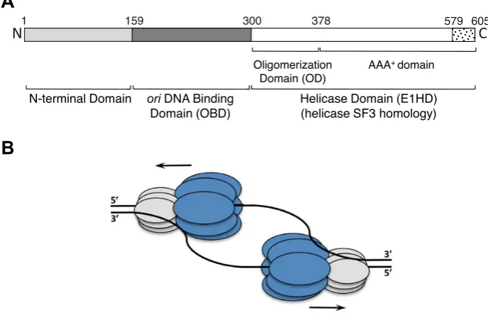
Figure 1. Schematic representation of the 605 amino acid BPV-1 E1 protein and helicase action. (A) The N-terminus (residues 1–300) contains the sequence-specific dsDNAoribinding domain (OBD) and an N-terminal regulatory domain. The C-terminal residues 301–605 form the helicase domain module (E1HD) comprising of the oligomerization domain (OD), AAA+ domain and acidic tail (residues 579–605). (B) Two hexameric helicases are thought to assemble atoriand drive bidirectional DNA replication. The helicase domain module of E1 is shown in dark grey (E1HD) and the N-terminal domains, including the OBD, in light grey.
tral ssDNA binding tunnel. From these data models for nu-cleotide dependent translocation on ssDNA have been pro-posed (7,17), but the mechanism of base pair separation re-mains unknown. The E1HD structure with ssDNA bound in the central tunnel (7) fits with a ‘steric exclusion’ model for dsDNA unwinding, where active translocation on one ssDNA strand displaces the second ‘passive’ strand. Bio-chemical and structural studies support a similar mecha-nism in prokaryotic DnaB (18) and the dsRNA helicase Rho (8), among others. However, one important inference that can be drawn from the E1HD/ssDNA/ADP structure (7) and its revelation of the protein orientation relative to the direction of ssDNA translocation (3–5) (19) is that the N-terminal half of E1 (∼residues 1–300) must face the RFJ. Accordingly, the N-terminal part of E1 that includes the double-stranded DNA (dsDNA) binding OBD could influ-ence base separation at the RFJ through interactions with the ss- and dsDNA (2). According to the currently accepted ‘steric exclusion’ mechanism, E1 moves in the 3–5 direc-tion along the active ssDNA strand, with the N-terminal half pushing on the fork to separate dsDNA strands outside the E1 complex (20). However, the evidence for 5-strand exclusion in this model is indirect and the role of individ-ual E1 domains in unwinding remains ill defined. The lack of structural data on complexes with DNA replication fork substrates could be explained by the expected highly dy-namic and mobile nature of such assemblies and by poten-tial symmetry perturbations and conformational variation induced by binding of such DNA, hindering crystalliza-tion and resolucrystalliza-tion EM reconstruccrystalliza-tion. Indeed, high-resolution structural data for hexameric helicases in general are available only for complexes with short single-stranded
nucleic acid segments bound (7,9), establishing that the ac-tive strand is pulled through the central tunnel of the C-terminal motor domain.
To advance understanding of the mechanism of dsDNA strand separation we have used electron microscopy (EM) and single particle analysis to obtain structures of the full length BPV helicase E1 and two complexes of E1 bound to a RFJ end-labelled with protein tags. We have used negative stain EM that allows imaging of samples with high contrast to accurately reveal positions of labels that are 20–60 kDa in size, as employed in this study. The combined approach of DNA end-tagging and negative stain EM indicated the dsDNA entrance and passive ssDNA strand exit points in the helicase–RFJ complex. Structural data correlate with DNA–protein contact points in RFJ DNA bound E1 com-plexes determined by nuclease footprinting. Notably, the data indicate that dsDNA enters the E1 complex through a tight side tunnel and not along the central axis of the hex-amer as commonly assumed for E1 and other hexhex-americ helicases (18,21–23), while the 5 ‘passive’ strand exits the complex via a channel opposite the dsDNA entry tunnel. DNA unwinding is therefore taking place at the entrance to the helicase domain, inside and not outside the hexamer.
MATERIALS AND METHODS
For the full experimental protocol, please see Supplemen-tary Data.
E1 proteins were purified as previously described (12,24). The monomers for assembling the monovalent tetrameric streptavidin complex (MTS) were expressed from plasmids Streptavidin-Dead and
at Birkbeck College, University of London on August 5, 2015
http://nar.oxfordjournals.org/
Streptavidin-Alive (25), which were supplied by Dr Alice Ting’s Laboratory, MIT, via Addgene.org. Helicase and OH•footprinting were performed as previously described (24,26), with minor modifications indicated in the sup-plementary experimental procedures. Protein–DNA com-plexes for EM were prepared by incubating protein and DNA at a 6:1 molar ratio before size exclusion chromatog-raphy (SEC) on a Superdex-200 (GE Healthcare) gel fil-tration column (10 mM Tris–Cl pH 8.0, 225 mM NaCl, 5% v/v glycerol, 2 mM dithiothreitol (DTT) , 0.1 mM phenylethanesulfonylfluoride (PMSF), 0.1 mM ethylenedi-aminetetraacetic acid).
Images were recorded using a Tecnai F20 electron mi-croscope operated at 200 keV, images were recorded using a Gatan Ultrascan 4000 4k ×4k CCD camera at a nom-inal magnification of 62 000. Image processing was per-formed with CTFIT (27) and IMAGIC-5 (28,29), with the alignment and classification of images performed as pre-viously described, and references therein. Angular orienta-tions of class averages were determined by angular reconsti-tution. Three-dimensional (3D) maps were calculated using the exact-filter back projection algorithm (29,30) (see Sup-plementary Table S1). Interpretation and illustrations were done using Chimera (31).
RESULTS
The full-length E1 helicase preferentially unwinds forked DNA substrates
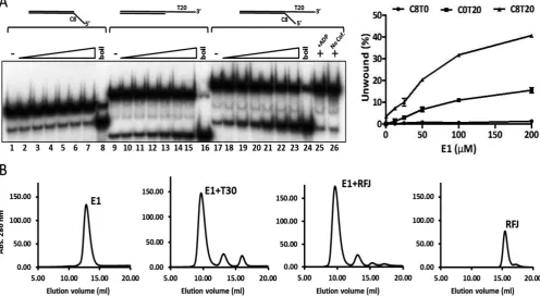
E1 is a 3–5 helicase that initiates unwinding of dsDNA substrates with 3 ssDNA tails (19). To establish the basic configuration of a substrate appropriate for structural anal-ysis we compared the unwinding of simple linear partially single- and double-stranded substrates to a forked substrate with 5and 3ssDNA tails using a radiometric strand dis-placement assay. In accord with previous observations, a short double stranded oligonucleotide substrate (30 bp ds-DNA) with a 3 T20 but not a 5C8 ssDNA tail was un-wound by E1 (Figure 2A, lanes 1–8 compared to 9–15). However, the extent of unwinding of a forked-substrate with both 3T20 and 5C8 ssDNA tails increased approximately three-fold compared to the substrate with only a 3 T20 tail (Figure2A, lanes 17–24 compared to 9–16). Strand dis-placement was observed in the presence of ATP/Mg2+, but not in reactions without nucleotide cofactor or with adeno-sine diphosphate (ADP) in place of ATP (lanes 25 and 26).
Formation of E1–DNA complexes
Like the E1 helicase domain residues 299–605 (12), puri-fied full length E1 (E1FL) formed a stable complex with ss-DNA (T30) in the absence of nucleotide cofactors and the peak fractions of the complex purified by gel-filtration chro-matography showed a homogeneous population of hexam-eric particles when examined by negative stain EM (Sup-plementary Figure S1A and E). Using RFJ substrates with dsDNA arms from 10–30 bp and ssDNA arms 3T20 and 5 C8–12, we observed similar stable complexes in gel filtration chromatography (complex formation with a substrate com-prising of 10 bp dsDNA and 3T20 and 3C10 ssDNA arms is shown in Figure2B). In all cases, the chromatograms did
not indicate the presence of intermediate species eluting be-tween the monomeric and hexameric populations that were effectively resolved by the chromatography.
Electron microscopy and 3D reconstruction of E1 helicase complexes
First, a structure of the purified E1FL–T30 complex ex-amined by negative stain EM (Supplementary Figure S1A and E) was determined with C6 rotational symmetry im-posed during the initial structure determination. Having this restraint we did not expect to reveal the position of the DNA relative to the individual subunits of the complex. The E1FL hexamer structure, determined at a resolution of 18
˚
A (Figure3A), has a maximum diameter of 130 ˚A and a height of 100 ˚A and can accommodate∼410 kDa of pro-tein mass, in agreement with the predicted 409 kDa. The shape of the complex resembles a triple-tiered ring, with the middle ring having the larger diameter and the upper ring the smallest (Figure3A, left). To determine the organiza-tion of the E1FL particle, as described further below, we used a comparison of the EM projections of E1HD (Sup-plementary Figure S2) and E1FL, and domain identifica-tion using antibody labelling. There is a central tunnel (‘a’) that varies in diameter along its length: at the bottom of the structure (C-terminus) the tunnel is∼20 ˚A in diameter; travelling upwards the tunnel ‘a’ forms a chamber within the HD area, which expands to 27 ˚A below sub-domain 3 (Figure3A, middle) and then it narrows to 23 ˚A in diame-ter at the entrance to the central tunnel formed by the upper ring. A second chamber is formed by the middle and upper rings and it is connected to the protein surface via twelve discernible tunnels (Figure3A, middle and Supplementary Figure S2).
After obtaining the structure with symmetry C6 imposed we verified whether the structure had the same appear-ance without applying any symmetry during the reconstruc-tion process. For the asymmetric reconstrucreconstruc-tion the same dataset was used and the initial model was obtained using mostly tilted views and a few end views of the complex. The structure demonstrated the same general features as described above for the symmetrized structure but was ob-tained at a lower resolution of∼23 ˚A. We observed the same triple-tiered organization with six clearly defined subunits in the bottom and middle rings (compare Figure3A–C). Strikingly however, in the upper ring there was clear asym-metry revealed by the presence of a bulk of density on the outer side of the upper ring (right panel, Figure3B and C). The size of this bulk, highlighted with the black oval in Fig-ure3C, is about 20×25 ˚A. The centre of this bulk of den-sity is located at a distance of∼36 ˚A from the centre of the opening to central tunnel ‘a’. As expected, the lower reso-lution of the asymmetrical structure did not allow as clear definition of the inner channels of the complex compared to those defined in the symmetrical reconstruction and did not reveal any densities inside of the inner chamber of the E1 hexamer that could be reliably identified as ssDNA.
Analysis of the symmetric and asymmetric structures (Figure3) has allowed us to identify a single E1 subunit in the hexameric E1FL structure. A monomer, extracted us-ing the segmentation function of the molecular modellus-ing
at Birkbeck College, University of London on August 5, 2015
http://nar.oxfordjournals.org/
Figure 2. Helicase activity of E1 and protein–DNA complex formation. (A) Full length E1 preferentially unwinds forked test substrates with 5and 3 ssDNA tails. The DNA substrate concentration used was 0.25 nM and the protein titration range was 6.25, 12.5, 25, 50, 100 and 200 nM E1. Heat denatured substrate is indicated as ‘boil’. No unwinding was observed in reactions containing ADP in place of ATP (lane 25) or without nucleotide cofactor (lane 26). The graph on the right shows the data from three independent repeats (mean and SD). (B) Formation and resolution of E1–DNA complexes by gel filtration chromatography. E1 (50M) and DNA substrates were mixed at a molar ratio of 6:1 and incubated at room temperature for 15 min before chromatography. Complex formation is shown for ssDNA T30 and the RFJ like substrate with 10 bp dsDNA, 3T20 and 5C10 ssDNA arms. Similar results were obtained with the forked substrate used in the helicase assays (not shown). All DNAs alone eluted late in the chromatogram as illustrated for the fork dsDNA 10, ssDNA 3T20 and 5C10 (‘RFJ’ trace on the right).
programme Chimera (31), is shown in Figure4to demon-strate the overall domain organization. Globular domain 4 shapes a dense ring at the base of the hexamer; domain 3 forms the inner ring that constrains the central tunnel. Do-main 2 creates the outer rim of the wide middle ring of the structure. Finally, small domain 1 is located on top, form-ing the narrowest rform-ing of the complex. Accordform-ingly, the two sets of six side openings, ‘b’ and ‘c’ to corresponding tun-nels observed in the E1FL hexamer (Figure 3A, middle) can be described as follows: tunnel ‘b’ is formed by E1 do-mains 1 and 2, has a length of∼25 ˚A and a cross-section of∼18×20 ˚A. Therefore, like the central axial tunnel ‘a’, it is wide enough to accommodate dsDNA. Tunnel ‘c’ is formed by domain 4 of one subunit and domains 2 and 4 of the neighbouring subunit and has a cross-section of∼13
×20 ˚A with a length of∼30 ˚A. Being smaller than tunnel ‘b’ it could accommodate ss- but not dsDNA. Significantly, the additional bulk of density observed on the upper tier of the E1FL–ssDNA complex after asymmetrical reconstruc-tion (Figure3B, right panel and Figure3C) coincides with a tunnel opening ‘b’.
E1 hexamers translocate 3–5on ssDNA and this ‘active’ 3ssDNA strand, also corresponding to the leading repli-cation strand in this case, is engaged by the ssDNA binding segments of the AAA+domain (7,24). The E1FL–ssDNA structures, revealing previously unidentified tunnels and the
additional asymmetrically positioned density in the upper tier (Figure 3), led us to determine directly the point of dsDNA entry into the complex and the path taken by the second lagging or ‘passive’ 5ssDNA strand of an engaged replication fork.
First, we constructed a synthetic DNA replication fork with 30 bp dsDNA and 3T20 and 5C8 ssDNA tails where the 5end of the dsDNA was labelled with biotin. After for-mation of protein–DNA complexes, MTS (25) was bound and complexes were purified by gel filtration. Although sodium dodecyl sulphate-polyacrylamide gel electrophore-sis analyelectrophore-sis of the hexameric E1FL fractions clearly showed the specific incorporation of MTS, we failed to clearly visu-alize the binding of MTS in the hexameric particles exam-ined by negative stain EM (not shown). Subsequently, we adopted a substrate with a shorter dsDNA sequence, 10 bp dsDNA and 3 T20 and 5 C10 ssDNA tails, labelled with biotin on the 5end of the dsDNA with or without digox-igenin on the 5ssDNA end. Hexameric E1FL complexes were first assembled on these substrates before MTS or anti-digoxigenin Fab were bound, as appropriate, and complexes purified by gel-filtration chromatography (Supplementary Figure S3). Purified complexes were then used for negative stain EM, single particles were aligned, classified and the 3D-structure of the complexes determined without impos-ing symmetry (Figure5, Supplementary Figures S1 and S3).
at Birkbeck College, University of London on August 5, 2015
http://nar.oxfordjournals.org/
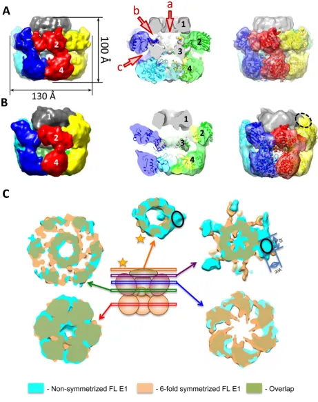
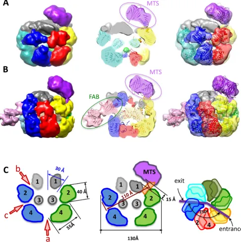
Figure 3. EM structures of full length E1 bound to ssDNA. The domains of E1FL are numbered: 1, N-terminal residues 1–158; 2, OBD; 3, OD and 4, AAA+ domain. Atomic structures of the E1HD (2V9P (17)) and the OBD (1KSX, (33)) are docked into the structures. (A) The symmetrized structure of E1FL–ssDNA. Arrowheads a, b and c indicate openings to tunnels within the E1FL structure. The opening to central tunnel ‘a’ is indicated at the N-terminus only. (B) The asymmetrical structure of E1FL–ssDNA. The same triple-tiered organization as in (A) was observed with six clear subunits in the bottom and middle rings. However, asymmetry was observed as the presence of a bulk of density on the outside of the upper ring, outlined by the blue ellipse. (C) Comparison of the symmetric and asymmetric reconstructions of the E1FL–ssDNA complex. Overlay of the symmetrized and asymmetrical structures. The upper tier corresponding to N-terminal domain 1 (indicated by a star) is located at the same height in both symmetrized and asymmetric structures. There are no distortions in that region except for the additional density on the outer side of the N-terminal tier in the asymmetric structure indicated by the black oval. Areas of overlap between the two structures are shown in green colour.
at Birkbeck College, University of London on August 5, 2015
http://nar.oxfordjournals.org/
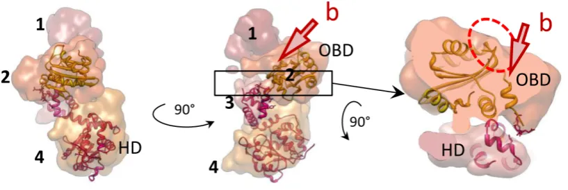
Figure 4.Structural organization of one E1 subunit. Atomic structures of E1HD (17) and the OBD (33) docked into one E1 subunit extracted from the hexamer. The domains are coloured in different shades and indicated with numbers as in Figure3. Two views (left and centre) are of a single subunit rotated by 90◦around the central (vertical) axis of the helicase. On the right, the segment indicated by the box in the central panel has been extracted and rotated 90◦around the horizontal axis. The DNA binding segments of the OBD are circled in red and their corresponding positions in the E1FL hexamer are near the opening to the tunnel ‘b’.
The final resolution of the structures obtained was 20 ˚A. The structure of the E1FL–RFJ complex with the ds-DNA end of the RFJ 5-labelled with MTS has the same overall triple ring organization as described above. An addi-tional density at the top of the complex located off the cen-tral axis of the hexamer was attributed to MTS (Figure5A, Supplementary Figures S1C and S1G). Subsequently, we determined the structure of the double-labelled E1FL–RFJ complex with MTS on the dsDNA arm (5-end) and anti-digoxigenin Fab on the 5 ssDNA arm (‘passive’ strand) (Figure5B, Supplementary Figure S1D and H). Only∼30% of the E1Fl–RFJ complex were labelled with MTS and Fab, so sorting of the particle images using 3D statistical analy-sis was necessary to extract dual-labelled complexes (30). The structure of this complex revealed two additional ex-ternal densities: one at the same position as in the complex with only the dsDNA labelled with MTS and the second extra mass located on the side of the complex, nearly op-posite to MTS, at the level between the high density lower ring and middle wide ring (Figure5B). Interestingly, both structures labelled with MTS on the dsDNA arm (with-out or with Fab on the 5ssDNA arm) were distorted, hav-ing the upper smallest rhav-ing pressed obliquely into the large ring of the OBD domains beneath the upper, extra, density (MTS). Overlay of these two structures demonstrated excel-lent overlapping of the density on the top of the complex, indicating unambiguously the position of MTS and its link to the E1 complex. The remaining extra density at the side of the dual-labelled complex was assigned to the Fab. In the structure, the Fab label disturbs the position of the OBD do-mains in the middle ring and appeared to be partially sub-merged into the complex. The distortions induced by MTS and Fab therefore demonstrate a degree of flexibility in the middle and upper tiers of E1 formed by domains 1 and 2.
Analysis of the E1FL structures
The structural organization of the E1FL hexamers was de-termined stepwise using three approaches. First we used an-tibodies (ab) specific to residues 1–129 and negative stain EM coupled with statistical analysis to determine the posi-tion of the corresponding N-terminal segment in the E1FL
complex, which was shown to correspond to domain 1 at the top of the complex (Figure3and Supplementary Fig-ure S2A).
Second, we aligned the E1FL EM structure with a 19 ˚A resolution EM structure of E1HD (residues 299–605) that we obtained using similar methods to the E1FL structure (Figure3 and Supplementary Figure S2B). A correlation analysis revealed that the best alignment was achieved with the bottom ring of the E1FL structure (cross correlation co-efficient, CCC of 0.84).
Third, available atomic coordinates of E1 domains were docked into the EM maps (Figure3A). Following the an-tibody labelling experiments and comparison with the EM projections of E1HD described above, an initial manual fit-ting of E1HD crystal structures (PDB IDs: 2GXA (7) and 2V9P (17)) in the lower ring (HD) was refined using Veda software (32) and Chimera (31). The fitting resulted in a CCC=0.72, which was reduced to∼0.2 for the upper tier of the complex indicating a better consistency between E1HD crystal structures and the lower tiers of the E1FL structure. An automated docking of the atomic structure of the OBD, residues 159–303 (PDB ID: 1KSX, (33)), into the E1FL map showed that the wide tier formed by domain 2 cor-responded well to six OBD domains (Figure3A, CCC = 0.76, Supplementary Table S2). Importantly, in this docked model the C-terminal linker connecting the E1 OBD and HD (amino acids 300–303) overlaps with the correspond-ing residues at the N-terminus of the E1HD X-ray structure fitted into the E1FL maps, confidently fixing one point of alignment. To further verify the assigned OBD orientation we inserted the HA-epitope sequence into a small surface loop (residues 225–228) after residue 226. The automated fitting of the OBD predicted that these residues would be exposed on the outer surface of E1FL. Again, antibody-labelling experiments demonstrated, in the presence of anti-HA antibodies, an additional bulk of density of the size and shape expected for IgG antibody (Supplementary Figures S5 and S6) at the position assigned to the OBD. Overall, the results of fitting the X-ray structure of the OBD into the EM maps were very similar for all the E1 complexes analysed, favouring the same orientation of the OBD (Sup-plementary Table S2). Interestingly, the location and
at Birkbeck College, University of London on August 5, 2015
http://nar.oxfordjournals.org/
Figure 5. EM structures of E1FL hexamers bound to protein tagged RFJ DNA. (A) Structures of E1FL complexes with monovalent tetrameric streptavidin (MTS) bound to the 5end of the dsDNA arm. (B) The structure with anti-digoxigenin Fab bound to the 5ssDNA arm in addition to MTS bound to the dsDNA of the RFJ. Subunits of structure are shown in different colours. Side views and cutaway views are shown with atomic models of E1HD and the OBD docked into EM maps (central column and a column on the right). (C) Cartoon representations of the unlabelled and MTS labelled E1 structures with distances and angles between tunnel openings indicated. There is a 15 ˚A gap present between upper part of the hexamer and MTS.
tation assigned to the OBD resulted in the dsDNA binding face of the OBD, circled with the dashed red line in Figure4, being positioned at the outer surface of the E1FL complex, at the entrance into the side tunnel ‘b’.
The footprint of E1FL bound to a RFJ DNA substrate
Our structural analysis suggests that the E1 N-terminal do-mains 1 and 2 adopt a configuration that could
accom-modate the RFJ DNA. We tested this by comparing the hydroxyl radical (OH•) nuclease protection ‘footprints’ of E1FL and E1HD bound to a RFJ-like DNA molecule (Fig-ure 6). The DNA binding substrate used was identical to the fork substrate used in the helicase assays shown in Fig-ure 2A, with 30 bp dsDNA, T20 3active ssDNA strand and a C8 5passive ssDNA strand. The OH•is a small, dif-fusible, nuclease and DNA is protected from cleavage only where there are very tight contacts with protein (34). We
at Birkbeck College, University of London on August 5, 2015
http://nar.oxfordjournals.org/
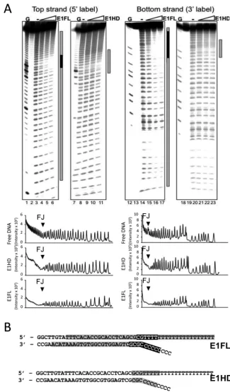
Figure 6. Hydroxyl radical footprinting of E1–RFJ complexes. (A) Lanes 1–11,32P 5-end labelled top strand and lanes 12–23 3-end labelled bottom strand. Lanes 1, 7, 12, 18, G ladder. Lanes 2, 13 and 19 show unreacted substrate. The protection patterns were analysed using the lane-profiling tool of the phosphorimaging software (shown below the gel images). A peak height reduction of 80% or more was scored as complete protection (indicated in black in the annotations on the right of each gel image; partial protections are shown in grey). (B) Projection of the OH•footprinting pattern on the fork sequence.
at Birkbeck College, University of London on August 5, 2015
http://nar.oxfordjournals.org/
ticipated that the OH•could diffuse into the protein com-plex through the well-defined openings and expected that only segments of RFJ-like DNA that interact tightly with protein would be protected.
Figure6A lanes 4–6 show the footprint of the E1FL com-plex bound to the top strand of the DNA fork (532P-end la-belled). Near complete protection of∼6 nt at the fork junc-tion (Figure6B) indicates the presence of close contacts be-tween E1FL and DNA. In addition, we observed a weaker and diminishing protection extending over ∼20 bp of ds-DNA from the junction point and over the entire 3and 5 ssDNA arms. Importantly, the protection pattern over the dsDNA lacks features such as periodic protection and sus-ceptibility to cleavage observed for DNA lying on a protein surface (34) and is best interpreted as encirclement of the dsDNA in a protein sheath. In comparison, for the E1HD we observed only partial protection over about 8 nt on each DNA strand centred over the fork junction (lanes 9–11 and Figure6B). Similar patterns of protection to the 5-end la-belled top strand were observed for the 3-end labelled bot-tom strand for both E1FL and E1HD complexes, respec-tively (lanes 15–17 and 21–23). These data therefore indicate that the E1HD makes a limited set of weak protein–DNA interactions with the RFJ, while in the intact E1FL complex there is a more extensive set of interactions with all arms of the fork.
dsDNA and the 5ssDNA strand enter and exit E1 through opposing side tunnels
The position of the MTS label attached to the dsDNA end of the DNA fork was identical in the single- and double-labelled E1 helicase particles. Figure5A shows the structure of the complex labelled with MTS on the 5 dsDNA end of the fork, docked with the atomic coordinates for E1 and MTS (PDB ID: 3RY1, (35)). Figure5B shows the structure of the double-labelled E1FL complex and the fitting of the atomic coordinates for E1, MTS and the anti-digoxigenin Fab X-ray models (PDB ID: 1IGJ, (36)). In each case, the electron density maps show that the position of the MTS is not on the top of the central tunnel. Moreover, there is a significant gap (∼15 ˚A) present between the upper part of the hexamer and MTS makes contact with the E1 surface at a point that is∼45 ˚A away on the upper side of the com-plex (Figure5C and Supplementary movie S1). The MTS contact with E1 is in the vicinity of the dsDNA-binding site of the OBD at the entrance into a side tunnel ‘b’. A clear region of electron density connects the MTS with the en-trance to tunnel ‘b’, while the opening to the central tunnel ‘a’ is unobstructed.
Significantly, the electron densities corresponding to the MTS and Fab labels are at fixed positions on opposing faces of the E1FL complex separated by a distance of ∼120 ˚A at an angle of ∼150◦ across the central axis of E1 (Fig-ure5B and C). The distance between the MTS and Fab la-bels (∼110 ˚A) measured across the protein complex is in accord with their length of separation in the fork construct (Figures5C and7). The best explanation for such an ar-rangement is that the dsDNA enters E1 so that the fork junction is above the helicase motor unit. Fab makes a con-tact with the E1 complex in the area of the second tier. It
is clear that the two single stranded ends of the unwound DNA, exiting the E1 hexamer through tunnels ‘a’(3) and ‘c’(5) would be separated by a path of at least 200 ˚A around the outside of E1.
DISCUSSION
The X-ray structure of the E1HD/ssDNA/ADP hexamer showed that the ‘active’ 3ssDNA strand, or leading repli-cation strand, exits along the central axis of the HD (7). The currently favoured ‘steric exclusion’ model of DNA un-winding for E1 (20), while incorporating 3exit at the end of a central tunnel, is based on exclusion of dsDNA and the 5ssDNA strand outside the helicase at the opposite end of the tunnel and similar steric exclusion models are proposed for other hexameric helicases (9,18,21–23,37,38). However, the steric exclusion model is based on indirect observation and until now has not been validated with structural infor-mation on complexes with complete replication forks con-taining dsDNA as well as two single-stranded segments. One possible reason for this is that these replication com-plexes are highly dynamic, mobile and demonstrate confor-mational diversity, so complicating detailed structural anal-ysis.
Here, we produced stable hexameric complexes of full length E1 with ssDNA and intact replication forks bound, determined structures at 18–23 ˚A resolution and localized DNA replication fork entrance and exit points at the hex-amer surface by direct visualization of DNA ends labelled with MTS and Fab. The atomic structures of the E1 helicase domain determined without (17) and with ssDNA and nu-cleotide cofactor bound (7) are virtually identical with the C-␣r.m.s. deviation of 0.35 ˚A. In these crystal structures, the E1 OD (residues 308–378) is highly symmetrical, while the ATP binding sites are found at the interfaces between adjacent AAA+ domains (residues 379–578) that show po-sitional variations of up to 7.5 ˚A. It is possible therefore that the helicase domain could impose asymmetry on the N-terminal portion of the molecule mediated through the AAA+ domain positional asymmetry, but it is unlikely that protein–nucleic acid interactions are significantly different within the N-terminal half (residues 1–307) of the complex, with and without nucleotide cofactor bound. Together with DNA footprinting experiments, our structural observations indicate, first, that at least 10 bp of dsDNA enters inside the intact helicase complex and that unwinding most likely takes place at the entrance to the helicase domain (Figure7). Second, labelling with MTS suggests, unexpectedly, that ds-DNA enters not along the central tunnel but through the side tunnel ‘b’. We favour this interpretation because we ob-serve clear density linking the MTS to the entrance of tun-nel ‘b’, while the entrance to tuntun-nel ‘a’ is unobstructed and there is a complete absence of density between E1 entrance ‘a’ and MTS in our models. However, we acknowledge that higher resolution data would be required to completely ex-clude an axial path for dsDNA entry. Third, labelling of the 5 ssDNA end with Fab indicates that the lagging (5 pas-sive) strand exits the E1 complex on an opposing side to ds-DNA entry via the narrower tunnel ‘c’. The negative stain EM benefits from excellent contrast, that is important for initial analysis of dynamic systems (39) and the resolutions
at Birkbeck College, University of London on August 5, 2015
http://nar.oxfordjournals.org/
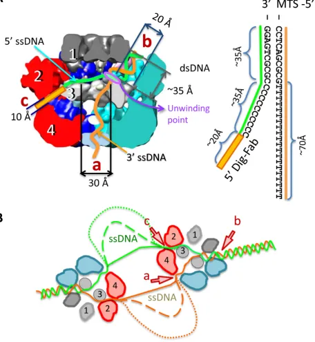
Figure 7. Diagram of the E1 DNA unwinding machine. (A) The model for replication fork engagement is shown with the sizes of length and diameters of tunnels. Schematic representation of the DNA fork used in experiments is shown to the right. The DNA lengths are dsDNA∼35 ˚A; 5ssDNA,∼35 ˚A and 3ssDNA,∼70 ˚A. The lengths of ssDNA are based on corresponding lengths in duplex DNA and therefore could be larger. The length of the link between the digoxigenin group and DNA is also indicated (∼20 ˚A). It is proposed that dsDNA enters into the hexameric assembly through tunnel ‘b’ where it faces domain 3 (the ‘wedge’) and base pairs are separated at the entrance into the HD. The ‘active’ ssDNA strand is pulled along the central tunnel (towards exit ‘a’) by the ATP-fuelled HD motor. The ‘passive’ ssDNA strand exits via tunnel ‘c’. (B) Model of DNA unwinding by a double hexamer of E1. dsDNA is shown entering the E1 assembly as in (A) and the dashed lines indicate the accumulation of increasing amounts of ssDNA.
at Birkbeck College, University of London on August 5, 2015
http://nar.oxfordjournals.org/
obtained are more than sufficient for deducing DNA end-label locations (MTS and Fab) on the molecular surface of E1, that are separated by at least 200 ˚A.
Our observations therefore suggest a revision of the steric exclusion model proposed for E1 (20) where, alternatively, DNA strands are wedged apart within the hexameric as-sembly and not outside. The E1 helicase domain (residues
∼300–608) forms stable hexamers (12) with the active ss-DNA strand bound in the central tunnel (7) and these com-plexes can unwind helicase substrates (12). In the absence of the N-terminal half of the protein (domains 1 and 2), and therefore the absence of additional channels that could accommodate DNA, these assemblies are likely to be op-erating by a strict steric exclusion mechanism. As such, the entrance to the helicase domain acts to wedge DNA strands apart while the passive ssDNA strand is excluded from the HD complex. Together with previous data locating the ac-tive ssDNA strand (7), our new structural data show that all DNA replication fork arms in the full length E1 hexamer are entering or leaving via well-defined openings that may be fixed for the duration of translocation. Thus, our model en-visions the same wedging mechanism at that entrance to the helicase domain (Figure7), but with all arms of the repli-cation fork passing through discrete conduits. This could modify our understanding of the mechanism of DNA un-winding, since direct interactions with the dsDNA ahead of the fork and also the passive ssDNA strand could influence the efficiency of the unwinding process (2). The anatomy of E1-RFJ DNA engagement deduced suggests that the E1 hexamer may be operating like the heterotrimeric E. coli RecBCD helicase whose structure has also been de-termined in complex with a fork-like DNA molecule (40). In RecBCD, several DNA base pairs enter a short tunnel formed between the RecB and C subunit and an ‘arm’ of the RecB subunit contacts dsDNA 12 bp from the fork junc-tion. DNA strands are split across a protein ‘pin’ provided by RecC, as the ssDNA tails are pulled through discrete channels by the RecB and D motors. It is likely that a spe-cific protein segment corresponding to a ‘pin’ is involved in wedging DNA strands apart at the entrance to the E1 heli-case domain.
It is unclear whether other hexameric helicases unwind dsDNA as proposed for E1. However, there are biochemical data widely supporting a steric exclusion model and struc-tural data indicating multiple channels for DNA entry and exit in other hexameric helicases including T7 gp4 (37), bac-terial DnaB (18), SV40 LTag (38), archaeal MCM (22,41) and the eukaryotic MCM2–7 complexes (42,43). Such data are not incompatible with our model incorporating inclu-sion of the RFJ within the hexamer, now proposed on the basis of the structural observations of E1FL–RFJ complex. Failure of these replicative hexameric helicases to displace a biotin-mediated streptavidin ‘roadblock’ on the passive ssDNA strand is frequently taken as evidence of its steric exclusion (e.g. refs.20and38). However, SV40 L-Tag hex-amers have been shown to bypass a covalently linked bulky adduct even on the active translocation strand, even though it is pulled through the motor domain of the toroid (38). A ring-opening mechanism was suggested, akin to that previ-ously demonstrated in T7 gp4 (44). Given the similarities between E1 and L-Tag it is likely that the hexameric E1
protein ring can also open in a dynamic engagement with the RFJ. Notably, opening of only the upper N-terminal ring would be needed to by-pass obstacles such as DNA secondary structure or bound proteins on the lagging (pas-sive) DNA replication strand. Indeed, the distortions ob-served in the protein-tagged RFJ complexes (Figure5) in close proximity to streptavidin and Fab in particular which is submerged into the complex, not only demonstrate that the bound replication fork is intact but indicate that N-terminal domains 1 and 2 (∼residues 1–300) are flexible. Two independent rings (HD and the N-terminal-OBD ring in the case of E1), each engaging DNA and that can each open and close, could facilitate by-pass while ensuring that the helicase remains stably associated with its substrate.
In a toroidal protein–DNA complex the nucleic acid can be internal and completely surrounded by protein or lie on its surface. In the latter case only one face of a dsDNA helix would contact protein leaving the outer surface sus-ceptible to nuclease cleavage. In OH• footprinting exper-iments such interactions produce very characteristic peri-odic cutting pattern progressing through protection to no protection with centres spaced 10–11 bases apart (one heli-cal turn), as illustrated by lambda repressor and Cro pro-teins (34). The densitometry traces of E1FL–RFJ com-plexes (Figure6A) show no evidence of periodicity but in-stead a uniform protection approaching two turns of the dsDNA helix. This is best interpreted as E1 completely en-circling the DNA, forming a ‘sheath’. Furthermore, since we only observe hexamers of E1FL (Figure 2) or E1HD (12) in the presence of DNA it is unlikely that the observed protections are representative of partial E1–DNA complex formation. A corollary to this and consistent with our struc-tural observations is that the RFJ is inside the complex in a chamber at the entrance to the helicase motor domain. The complete protection observed over the first five bases of the 5’ passive ssDNA are also consistent with their inclusion within the complex and exit most likely via a side tunnel ‘c’. Likewise, the DNA protection pattern for the 3‘active’ strand is in agreement with its known passage through the central tunnel of the helicase motor domain bearing the ss-DNA binding sites (7,24). Direct visualization of the paths taken by the replication fork strands will ultimately require higher resolution structures of intact E1–RFJ complexes that have thus far proven difficult to obtain for this class of helicase and, indeed, small asymmetric structures in general that do not lend themselves readily to cryo-EM (39,45,46). However, our structural data identifying the positions of the DNA ends with surface labels are in accord with the arrangement and dimensions of the tunnels in E1 with re-spect to the proposed path of the occupying DNA and the DNA protections we observe in footprinting experiments. Furthermore, our data do not exclude the possibility that an extended part of the lagging strand ssDNA is wrapped on the outer surface of E1, as inSulfolobus solfataricusMCM, to enhance the unwinding process (22).
Our results, defining the structural arrangement of an in-tact E1 helicase complex and its orientation with respect to the RFJ, are consistent with recent FRET data that suggest the RFJ is located nearer the OBD and away from the HD (20). Furthermore, while we do not consider the continu-ity of the unwinding process here, our proposition that the
at Birkbeck College, University of London on August 5, 2015
http://nar.oxfordjournals.org/
plain the unevenness of unwinding of dsDNA observed for E1HD, which is ameliorated by the presence of the OBD domain (20), and is likely to be unperturbed in the case of full length E1 at the RFJ in vivo. Our model for dsDNA unwinding shown in Figure 7B also incorporates two E1 helicases acting in unison, consistent with the assembly of double hexamers at the origin of DNA replication (47).
The helicase-catalyzed DNA processing events in DNA replication are conserved and, like E1 (47), highly dynamic particles ofDrosophila melanogasterand yeast MCM com-plexes, that incorporate ring opening and closing mecha-nisms, have also been observed during establishment of bi-directional DNA replication (39,48–51). Observations in ar-chaeal systems also indicate that DNA can take alternative paths in associated hexameric MCM complexes, exempli-fied by the wrapping of dsDNA around the external sur-face of the putativeMethanothermobacter thermoautotroph-icusMCM pre-replication complexes (52). The new insights into E1 and how it engages the RFJ presented here there-fore have broader implications for understanding how DNA processing machines work.
ACCESSION NUMBERS
The EM density maps have been deposited in the EMDB with accession codes EMD-3087 for E1HD. EMD-3088, for E1FL asymmetrical structure, EMD-3089 for the E1FL symmetrical structure, EMD-3090 for the complex E1FL-MTS and EMD-3091 for the complex E1-E1FL-MTS-FAB.
SUPPLEMENTARY DATA
Supplementary Dataare available at NAR Online.
ACKNOWLEDGEMENTS
We thank Luchun Wang for the help with EM, David Houl-dershaw and Richard Westlake for the computer support. Plasmids for generation of monovalent, tetrameric, strep-tavidin (MTS) were provided under a Material Transfer Agreement (MTA) via Addgene.org.
FUNDING
Yorkshire Cancer Research [S302 to C.M.S., A.A..; in part]; BBSRC [BB/J008648/1 to E.V.O., C.M.S.]; Wellcome Trust (to E.V.O.). Funding for open access charge: BB-SRC [BB/J008648/1 to E.V.O., C.M.S.]; Wellcome Trust 086018/Z/08/Z (to E.V.O.).
Conflict of interest statement.None declared.
REFERENCES
1. Singleton,M.R., Dillingham,M.S. and Wigley,D.B. (2007). Structure and mechanism of helicases and nucleic acid translocases.Annu. Rev. Biochem.,76, 23–50.
2. Wu,C.G. and Spies,M. (2013) Overview: What are helicases?Adv. Exp. Med. Biol.,767, 1–16.
3. Labib,K., Tercero,J.A. and Diffley,J.F. (2000) Uninterrupted MCM2-7 function required for replication fork progression.Science, 288, 1643–1647.
5. Medagli,B. and Onesti,S. (2013) Structure and mechanism of hexameric helicases.Adv. Exp. Med. Biol.,767, 75–95.
6. Lyubimov,A.Y., Strycharska,M. and Berger,J.M. (2011) The nuts and bolts of ring-translocase structure and mechanism.Curr. Opin. Struct. Biol.,21, 240–248.
7. Enemark,E.J. and Joshua-Tor,L. (2006) Mechanism of DNA translocation in a replicative hexameric helicase.Nature,442, 270–275.
8. Thomsen,N.D. and Berger,J.M. (2009) Running in reverse: The structural basis for translocation polarity in hexameric helicases.Cell, 139, 523–534.
9. Itsathitphaisarn,O., Wing,R.A., Eliason,W.K., Wang,J. and Steitz,T.A. (2012) The hexameric helicase DnaB adopts a nonplanar conformation during translocation.Cell,151, 267–277.
10. Einstein,M.H. and Goldberg,G.L. (2002) Human papillomavirus and cervical neoplasia.Cancer Invest.,20, 1080–1085.
11. Stenlund,A. (2003) Initiation of DNA replication: lessons from viral initiator proteins.Nat. Rev. Mol. Cell Biol.,4, 777–785.
12. Castella,S., Burgin,D. and Sanders,C. (2006a) Role of ATP hydrolysis in the DNA translocase activity of the bovine papillomavirus (BPV-1) E1 helicase.Nucleic Acids Res.,34, 3731–3741.
13. Wilson,V.G., West,M., Woytek,K. and Rangasamy,D. (2002) Papillomavirus E1 proteins: form, function and features.Virus Genes, 24, 275–290.
14. Whelan,F., Stead,J.A., Shkumatov,A.V., Svergun,D.I., Sanders,C.M. and Antson,A.A. (2011) A flexible brace maintains the assembly of a hexameric replicative helicase during DNA unwinding.Nucleic Acids Res.,40, 2271–2283.
15. Hickman,A.B. and Dyda,F. (2005) Binding and unwinding: SF3 viral helicases.Curr. Opin. Struct. Biol.,15, 77–85.
16. Clertant,P. and Seif,I. (1984) A common function for polyoma virus large-T and papillomavirus E1 proteins?Nature,311, 276–279. 17. Sanders,C.M., Kovalevskiy,O.V., Sizov,D., Lebedev,A.A.,
Isupov,M.N. and Antson,A.A. (2007) Papillomavirus E1 helicase forms an asymmetric assembly in the absence of DNA and nucleotide cofactors.Nucleic Acids Res.,35, 6451–6457.
18. Kaplan,D.L. and O’Donnell,M. (2004) Twin DNA pumps of a hexameric helicase provide power to simultaneously melt two duplexes.Mol. Cell,15, 453–465.
19. Seo,Y-S., Muller,F., Lusky,M. and Hurwitz,J. (1993) Bovine papilloma virus (BPV)-encoded E1 protein contains multiple activities required for BPV DNA replication.Proc. Natl. Acad. Sci. U.S.A.,90, 702–706.
20. Lee,S-J., Syed,S., Enemark,E.J., Schuck,S., Stenlund,A., Ha,T. and Joshua-Tor,L. (2014) Dynamic look at DNA unwinding by a replicative helicase.Proc. Natl. Acad. Sci. U.S.A.,111, E827–E835. 21. Patel,S.S. and Picha,K.M. (2000). Structure and function of
hexameric helicases.Annu. Rev. Biochem.,69, 651–697.
22. Graham,B.W., Schauer,G.D., Leuba,S.H. and Trakselis,M.A. (2011) Steric exclusion and wrapping of the excluded DNA strand occurs along discrete external binding paths during MCM helicase unwinding.Nucleic Acids Res.,39, 6585–6595.
23. Takahashi,T.S., Wigley,D.B. and Walters,J.C. (2005) Pumps, paradoxes and ploughshares: mechanism of the MCM2-7 DNA helicase.Trends Biochem. Sci.,30, 437–444.
24. Castella,S., Bingham,G. and Sanders,C.M. (2006b) Common determinants in DNA melting and helicase-catalysed DNA unwinding by papillomavirus replication protein E1.Nucleic Acids Res.,34, 3008–3019.
25. Howarth,M., Chinnapen,D.J., Gerrow,K., Dorrestein,P.C.,
Grandy,M.R., Kelleher,N.L., El-Husseini,A. and Ting,A.Y. (2006) A monovalent streptavidin with a single femtomolar biotin binding site. Nat. Methods,3, 267–273.
26. Dixon,W.J., Hayes,J.J., Levin,J.R., Weidner,M.F., Dombroski,B.A. and Tullius,T.D. (1991) Hydroxyl radical footprinting.Methods Enzymol.,208, 380–413.
27. Tang,G., Peng,L., Baldwin,P.R., Mann,D.S., Jiang,W., Rees,I. and Ludtke,S.J. (2007) EMAN2: an extensible image processing suite for electron microscopy.J. Struct. Biol.,157, 38–46.
28. van Heel,M., Harauz,G., Orlova,E.V., Schmidt,R. and Schatz,M. (1996) A new generation of the IMAGIC image processing system.J. Struct. Biol.,116, 17–24.
at Birkbeck College, University of London on August 5, 2015
http://nar.oxfordjournals.org/
29. van Heel,M., Gowen,B., Matadeen,R., Orlova,E.V., Finn,R., Pape,T., Cohen,D., Stark,H., Schmidt,R., Schatz,M.et al.(2000)
Single-particle electron cryo-microscopy: towards atomic resolution. Q. Rev. Biophys.,33, 307–369.
30. Orlova,E.V. and Saibil,H.R. (2010) Methods for three-dimensional reconstruction of heterogeneous assemblies.Methods Enzymol.,482, 321–341.
31. Goddard,T.D., Huang,C.C. and Ferrin,T.E. (2007) Visualizing density maps with UCSF Chimera.J. Struct. Biol.,157, 281–287. 32. Navaza,J., Lepault,J., Rey,F.A., Alvarez-R ´ua,C. and Borge,J. (2002)
On the fitting of model electron densities into EM reconstructions: a reciprocal-space formulation.Acta. Crystallogr. D. Biol. Crystallogr., 58, 1820–1825.
33. Enemark,E.J., Stenlund,A. and Joshua-Tor,L. (2002) Crystal structures of two intermediates in the assembly of the papillomavirus replication initiation complex.EMBO J.,21, 1487–1496.
34. Tullius,T.D. and Dombrowski,B.A. (1986) Hydroxyl radical ‘footprinting’: high-resolution information about DNA-protein contacts and application torepressor and Cro protein.Proc. Natl. Acad. Sci. U.S.A.,83, 5469–5473.
35. Le Trong,I., Wang,Z., Hyre,D.E., Lybrand,T.P., Stayton,P.S. and Stenkamp,R.E. (2011) Streptavidin and its biotin complex at atomic resolution.Acta Crystallogr. D. Biol. Crystallogr.,67, 813–821. 36. Jeffrey,P.D., Strong,R.K., Sieker,L.C., Chang,C.Y., Campbell,R.L.,
Petsko,G.A., Haber,E., Margolies,M.N. and Sheriff,S. (1993) 26–10 Fab-digoxin complex: affinity and specificity due to surface complementarity.Proc. Natl. Acad. Sci. U.S.A.,90, 10310–10314. 37. Egelman,E.H., Yu,X., Wild,R., Hingorani,M.M. and Patel,S.S.
(1995) Bacteriophage T7 helicase/primase proteins form rings around single-stranded DNA that suggests a general structure for hexameric helicases.Proc. Natl. Acad. Sci. U.S.A.,92, 3869–3873.
38. Yardimci,H., Wang,X., Loveland,A.B., Tappin,I., Rudner,D.Z., Hurwitz,J., van Oijen,A.M. and Walter,J.C. (2012) Bypass of a protein barrier by a replicative helicase.Nature,492, 205–209. 39. Costa,A., Renault,L., Swuec,P., Petojevic,T., Pesavento,J.J., Ilves,I.,
MacLellan-Gibson,K., Fleck,R.A., Botchan,M.R. and Berger,J.M. (2014) DNA binding polarity, dimerization, and ATPase ring remodeling in the CMG helicase of the eukaryotic replisome.Elife,3, e03273.
40. Singleton,M.R., Dillingham,M.S., Gaudler,M., Kowalczykowski,S.C. and Wigley,D.B. (2004) Crystal structure of RecBCD enzyme reveals a machine for processing DNA breaks.Nature,432, 187–193.
41. Brewster,A.S., Wang,G., Yu,X., Greenleaf,W.B., Carazo,J.M., Tjajadi,M., Klein,M.G. and Chen,X.S. (2008) Crystal structure of a near-full-length archaeal MCM: Functional insights for an AAA+ hexameric helicase.Proc. Natl. Acad. Sci. U.S.A.,105, 20191–20196. 42. Kaplan,D.L., Davey,M.J. and O’Donnell,M. (2003) Mcm4, 6, 7 uses a ‘pump in ring’ mechanism to unwind DNA by steric exclusion and actively translocates along a duplex.J. Biol. Chem.,278, 49171–49182. 43. Fu,Y.V., Yardimci,H., Long,D.T., Ho,T.V., Guainazzi,A.,
Bermudez,V.P., Hurwitz,J., van Oijen,A., Sch¨arer,O.D. and Walter,J.C. (2011) Selective by pass of a lagging strand roadblock by the eukaryotic replicative DNA helicase.Cell,146, 931–941. 44. Ahnert,P., Picha,K.M. and Patel,S.S. (2000) A ring-opening mechanism for DNA binding in the central channel of the T7 helicase-primase protein.EMBO J.,19, 3418–3427.
45. Henderon,R. (2013) Avoiding the pitfalls of single particle
cryo-electron microscopy: Einstein from noise.Proc. Natl. Acad. Sci. U.S.A,110, 18037–18041.
46. Orlova,E.V. and Saibil,H.R. (2011) Structural analysis of
macromolecular assemblies by electron microscopy.Chem. Rev.,111, 7710–7748.
47. Schuck,S. and Stenlund,A. (2011) Mechanistic analysis of local ori melting and helicase assembly by the papillomavirus E1 protein.Mol. Cell,43, 776–787.
48. Evrin,C., Clarke,P., Zech,J., Lurz,R., Sun,J., Uhle,S., Li,H., Stillman,B. and Speck,C. (2009) A double-hexameric MCM2-7 complex is loaded onto origin DNA during licensing of eukaryotic DNA replication.Proc. Natl. Acad. Sci. U.S.A.,106, 20140–20245. 49. Costa,A., Ilves,I., Tamberg,N., Petojevic,T., Nogales,E.,
Botchan,M.R. and Berger,J.M. (2011) The structural basis of MCM2–7 helicase activation by GINS and Cdc45.Nat. Struct. Mol. Biol.,18, 471–477.
50. Chen,Z., Speck,C., Wendel,P., Tang,C., Stillman,B. and Li,H. (2008) The architecture of the DNA replication origin recognition complex inSaccharomyces cerevisiae.Proc. Natl. Acad. Sci. U.S.A.,105, 10326–10331.
51. Samel,S.A., Fern´andez-Cid,A., Sun,J., Riera,A., Tognetti,S., Herrera,C., Li,H. and Speck,C. (2014) A unique DNA entry gate serves for regulated loading of the eukaryotic replicative helicase MCM2-7 onto DNA.Genes Dev.,28, 1653–1666.
52. Costa,A., van Duinen,G., Medagli,B., Chong,J., Sakakibara,N., Kelman,Z., Nair,S.K., Patwardhan,A. and Onesti,S. (2008) Cryo-electron microscopy reveals a novel DNA-binding site on the MCM helicase.EMBO J.,27, 2250–2258.
at Birkbeck College, University of London on August 5, 2015
http://nar.oxfordjournals.org/