Intestinal epithelial cells use two distinct
pathways for HLA class II antigen processing.
R M Hershberg, … , J S Blum, G T Nepom
J Clin Invest.
1997;
100(1)
:204-215.
https://doi.org/10.1172/JCI119514
.
Intestinal epithelial cells express a low level of HLA class II molecules constitutively, with
elevated levels seen in the setting of mucosal inflammation including inflammatory bowel
disease. The ability of intestinal epithelial cells to act as antigen presenting cells for
alphabeta CD4(+) T lymphocytes was examined through a molecular analysis of the HLA
class II antigen processing pathway. We have shown that intestinal epithelial cells contain
abundant constitutive levels of the cathepsin proteases proven to function in HLA class II
mediated antigen presentation. Activation of these cells by gamma-IFN induced the
expression of invariant chain and HLA-DM alphabeta, thus facilitating the formation of
compact, SDS-stable HLA- DR alphabeta heterodimers. Using HLA-DR-restricted T cells
and retroviral mediated gene transfer of HLA-DR alleles into the intestinal epithelial cell
lines HT-29 and T84, we demonstrated efficient antigen processing and presentation to
CD4(+) T lymphocytes in the presence of the proinflammatory cytokine gamma-IFN. The
class II processing pathway and presentation in the presence of gamma-IFN was
indistinguishable from that observed with a conventional antigen presenting cell. Antigen
processing also occurred in intestinal epithelial cells in the absence of gamma-IFN, and in
contrast to that seen after stimulation with gamma-IFN, required high concentration of
antigen and was not inhibited by the protease inhibitor leupeptin. These data suggest the
use of two distinct pathways of HLA […]
Research Article
Find the latest version:
J. Clin. Invest.
© The American Society for Clinical Investigation, Inc. 0021-9738/97/07/0204/12 $2.00
Volume 100, Number 1, July 1997, 204–215
Intestinal Epithelial Cells Use Two Distinct Pathways for HLA Class II
Antigen Processing
Robert M. Hershberg,*‡ Paul E. Framson,* Diane H. Cho,* Lawrence Y. Lee,* Susan Kovats,‡ Jill Beitz,§ Janice S. Blum,§
and Gerald T. Nepom*‡
*Immunology and Diabetes Programs, Virginia Mason Research Center, Seattle, Washington 98101; ‡Department of Immunology,
University of Washington School of Medicine, Seattle, Washington 98195; and §Department of Microbiology and Immunology, Indiana
University School of Medicine, Indianapolis, Indiana 46202
Abstract
Intestinal epithelial cells express a low level of HLA class II molecules constitutively, with elevated levels seen in the set-ting of mucosal inflammation including inflammatory bowel disease. The ability of intestinal epithelial cells to act as an-tigen presenting cells for ab CD41 T lymphocytes was ex-amined through a molecular analysis of the HLA class II antigen processing pathway. We have shown that intestinal epithelial cells contain abundant constitutive levels of the cathepsin proteases proven to function in HLA class II medi-ated antigen presentation. Activation of these cells by g-IFN induced the expression of invariant chain and HLA-DM ab, thus facilitating the formation of compact, SDS-stable HLA-DR ab heterodimers. Using HLA-DR–restricted T cells and retroviral mediated gene transfer of HLA-DR alleles into the intestinal epithelial cell lines HT-29 and T84, we dem-onstrated efficient antigen processing and presentation to CD41 T lymphocytes in the presence of the proinflamma-tory cytokine g-IFN. The class II processing pathway and presentation in the presence of g-IFN was indistinguishable from that observed with a conventional antigen presenting cell. Antigen processing also occurred in intestinal epithelial cells in the absence of g-IFN, and in contrast to that seen af-ter stimulation with g-IFN, required high concentration of antigen and was not inhibited by the protease inhibitor leu-peptin. These data suggest the use of two distinct pathways of HLA class II antigen processing in enterocytes with dif-ferential immunomodulatory properties in the presence or absence of mucosal inflammation. (J. Clin. Invest. 1997. 100:204–215.) Key words: intestinal mucosa •
inflamma-tory bowel disease • antigen presentation • T lymphocytes •
human
Introduction
The epithelium of the intestine is a single cell layer that sepa-rates the highest concentration of foreign antigen from the largest population of lymphocytes in the body. Numerous
re-ports have described the expression of a low level of HLA class II antigens on the surface of normal intestinal epithelial cells (IECs)1 (1) and have demonstrated that increased
expres-sion of these molecules is associated with a diverse group of pathological conditions including inflammatory bowel disease (IBD) (1), graft versus host disease (GVHD) (2) and celiac disease (3). Since the expression of HLA class II molecules is a prerequisite for cells that function as antigen presenting cells (APCs) to CD41 T lymphocytes (for review see reference 4),
these observations suggested that the intestinal epithelium might function in the initiation and/or regulation of CD41 T
cell responses in the mucosa of the intestinal tract. Impor-tantly, mucosal CD41 T cells are implicated in the
pathogene-sis of IBD (5) and are required for the establishment of oral tolerance (6–8).
Efficient processing and presentation of antigens to CD41
T cells requires not only the expression of HLA class II mole-cules, but also the invariant chain (Ii), the HLA-DM ab het-erodimer, and a series of proteases that facilitate partial pro-teolysis of both internalized antigen and class II biosynthetic intermediates (for review see references 9 and 10). In most cells with known APC function (e.g., B cells, macrophages, and dendritic cells), the expression of these molecules is coor-dinately regulated (11). The intestinal epithelium illustrates an interesting exception, where the expression of HLA class II molecules has been observed in the absence of Ii (12). In fact, some have speculated that the HLA class II molecules ex-pressed constitutively on the gut epithelium may have a differ-ent function from those induced by mucosal inflammation (13). Several groups have described the presentation of anti-gens by IECs in human (14), rat (15), and mouse (16). How-ever, no studies, to date, have provided a detailed molecular analysis of the HLA class II processing pathway in IECs to ad-dress potential differences in the structure and function of HLA class II molecules in these cells before and after cytokine stimulation.
Using well-characterized human IECs engineered to ex-press constitutive class II molecules by retrovirus-mediated gene transfer, we have investigated the ability of IECs to both process and present peptide antigens to human CD41 T
lym-phocytes in the presence or absence of g-IFN. In the presence of g-IFN, IECs used a conventional class II pathway that in-volves Ii and HLA-DM, was inhibited by leupeptin, and facili-tated efficient antigen processing and presentation even at low
Address correspondence to Robert M. Hershberg, M.D., Ph.D., Vir-ginia Mason Research Center, 1000 Seneca Street, Seattle, WA 98101. Phone: 206-223-6391; FAX: 206-223-7543; E-mail: [email protected] ton.edu
Received for publication 26 November 1996 and accepted in re-vised form 9 April 1997.
antigen concentrations. Without g-IFN, less efficient antigen presentation, which required a higher concentration of antigen and was not sensitive to leupeptin, was observed. These data suggest that IECs use two distinct pathways for class II–medi-ated antigen presentation to CD41 T lymphocytes, and that
the relative use of these respective pathways will vary depend-ing on the degree of mucosal inflammation.
Methods
Cell lines and human IEC. The human colon carcinoma cell lines T84 and HT-29 and SW620 were obtained from the American Type Cul-ture Collection (Rockville, MD). T84, SW620, and the fibroblast ret-rovirus-producing lines PE-501 and PG-13 were grown in DME sup-plemented with 10% vol/vol FBS (Hyclone, Logan, UT), 20 mM supplemental glutamine, nonessential amino acids, and penicillin/ streptomycin. HT-29 cells were grown under undifferentiated condi-tions in McCoy’s medium supplemented as above. The B lymphocyte cell line (B-LCL) Priess (homozygous for HLA-DRB1*0401), the HLA class II and HLA-DM ab negative Bare Lymphocyte Syn-drome (BLS)-1 cell line transfected with HLA-DRB1*0401 (17), the B-LCL MAT, and the T2 cell line containing a HLA class II homozy-gous deletion were grown in RPMI with 10% vol/vol FBS. The HLA-DRB1*0401 restricted T cell hybridoma cell lines described below were passaged twice weekly in RPMI with 10% vol/vol FBS and 50 mM
b-mercaptoethanol.
The IEC lines were HLA-DR and HLA-DQ genotyped by re-verse dot blot hybridization (18). The following HLA haplotypes were identified: T84 (DRB1*0101/09012, DRB4*0101, DQA*0101/ 0501, DQB*03/0303), and HT-29 (DRB1*0402/0701, DRB4*0101, DQA*03/0201, DQB*0302/0201).
Human IEC were prepared from surgical specimens essentially as described (19–21). Each preparation of cells was stained with various mAb, analyzed by flow cytometry, and found to be , 2% CD31 and
. 95% B91 (an IEC specific mAb, kindly provided by L. Mayer, Mount Sinai Medical Center, New York). Chloroquine, bafilomycin A1 brefeldin A were obtained from Sigma Chemical Co. (St. Louis,
MO), leupeptin and pepstatin from Boehringer Mannheim Biochemi-cals (Indianapolis, IN), hygromycin from Calbiochem Corp. (La Jolla, CA), G418 (Geneticin) from GIBCO BRL (Gaithersburg, MD),
g-IFN and GM-CSF from Genzyme, Corp. (Cambridge, MA), and TNFa from R & D Systems (Minneapolis, MN).
Flow cytometry. Adherent cells were trypsinized and washed one time in cold PBS. Approximately 106 live cells were stained with
satu-rating concentrations of monoclonal antibody in PBS with 0.02% so-dium azide and 0.5% FBS (staining buffer) at 48C for 30 min. Surface expression of HLA-DR was detected with the monoclonal Ab L243 (22). The secondary reagent used to detect surface staining was phy-coerythrin-conjugated goat anti–mouse IgG (Jackson ImmunoRe-search Labs Inc., West Grove, PA). A nonspecific isotype matched antibody was used in each experiment to determine background staining. Cells were analyzed and sorted on a FACSCAN™ flow
cy-tometer.
Generation of retroviral transfectants. The HLA-DRA and -DRB containing retroviral vectors used have been described previously (17, 23). Briefly the HLA-DRA or -DRB cDNA was expressed under the control of the CMV-IE promoter and the cDNA for the dominant selectable marker (hygromycin-resistance for the DRA vector and neomycin-resistance for the DRB vectors) was expressed under the control of the viral LTR. 106 epithelial cells were plated 24 h before
infection. The cells were washed once, then 3–4 ml of fresh, 0.45 ml filtered, viral supernatent supplemented with 4 mg/ml polybrene were added to the adherent cells. After z 8 h, 5 ml of fresh complete
me-dium was added and the cells were cultured for 48 h before drug se-lection. The cells were first infected with the DRA vector, selected as pools and then infected with the DRB vectors. For HT-29 cells, after the second infection, clones resistant to hygromycin and neomycin
were identified, isolated using standard glass cloning rings and ex-panded and analyzed as clones. T84 cells were resistant to cloning and pools of hygromycin and neomycin resistant T84 cells were expanded and analyzed.
Immunoblotting and analysis of HLA class II dimers. The anti-bodies used in these studies were: anti-Ii, Pin-1, and anti–HLA-DMb
rabbit serum (24) (both kindly provided by Dr. Peter Cresswell, Yale University, New Haven, CT); anti–HLA-DMa rabbit serum derived after immunization of rabbits with a peptide (amino acids 73–95) de-rived from HLA-DMa; anti–HLA-DRa, DA6.147 (kindly provided by Dr. Veronica Van Heyningen, MRC Human Genetics Unit, West-ern General Hospital, Edinburgh, United Kingdom).
Cells were trypsinized, as necessary, and washed once with PBS at 48C. The cells were then lysed at 5 3 107/ml in lysis buffer (1% NP-40,
0.15 M NaCl, 5 mM EDTA, 50 mM Tris-Cl, pH 7.2, 1.0 mM PMSF, 1.0 mg/ml leupeptin, and 1.0 mg/ml pepstatin) for 30 min on ice; pro-tease inhibitors were obtained from Boehringer Mannheim Biochem-icals. 5–10 3 106 cell equivalents of a cleared lysate were mixed with
the appropriate volume of 23 sample buffer (13 concentration: 62.5 mM Tris-Cl, pH 6.8, 2% SDS, 10% glycerol) with 100 mM DTT added fresh. The samples were boiled for 10 min, electrophoresed on a 10% polyacrylamide gel, transferred to nitrocellulose, and then im-munoblotting was performed. Binding of primary antibodies was de-tected using peroxidase-conjugated F(ab9)2 goat anti–mouse IgG 1
IgM (Jackson ImmunoResearch Laboratories, Inc.) and a chemilumi-nescent substrate for peroxidase, ECL, (Amersham Corp., Arlington Heights, IL) followed by exposure to film.
To detect HLA class II ab dimers, 5–10 3 106 cell equivalents were
mixed with the appropriate volume of 23 sample buffer with or with-out the addition of DTT. The nonreduced samples were incubated at room temperature for 30 min, and the reduced samples were boiled for 10 min before loading the samples on a 10% polyacrylamide gel. Immunoblotting with the DA6.147 was performed as outlined above. The DA6.147 mAb detects both HLA-DR ab heterodimers and HLA-DR a monomers.
Reverse transcriptase (RT)-PCR analysis. RNA was prepared from cells using the Quickprep Micro® mRNA purification kit
(Phar-macia LKB Biotechnology, Inc., Piscataway, NJ) and converted to cDNA using the Superscript™ preamplification system (GIBCO BRL).
Primers used for glyceraldehyde-3-phosphate dehydrogenase (GAPDH) were 59 TGA TGA CAT CAA GAA GGT GGT 39, 59 CAG TGA GGG TCT CTC TCT TCC 39; for HLA-DMA were 59 ACT TTT CCC AGA ACA CTC GG 39, 59 CTG GAA GCT GAG TCC ATC G 39; and for HLA-DMB were 59 ACA GCA CCT CAA CCA AAA AGA 39, 59 GGG GTT AAG GCT AAA TGG GA 39. Reactions were performed using 1.25 U Taq polymerase, and 30 cycles of 1 min each at 94, 53, and 72 degrees. This resulted in the generation of a 301-bp, a 341-bp, and a 321-bp for GADPH, DMA, and DMB, re-spectively. Products of the individual reactions were electrophoresed on 2% agarose gels and stained with ethidium bromide.
Antigen presentation assays. Antigen specific, HLA-DRB1*0401 restricted T cell hybridomas were kindly provided by D. Zaller and L.Wicker (Merck Research Labs, Rahway, NJ). The tetanus-specific T cell hybrid 49.23.2, and the HSA-specific T cell hybrid 17.9 were generated after immunization of a DRA*DRB1*0401 transgenic mouse with tetanus or HSA, respectively (25). Protein antigens used were tet-anus toxoid (TT) (either Wyeth Refined Concentrate; Wyeth-Ayerst, Swiftwater, PA, or #0054-04-2; Connaught, Marietta, PA) or HSA (protease free, #A4327; Sigma Chemical Co.). Before use, they were extensively dialyzed at 48C against PBS.
For the antigen presentation assays using the adherent epithelial cell lines, the cells were cultured as indicated in the presence of 500 U/ml of g-IFN for 48 h. Cells were then trypsinized and plated at 40 3
1023 cells per well in a flat bottom 96-well plate in the presence of g-IFN as indicated, and allowed to adhere overnight. The cells were pulsed with antigen at the indicated concentration for 6 h at 378C and free antigen was removed. 105 T cells were then added and cocultured
col-lected. For the assays using the B-LCL Priess and the BLS-1 HLA-DRA, DRB1*0401 transfectant, the cells were pulsed with antigen in suspension for 6 h and washed several times. Fixation, where indi-cated, was performed by washing cells once with PBS and incubation for 10 min with PBS containing 1% paraformaldehyde followed by two washes in PBS. T cell IL-2 and IL-4 production was determined by survival of the IL-2/IL-4–dependent cell line HT-2 by measuring [3H]thymidine incorporation (1.0 mCi per well). All assays shown were
done in triplicate and repeated at least three times with similar re-sults.
For the drug inhibition studies, cell lines were pulsed with the drug for 1 h before the addition of antigen. The 6 h incubation with antigen was in the presence of drug, and the cells were washed to re-move drug and antigen before addition of the T cell hybridoma cells. The doses used were determined to be noncytopathic to the epithelial cells by light microscopy.
TT-specific DRB1*0401–restricted human T cell clones were de-rived as in (26) from PBMC obtained from a DRB1*0401/DRB1* 0101 donor and enriched for T cells by passage over nylon wool. These cells were stimulated in the presence of 20 U/ml IL-2 by autologous PBMC which had been pulsed with 2 mM TT for 4 h and irradiated. This T cell culture was restimulated weekly. Clones were obtained by limiting dilution at ratios of 0.3, 3, and 10 cells per well in Terasaki
plates performed at the beginning of the third week. These clones were screened for DRB1*0401 restriction and TT specificity with a panel of B-LCL of known homozygous HLA haplotype. For prolifer-ation assays, the epithelial cells used as APCs were g-irradiated with 15,000 rad before plating at 40 3 103 cells per well in 96-well
microti-ter plates. Afmicroti-ter allowing the cells to adhere overnight, the APCs were pulsed with TT for 4 h and washed to remove free antigen. 50,000 human T cells were then added to each well, and the T cells and epithelial cells were coincubated for 72 h, with 1.0 mCi per well [3H]thymidine present during the last 16 h. The cultures were
har-vested onto glass fiber filters using an LKB cell harvester and radiola-bel incorporation measured by scintillation spectroscopy and re-corded as counts per minute. All data points were obtained using triplicate cultures.
Cathepsin analyses. 2–5 3 107 cells were lysed in 20 mM Hepes,
1% Triton X-100, pH 6.8, on ice and the nuclei removed by 5 min centrifugation in the cold at 1,000 rpm. The lysates were assayed for protein using the Bio-rad protein assay reagent (Bio-Rad Laboratories, Richmond, CA) (27) and several dilutions of each cell lysate were tested in the protease assays to ensure the linear nature and reproducibility of enzyme activity measurements. Cathepsins B, L, and H were detected using a fluorometric assay with the following substrates: for cathepsin B: ZArg ArgNMec; cathepsin L: ZpheArgNMec; and cathepsin H:
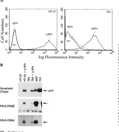
Arg-Figure 1. Intestinal epithe-lial cells express HLA class II molecules, Ii, and HLA-DM a and b molecules after stimulation with g-IFN. (A) Flow cytometric analysis of surface class II expression in T84 and HT-29. Primary an-tibody was HLA-DR spe-cific mAb L243. ISO, non-specific isotype control with identical secondary reagent. The cells were analyzed on a FACSCAN™ flow cytometer
without gating. (B) Immuno-blot analysis of Ii, HLA-DMb, and HLA-DMa in T84 and HT-29. Arrows, 33 kD core Ii chain, and the predicted MW HLA-DM a
[image:4.612.57.457.313.758.2]NMec (28, 29). Assays were run for 30 min at 378C in duplicate and en-zyme activity calculated as m units or n moles product formed per minute. Aspartyl protease activity was measured using 1.6% hemoglo-bin as a substrate in 0.2 M sodium acetate, 0.2 M KCl, 0.1% Triton X-100, pH 4.5, in the presence and absence of 5 mg/ml pepstatin A. These assays were run for 1 h at 378C, followed by precipitation of undigested substrate with trichloroacetic acid and detection of the soluble peptide products with ninhydrin reagent at OD 570 nm (30). Aspartyl protease activity was calculated such that one unit is equal to the amount of peptide formation inhibitable by pepstatin per mi-crogram cell protein per hour. All assays were repeated twice and the SEM was , 10%.
Results
g-IFN induces HLA class II antigens, Ii, and HLA-DM ab, and modulates protease activity in IECs. The IEC lines T84 and HT-29 were analyzed by flow cytometry for expression of en-dogenous HLA-DR molecules on the cell surface before and after stimulation with g-IFN. Despite the presence of HLA-DRA and HLA-DRB message detectable by RT-PCR (HT-29 and T84) and intracellular HLA-DR observed by confocal mi-croscopy (T84) in the absence of cytokine stimulation (data not shown), constitutive surface expression of HLA-DR was not detectable by flow cytometry (Fig. 1 A). Surface expression of HLA-DR molecules was induced by g-IFN , with z 50%
maximal surface expression of HLA-DR detected 24 h after adding g-IFN and maximal expression seen after 48 h. HLA-DQ expression was also detected after stimulation with g-IFN, but with very different kinetics. Surface expression was not seen until day 7 after stimulation, with maximal levels at day 9 (data not shown).
The Ii and the HLA-DM ab heterodimer are required for optimal antigen processing via the HLA class II pathway (for review see references 9 and 10). In general, these molecules are coordinately regulated with HLA-DR expression, al-though the expression of the HLA class II ab heterodimer in the absence of Ii has been reported in IECs (12). However, other investigators have demonstrated the constitutive expres-sion of Ii in IEC in human (31) and mouse (32). We performed Western blots using antibodies specific for Ii, HLA-DM a, and HLA-DM b to assess whether the epithelial cell lines express these molecules constitutively or after induction with g-IFN. For these experiments, we included the T2 cell line (which has a deletion on chromosome 6 that spans the class II region of the MHC including the genes encoding HLA-DM a and b), as a negative control. The expression of the Ii gene (located on human chromosome 5) is unaffected in this mutant cell line. As seen in Fig. 1 B, no constitutive Ii was detectable in the IEC lines, but the p33 species of Ii was readily inducible in the
g-IFN treated cells. As seen in Fig. 1 B, HLA-DM a and b pro-tein was detectable only after treatment with g-IFN. The ex-pression of both the a and b subunits of HLA-DM is required for the accessory role of these proteins in facilitating antigen presentation.
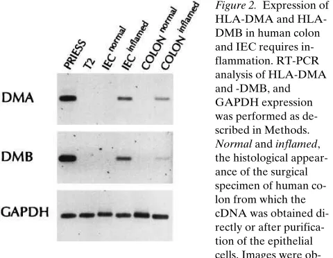
To extend these findings both to normal human IEC and to IBD, we performed RT-PCR using primers specific for either HLA-DMA or -B on cDNA prepared from both intact tissue and purified IEC from normal or inflamed colon tissue. The T2 cell line was used to ensure specificity of the PCR for DMA and DMB. As shown in Fig. 2, expression of HLA-DMA and -B was only seen in the inflamed colon and IEC. Overnight culture of both noninflamed or inflamed IEC in the presence
of g-IFN resulted in further induction of both HLA-DM genes, without affecting the expression of GAPDH (data not shown).
The expression of cysteine proteases (cathepsins B, L, and H) has been described in the mucosa of the gastrointestinal tract, and these enzymes were histochemically localized to en-terocytes of the rat duodenum (33). We performed an analysis of both the cysteine and aspartyl-protease activity in our hu-man IEC lines. The results of the protease assays are shown in Table I, and are representative of three independent assays on each cell line. In all instances, the constitutive activity of cathep-sins B, L, and H, as well as the aspartyl-proteases equaled or exceeded that seen in the human B-LCL Priess. The activity of cathepsin B in the T84 cell line showed a modest (2–3-fold) in-crease after g-IFN stimulation, similar to the increase that is seen in macrophages (Blum, J., unpublished data). Interest-ingly, the aspartyl-protease activity (and to a lesser extent, the cathepsin L and H activity) was lower after g-IFN stimulation of T84 cells in each experiment. No significant patterns or changes in protease activity were observed in the HT-29 cell line after stimulation with g-IFN.
In the absence of g-IFN, expression of a transfected HLA class II allele in IEC lines results in HLA molecules with an al-tered conformation. To permit the study of the HLA class II molecules and the processing pathway in IECs in the presence or absence of proinflammatory cytokines, we used retroviral gene transfer to constitutively express a specific HLA-DR
[image:5.612.315.555.60.246.2]al-Figure 2. Expression of DMA and HLA-DMB in human colon and IEC requires in-flammation. RT-PCR analysis of HLA-DMA and -DMB, and GAPDH expression was performed as de-scribed in Methods. Normal and inflamed, the histological appear-ance of the surgical specimen of human co-lon from which the cDNA was obtained di-rectly or after purifica-tion of the epithelial cells. Images were ob-tained on an Eagle Eye II Imager. Quantitation of band intensity was determined to be in the linear range.
Table I. Quantitation of Cellular Protease Activity
Cells Cathepsin B* Cathepsin L* Cathepsin H*
Aspartyl protease‡
Priess 7.57 3 1022 1.15 3 1022 6.38 3 1022 7.5 3 1026
HT-29 1.75 3 1021 3.20 3 1021 2.29 3 1022 6.6 3 1026
HT-29 1 IFNg 1.56 3 1021 3.92 3 1021 2.15 3 1022 7.2 3 1026
T84 4.73 3 1021 7.92 3 1021 2.77 3 1022 13.5 3 1026
T84 1 IFNg 9.84 3 1021 3.22 3 1021 1.34 3 1022 3.8 3 1026
*Cathepsins B, L, and H activity are given as milliunits per mg protein.
[image:5.612.315.557.620.711.2]lele in human IEC lines. Few HLA-DR restricted, antigen-specific T cell hybridomas have been reported. We had de-scribed previously studies using a panel of HLA-DRB1*0401 restricted T cell hybridomas that were generated in a HLA-DRB1*0401 transgenic mouse (22, 25). As determined by HLA genotyping, neither of the cell lines contained the HLA-DRB1*0401 allele. Accordingly, we chose the HLA-HLA-DRB1*0401 allele for our transfection studies.
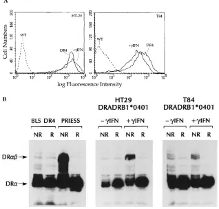
The IEC lines were infected with recombinant retroviruses that directed the surface expression of the HLA-DRA/HLA-DRB1*0401 (DR4) heterodimer. This was accomplished by serial infection first with a retroviral vector expressing the non-polymorphic DRA allele, followed by infection with a second recombinant virus expressing either the DRB1*0401 allele, or the DRB1*1101 (DR5) allele to be used as a negative control in the processing experiments. As shown in Fig. 3 A, levels of surface expression similar to that after g-IFN induction could be attained in clones (in HT-29) or pools (in T84) of infected cells. In all of the transfectants, we verified the specificity of the expressed HLA-DR using allele specific antibodies (data not shown). The expression seen was stable over 2–3 mo, and fresh aliquots of cells were thawed after every 10–12 passages for use in the antigen processing studies.
In the absence of HLA-DM ab, HLA class II antigens dis-play an altered conformation, evidenced by a marked reduction in their ability to form stable ab heterodimers in the presence of SDS (22, 34). This reflects a relatively inefficient removal of Ii peptides from the peptide binding groove of HLA-DR and a less efficient loading of peptides that would result in a compact form of the HLA-DR ab heterodimers that is stable in SDS (35). To determine whether (a) HLA class II antigens differ
biochemically when expressed in B lymphocytes as compared to IECs, and (b) Ii and HLA-DM ab can alter the conforma-tion of the HLA class II antigens, we looked for SDS-stable HLA class II ab heterodimer in the IECs transfected with HLA-DRB1*0401. As a negative control, we used a BLS-1 cell line expressing the same transfected HLA-DRB1*0401 allele. We have shown previously that BLS cells, which do not ex-press HLA-DM ab because of a defect in the transcription factor RF-X, do not generate SDS-stable dimers (Fig. 3 B and reference 22). Similarly, SDS-stable HLA class II ab hetero-dimers are not observed in either the HT-29 and T84 HLA-DRB1*0401 transfectants (Fig. 3 B, 2g-IFN). However, the same transfectants treated with g-IFN (resulting in the induc-tion of both Ii and HLA-DM ab) contain SDS-stable dimers. These data suggest that IECs display different MHC–peptide complexes in the presence or absence of inflammatory me-diators.
Distinct antigen processing pathways in the epithelial cell HLA class II transfectants in the absence or presence of g-IFN.
The presence of stable class II dimers only after treatment with
g-IFN suggested a difference in the processing of class II mole-cules in the presence or absence of inflammatory cytokines. To determine if these differences were functionally relevant with respect to T cell stimulation, we then tested the HLA-DRB1*0401 transfectants for their ability to process and present peptide antigens to T lymphocytes. In the first series of experi-ments, the responder T cells were CD41 T cell hybridomas
[image:6.612.54.372.439.742.2]generated after immunization of HLA-DRB1*0401 transgenic mice with (a) whole TT or (b) HSA (see Methods). The pre-cise HLA-DR restriction of the T cell hybrids to HLA-DRB1* 0401 was confirmed using a large panel of HLA-DR typed
Figure 3. Transfected HLA class II molecules can be expressed at high levels in IEC lines, but are only stable in SDS after treatment with g -inter-feron. (A) Flow cytometric analysis of class II ex-pression using the mAb L243, as in Fig. 1 A. DR4 corresponds to the HLA-DRB1*0401 transfec-tants generated using retroviral vectors without the addition of g-IFN. 1g-IFN corresponds to the wild-type cell treated with 500 U/ml g-IFN for 48 h before staining. WT, the staining of wild-type cells with L243. (B) Detection of SDS stable class II ab dimers in the HLA-DRB1*0401 transfec-tants by immunoblot analysis using the mAb DA6.147 which detects both the HLA-DR a
monomer and the HLA-DR ab dimer. NR, the nonreduced, nonboiled lysates prepared in 2% SDS as outlined in Methods. R, identical samples boiled for 10 min in the presence of DTT. Arrow DRab, the 55–60-kD reactivity of the HLA-DR
ab heterodimer. Arrow DRa, the z 30-kD
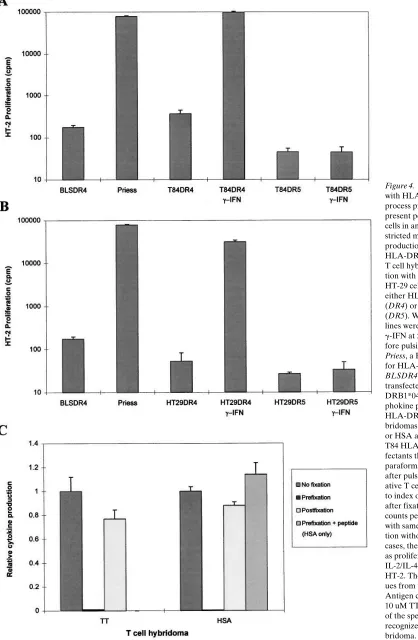
Figure 4. IEC lines transfected with HLA class II alleles can process protein antigens and present peptides to CD41 T cells in an MHC class II re-stricted manner. Lymphokine production by a TT-specific, HLA-DRB1*0401–restricted T cell hybridoma after incuba-tion with (A) T84, or (B) HT-29 cells transfected with either HLA-DRB1*0401 (DR4) or HLA-DRB1*1101 (DR5). Where indicated, IEC lines were stimulated with
B-LCLs (data not shown). Epithelial cells transfected with and expressing a HLA-DRB1*1101 (DR5) allele and HLA-DM deficient BLS cells transfected with HLA-DRB1*0401 were used as negative controls in the antigen processing experiments. Fig. 4 A shows data from a representative processing experi-ment using the TT-specific T cell hybridoma. T84 (Fig. 4 A) and HT-29 (Fig. 4 B) HLA-DRB1*0401 transfectants treated with g-IFN for 48 h processed intact TT and presented peptide in an HLA-DR restricted manner. Identical results were ob-tained using HSA and the HSA-specific T cell hybridoma (data not shown). Maximal values for the T84 transfectants treated with g-IFN were consistently as high or higher than the
B-LCL expressing the HLA-DRB1*0401 molecule (Priess). The maximal values seen in the HT-29 transfectants were con-sistently 20–40% of those seen in similarly treated T84 trans-fectants. In no instance was T cell activation observed within either cell line transfected with HLA-DRB1*1101.
Fixation experiments were performed to formally exclude the possibility that IECs merely were presenting contaminat-ing antigenic peptides rather than processcontaminat-ing whole antigen. Epithelial cells were treated with 1% paraformaldehyde be-fore, or 4 h after, the addition of antigen. Additionally, with the HSA-specific T cell hybridoma (where the peptide speci-ficity has been determined) peptide was added after
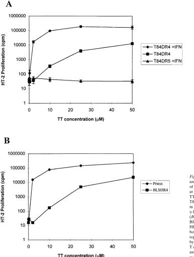
prefix-Figure 5. IEC lines can process and present antigen in the absence of g-IFN when exposed to antigen at high concentration. (A) TT dose–response curves for the T84 DR4 and DR5 transfectants, in the presence or absence of
[image:8.612.58.444.223.734.2]ation (prefix 1 peptide). As seen in Fig. 4 C, prefixation of APC before antigen addition abrogated T cell stimulation. The lack of inhibition in the postfix condition and the complete res-toration of the response by adding peptide after prefixation suggest that the effects seen were not the result of the destruc-tion of the HLA class II molecules on the cell during the fixa-tion process. These data (and the drug inhibifixa-tion data that fol-low) suggest intracellular processing of antigen is required in these assays to stimulate the T cell hybridomas.
Interestingly, the HLA-DRB1*0401 transfected IEC lines were able to process antigen and stimulate the T cells even in the absence of g-IFN when exposed to a relatively high concentra-tion of antigen. The observed processing was dose-dependent (Fig. 5, and similar data with HSA not shown). The responses observed at concentrations above 10 mM were 10–20% of those seen at the same concentration of antigen in the presence of
g-IFN. The dose response curves comparing antigen pro-cessing in the BLS cell line transfected with HLA-DRB1*0401 to the normal B-LCL Priess (homozygous for HLA-DRB1*0401) showed a similar pattern to that seen with the epithelial cell transfectants with and without g-IFN (Fig. 5 B).
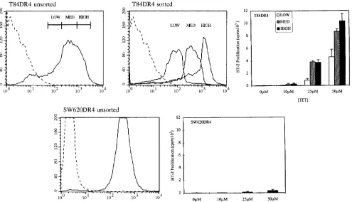
To investigate whether the antigen presentation observed in the absence of g-IFN required high levels of surface class II expression, we sorted the pools of T84 DR4 transfectants using FACS® into high (mean channel fluorescence [mcf] 5 965),
medium (mcf 5 383), and low (mcf 5 95) fractions and ana-lyzed them independently for their ability to process and present
antigen at various antigen concentrations in the absence of
g-IFN. As seen in Fig. 6, although the difference between the surface class II expression in the high and low fractions is more than 10-fold, only modest differences in processing were seen. It is difficult to directly compare the low expression seen in T84 to the low level of expression seen in normal enterocytes in situ. Still, these data suggest that IEC do not require the ex-tremely high levels of surface class II seen in the context of in-flammation to stimulate CD41 T cells. In marked contrast,
SW620 cells transfected with the identical HLA-DRB1*0401 allele and expressing this molecule at a high level on its surface were observed to be essentially incapable of processing and presenting antigen (Fig. 6). Hence, overloading any cell line with class II alone is insufficient to confer the ability to process and present antigen to CD41 T cells.
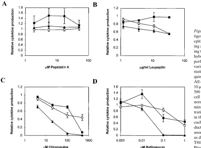
[image:9.612.61.556.386.670.2]Next, we tested several drugs known to block antigen pre-sentation in other systems (a) to determine whether antigen processing by IECs was inhibited in a similar manner to con-ventional APCs, and (b) to determine whether one could phar-macologically distinguish the g-IFN–dependent from the g-IFN– independent processing seen in the IEC lines. We compared the response of the drugs in the T84 HLA-DRB1*0401 trans-fectants with and without g-IFN to the response of the HLA-DRB1*0401 B-LCL Priess. The data are normalized to the T cell stimulation observed in the absence of drug. The transfectants were analyzed by flow cytometry after treatment with the highest concentration of the individual drugs and no decrease
Figure 6. High levels of class II are neither necessary nor sufficient for efficient stimulation of CD41 T cells in the absence of g-IFN. T84 HLA-DRB1*0401 transfectants, initially selected and expanded as pools, were sorted into high, medium, and low expressing fractions using the mAb L243 and a FACSCAN™ cell sorter. Mean channel fluorescence of these populations are detailed in the text. The three populations were tested
in the surface expression of HLA class II molecules was ob-served (data not shown).
Several distinct patterns were observed. Some drugs, as is seen with chloroquine (Fig. 7 C), bafilomycin A1 (Fig. 7 D),
and brefeldin A (not shown), inhibited the antigen processing by the B-LCL, as well as the T84 HLA-DRB1*0401 transfec-tants with or without stimulation by g-IFN. Interestingly, leu-peptin consistently inhibited the antigen processing seen in B cells and in the g-IFN treated epithelial cell transfectants, but showed no inhibition of the g-IFN–independent processing and presentation (Fig. 7 B). Conversely, pepstatin modestly stimulated the g-IFN–independent antigen processing in the epithelial transfectants, but not that seen in the B-LCL– or
g-IFN–treated epithelial cell transfectants (Fig. 7 A). These data suggest that the intrinsic HLA-DM and Ii independent path-way in the epithelial cells differs biochemically from the anti-gen processing seen in the presence of inflammatory media-tors.
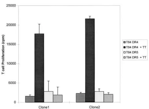
IEC HLA-DR transfectants stimulated the proliferation of normal human CD41 T cell clones. To extend our
observa-tions on the T cell hybridomas, we investigated whether the epithelial cell transfectants could stimulate the antigen-spe-cific, HLA-DR–restricted proliferation of human T cell clones. TT-specific, HLA-DRB1*0401–restricted human T cell clones were generated and tested in antigen processing assays using IECs as APCs. As shown in Fig. 8 A, g-IFN–stimulated T84 HLA-DRB1*0401 cells pulsed with TT were capable of stimu-lating the proliferation of two independent T cell clones in an antigen-specific and HLA-DR–restricted manner.
CD80 and CD86 were not expressed in any of the epithelial cell lines tested and were not induced by g-IFN alone or in combination with GM-CSF or TNFa (data not shown).
Discussion
IECs express a low level of HLA class II antigens constitu-tively, and their expression is enhanced in inflammatory condi-tions including IBD (1). In this study, we describe two distinct pathways for antigen processing by IECs that distinguish be-tween the activated and nonactivated states. One pathway is similar to that seen in conventional APCs, uses similar pro-teases, Ii, HLA-DM ab, and facilitates efficient antigen presen-tation even at low antigen concentrations. The other pathway functions independent of Ii and HLA-DM ab, and requires a relatively high concentration of antigen to elicit T cell stimula-tion.
The g-IFN stimulated IEC lines express SDS-stable HLA-DR ab heterodimers, Ii, HLA-DM ab, and contain sufficient protease activity to efficiently process and present exogenous protein antigens to CD41 T cells. The observation is unlikely
to be unique to the IEC lines as enterocytes from inflamed co-lon (in contrast to those from uninflamed specimens) were also shown to express HLA-DMA and -B, and the expression could be augmented further with exogenous g-IFN. In our ex-periments, the g-IFN–dependent antigen processing seen in the T84 cell line was inhibited by chloroquine, bafilomycin, and leupeptin, suggesting the use of an acidic cellular compart-ment and cysteine proteases in class II processing. In fact, the
g-IFN stimulated T84 cells were as effective in stimulating the T cell hybridomas as a B-LCL over a wide range of antigen concentrations. These data suggest that in the context of mucosal inflammation, IECs use the conventional class II pro-cessing pathway and can function as efficient APCs to CD41
[image:10.612.57.455.443.735.2]T cells in the lamina propria. g-IFN also effects epithelial bar-rier function and facilitates paracellular transport of luminal
Figure 7. Drug sensitivity of an-tigen presentation by intestinal epithelial cells. Antigen process-ing and presentation assays us-ing the HLA-DRB1*0401 T cell hybridomas specific for TT were performed in the presence of various concentrations of pep-statin (A), leupeptin (B), chloro-quine (C), and bafilomycin (D). All data were obtained using 10 mM TT, and, where indicated, 500 U/ml of g-IFN. Relative T cell stimulation is an index that normalizes the counts per minute at a specific drug concen-tration to the counts per minute in the absence of any drug, for each cell line. Lymphokine pro-duction by the hybridoma was assayed using the HT-2 cell line, as described in Fig. 4. s,
T84DR4 1IFN; j, T84DR4; m,
antigens (36). Hence, both the basolateral and apical surfaces of the enterocyte would be exposed to high concentrations of antigen and could participate in the endocytosis of foreign an-tigens to initiate processing via class II molecules.
We also demonstrated that IEC lines possess a functional class II pathway that operates in the absence of g-IFN (and therefore in the absence of Ii and HLA-DM) and requires a relatively high concentration of antigen. The presentation of exogenous foreign antigens by an APC requires the endocyto-sis, proteolyendocyto-sis, and loading of the processed peptides onto newly synthesized HLA class II ab heterodimers. Ii, encoded by a non-MHC–linked gene, physically associates with nascent HLA class II molecules in the endoplasmic reticulum and di-rects the complex to an acidic intracellular compartment where peptide loading occurs (for review see reference 9). The prod-ucts of the MHC-linked HLA-DM a and b genes facilitate the removal of residual portion (CLIP) of the Ii from the peptide binding groove of the HLA class II molecules and its replace-ment with an antigenic peptide. Without the leucine-based en-docytic targeting of the newly synthesized class II molecules normally provided by the cytoplasmic tail of Ii (37), or cyto-plasmic tail of the HLA-DRA (38) or -DRB chain (38, 39), the mechanism of antigen presentation is likely to involve the re-cycling of class II molecules from the surface to an endocytic compartment where loading of endocytosed, partially de-graded peptides can occur. The g-IFN–independent pathway in IEC is inhibited by bafilomycin, a specific inhibitor of vacu-olar [H1]ATPase activity (40, 41), suggesting the involvement
of an acidic late endosomal or lysosomal cellular compartment where degradation of internalized protein antigen would oc-cur. Our data from the BLS HLA-DR transfectants, and data from several groups demonstrating Ii-independent antigen processing indicate that this pathway is not specific to IECs and is likely to be peptide epitope specific (39, 42; for review see reference 43). However, this pathway may be especially
relevant to the physiology of the IEC for two reasons. First, IECs are constantly exposed on their apical surface to the highest concentration of foreign antigen in the body and there-fore may sample and present antigens even in the absence of inflammation. Second, unlike conventional APCs, a subset of IECs may normally express class II antigens in the absence HLA-DM (and possibly Ii). Several recent reports have under-scored the importance of HLA-DM in editing the peptides that are presented by class II molecules (44). Hence, the HLA-DM–independent pathway would be likely to result in the pro-cessing and loading of peptide epitopes that differ from those seen in the context of mucosal inflammation.
The inhibitor studies point to a important distinction be-tween the two processing pathways. Intracellular protease ac-tivity is required both for the proteolytic degradation of en-docytosed proteins to antigenic peptides, and for the normal processing of the HLA class II/Ii complex to an intermediate that is a substrate for the HLA-DM ab heterodimer. In con-trast to the g-IFN–stimulated cells and the B-LCL, the antigen processing seen in the untreated HLA-DR transfectants was not inhibited by the cysteine protease inhibitor leupeptin. These data suggest that this pathway is less dependent on the cathepsin proteases B, D, and L that are normally implicated in class II antigen processing (45–47) and are consistent with the lack of requirement for Ii in this pathway. Interestingly, only the g-IFN–independent processing was modestly stimu-lated by the aspartyl protease inhibitor pepstatin. This corre-lates with the decreased aspartyl protease activity and the en-hanced processing seen in the T84 transfectants stimulated with g-IFN. Moreover, these findings are consistent with the observations of Vidard et al., which detailed the enhancement of antigen processing and presentation of certain peptide epitopes of ovalbumin after treatment of APCs with protease inhibitors (48). Hence, an important aspect to the regulation of antigen processing in tissues such as epithelia that are rich in aspartyl-proteases may also involve the specific downregula-tion of protease activity to prevent the degradadownregula-tion of protein antigens to peptide fragments too short to serve as peptide ligands for HLA-DR mediated antigen presentation.
The early reports describing antigen presentation by IECs in human (14) and rat (15) reported the proliferation of CD81
T lymphocytes with suppressor activity after stimulation of primed T cells with antigen pulsed, class II expressing IECs. However, HLA class II molecules are unlikely to stimulate CD81 T cell responses. Indeed, recent data in humans suggest
that the HLA class I like CD1d molecule, which is expressed on IECs (49) is involved in the stimulation of CD81 T cells in
the mucosa (50; for review see reference 51). Subsequent stud-ies suggested that rat enterocytes were unable to efficiently process protein antigens when compared to conventional APCs, and that antigen presentation by IECs was not sensitive to leupeptin (52). These findings are consistent with the leupep-tin independence and relatively inefficient processing we ob-served in the IEC transfectants not treated with g-IFN. Parallel studies in murine systems are somewhat conflicting. Kaiserlian et al. have demonstrated efficient class II restricted processing and presentation of antigen to CD41 T cell hybridomas using
[image:11.612.55.298.54.232.2]purified murine villous IECs (16). Hoyne et al. subsequently described the inability of murine IECs to process protein anti-gens, despite their ability to present peptides to T cells (53). Whether differences in the protocols used to isolate the IECs and/or differences in baseline levels of intestinal inflammation
Figure 8. Stimulation of human CD41 T cell clones by intestinal epi-thelial cells. g-IFN–stimulated, g-irradiated T84 HLA-DRB1*0401 (DR4) or HLADRB1*1101 (DR5) transfectants were used to restim-ulate HLA-DRB1*0401–restricted, TT-specific T cell clones. Data presented are [3H]thymidine incorporation by the T cell clones after a
can reconcile these differences remains to be seen. In this re-gard, variability in class II expression is observed along both the crypt-villous (1) and rostral-caudal (54) axes, probably un-derscoring important regional distinctions in the ability of the intestinal epithelium to process and present antigen to T cells.
In summary, IECs have extensive contact with T lympho-cytes both within the epithelium and in the underlying lamina propria (55). IECs are capable of expressing HLA class II mol-ecules in a variety of immunopathological contexts character-ized by mucosal inflammation with the associated production of inflammatory cytokines. In the presence of g-IFN, IECs be-come competent, efficient APCs, and are likely to contribute to the stimulation of CD41 T cells that accumulate in the
intes-tinal mucosa. In the absence of g-IFN, IECs can use a distinct class II pathway that requires higher concentrations of antigen and functions in the absence of HLA-DM. The polarized mor-phology of IECs underscores the functional segregation be-tween apical and basolateral events. It is possible that the
g-IFN–dependent and –independent pathways will correspond to antigen processing after endocytosis from the apical and ba-solateral surface, respectively. In addition, oral tolerance to fed antigens, which does not occur in mice after administration of g-IFN (56), may be associated with the selective use of the nonconventional class II pathway in enterocytes.
Acknowledgments
We gratefully acknowledge Dr. William Kwok for providing help with retroviral infections; Dr. Linda Wicker and Dennis Zaller (Merck Research Labs) for assistance with the T cell hybridomas; Su-san Masewicz for help in establishing the human T cell clones; Dr. Al-lan Mowat for helpful discussions and critical review of the manu-script; and Holly Chase for assistance in preparing the manuscript. R.M. Hershberg would like to dedicate this manuscript to Dr. John A. Ryan, Jr.
The work was supported from the Virginia Mason Medical Cen-ter Department of Surgery (R.M. Hershberg), by grant #AI38913 from the NIH (G.T. Nepom), a National Research Service Award from NIH (S. Kovats), and grant #AI33418 and #DK94017 from the NIH (J.S. Blum).
References
1. Mayer, L., D. Eisenhardt, P. Salomon, W. Bauer, R. Plous, and L. Picci-nini. 1991. Expression of class II molecules on intestinal epithelial cells in hu-mans. Differences between normal and inflammatory bowel disease. Gastroen-terology. 100:3–12.
2. Bland, P.W., and C.V. Whiting. 1992. Induction of MHC class II gene products in rat intestinal epithelium during graft-versus-host disease and effects on the immune function of the epithelium. Immunology. 75:366–371.
3. Ciclitira, P.J., J.M. Nelufer, H.J. Ellis, and D.J. Evans. 1986. The effect of gluten on HLA-DR in the small intestinal epithelium of patients with Coeliac disease. Clin. Exp. Immunol. 63:101–104.
4. Germain, R.N. 1994. MHC-dependent antigen processing and peptide presentation: providing ligands for T lymphocyte activation. Cell. 76:287–299.
5. Powrie, F. 1995. T cells in inflammatory bowel disease: protective and pathogenic roles. Immunity. 3:171–174.
6. Chen, Y., J. Inobe, and H.L. Weiner. 1995. Induction of oral tolerance to myelin basic protein in CD8-depleted mice: both CD41 and CD81 cells medi-ate active suppression. J. Immunol. 155:910–916.
7. Garside, P., M. Steel, F.Y. Liew, and A.M. Mowat. 1995. CD41 but not CD81 T cells are required for the induction of oral tolerance. Int. Immunol. 7: 501–504.
8. Barone, K.S., S.L. Jain, and J.G. Michael. 1995. Effect of in vivo deple-tion of CD41 and CD81 cells on the induction and maintenance of oral toler-ance. Cell. Immunol. 163:19–29.
9. Wolf, P.R., and H.L. Ploegh. 1995. How MHC class II molecules acquire peptide cargo: biosynthesis and trafficking through the endocytic pathway. Annu. Rev. Cell Dev. Biol. 267–306.
10. Cresswell, P. 1994. Assembly, transport, and function of MHC class II molecules. Annu. Rev. Immunol. 12:259–293.
11. Chang, C.H., and R.A. Flavell. 1995. Class II transactivator regulates the expression of multiple genes involved in antigen presentation. J. Exp. Med. 181:765–767.
12. Vidal, K., C. Samarut, J.-P. Magaud, J.-P. Revillard, and D. Kaiserlian. 1993. Unexpected lack of reactivity of allogeneic anti-Ia monoclonal antibodies with MHC class II molecules expressed by mouse intestinal epithelial cells. J. Immunol. 151:4642–4650.
13. Bland, P.W., and D.M. Kambarage. 1991. Antigen handling by the epi-thelian and lamina propria macrophages. Gastroenterol. Clin. North Am. 20: 577–596.
14. Mayer, L., and R. Shlien. 1987. Evidence for function of Ia molecules on gut epithelial cells in man. J. Exp. Med. 166:1471–1483.
15. Bland, P.W., and L.G. Warren. 1986. Antigen presentation by epithelial cells of the rat small intestine. I. Kinetics, antigen specificity and blocking by anti-Ia antisera. Immunology. 58:1–7.
16. Kaiserlian, D., K. Vidal, and J.-P. Revillard. 1989. Murine enterocytes can present soluble antigen to specific class II-restricted CD41 T cells. Eur. J. Immunol. 19:1513–1516.
17. Kovats, S., S. Drover, W. Marshall, D. Freed, P. Whiteley, G.T. Nepom, and J.S. Blum. 1994. Coordinate defects in HLA class II expression and antigen presentation in bare lymphocyte syndrome. J. Exp. Med. 179:2017–2022.
18. Leech, N., W. Hagopian, J.A. Hansen, G.T. Nepom, and A. Kitabchi. 1997. GAD65 and islet cell autoantibodies are associated with HLA class II al-leles in Black Americans. Autoimmunity. In press.
19. MacDonald, M.J., J. Gottschall, J.B. Hunter, and K.L. Winter. 1986. HLA-DR4 in insulin-dependent diabetic parents and their diabetic offspring: a clue to dominant inheritance. Proc. Natl. Acad. Sci. USA. 83:7049–7053.
20. Inoko, H., A. Ando, K. Tsuji, K. Matsuki, T. Juji, and Y. Honda. 1986. HLA-DQ beta chain DNA restriction fragments can differentiate between healthy and narcoleptic individuals with HLA-DR2. Immunogenetics. 23:126–128. 21. Cohen-Haguenauer, O., E. Robbins, C. Massart, M. Busson, I. Des-champs, J. Hors, J.M. Lalouel, J. Dausset, and D. Cohen. 1985. A systematic study of HLA class II-beta DNA restriction fragments in insulin-dependent di-abetes mellitus. Proc. Natl. Acad. Sci. USA. 82:3335–3339.
22. Kovats, S., G.T. Nepom, M. Coleman, B. Nepom, W.W. Kwok, and J.S. Blum. 1995. Deficient antigen presenting cell function in multiple genetic com-plementation groups of type II bare lymphocyte syndrome. J. Clin. Invest. 96: 217–223.
23. Kwok, W.W., D. Schwarz, B.S. Nepom, P.S. Thurtle, R.A. Hock, and G.T. Nepom. 1988. HLA-DQ molecules form a-b heterodimers of mixed allo-type. J. Immunol. 141:3123–3127.
24. Denzin, L.K., N.F. Robbins, C. Carboy-Newcomb, and P. Cresswell. 1994. Assembly and intracellular transport of HLA-DM and correction of the class II antigen-processing defect in T2 cells. Immunity. 1:595–606.
25. Woods, A., H.Y. Chen, M.E. Trumbauer, A. Sirotina, R. Cummings, and D.M. Zaller. 1994. Human major histocompatibility complex class II-restricted T cell responses in transgenic mice. J. Exp. Med. 180:173–181.
26. Tuosto, L., R.W. Karr, X.T. Fu, R.R. Olson, E. Cundari, E. Piccolella, R. Lechler, and G. Lombardi. 1994. Different regions of the N-terminal do-mains of HLA-DR1 influence recognition of individual peptide-DR1 com-plexes. Hum. Immunol. 40:312–322.
27. Weiner, G.J., and M.S. Kaminski. 1990. Anti-idiotypic antibodies recog-nizing stable epitopes limit the emergency of idiotype variants in a murine B cell lymphoma. J. Immunol. 144:2436–2445.
28. Barrett, A.J. 1980. Fluorimetric assays for Cathepsin B and Cathepsin H with methylcoumarylamide substrates. Biochem. J. 187:909–912.
29. Barrett, A.J., and H. Kirschke. 1981. Cathepsin B, Cathepsin H, and Cathepsin L. Methods Enzymol. 80:535–561.
30. Diment, S., M.S. Leech, and P.D. Stahl. 1988. Cathepsin D is membrane-associated in macrophage endosomes. J. Biol. Chem. 263:6901–6907.
31. Momburg, F., K. Koretz, and A. Von Herbay. 1988. Nonimmune human cells can express MHC class II antigens in the absence of invariant chain—an immunohistological study on normal and chronically inflamed small intestine. Clin. Exp. Immunol. 72:367–372.
32. Sanderson, I.R., A.J. Ouellette, E.A. Carter, and P.R. Harmatz. 1992. Ontogeny of class II MHC mRNA in the mouse small intestinal epithelium. Mol. Immunol. 29:1257–1263.
33. Furuhashi, M., A. Nakahara, H. Fukutomi, E. Kominami, D. Grube, and Y. Uchiyama. 1991. Immunocytochemical localization of cathepsins B, H, and L in the rat gastro-duodenal mucosa. Histochemistry. 95:231–239.
34. Mellins, E., L. Smith, B. Arp, T. Cotner, E. Celis, and D. Pious. 1990. Defective processing and presentation of exogenous antigens in mutants with normal HLA class II genes. Nature (Lond.). 343:71–74.
35. Roche, P.A. 1995. HLA-DM: an in vivo facilitator of MHC class II pep-tide loading. Immunity. 3:259–262.
36. Madara, J.L., and J. Stafford. 1989. Interferon-gamma directly affects barrier function of cultured intestinal epithelial monolayers. J. Clin. Invest. 83: 724–727.
38. Pinet, V., M. Vergelli, R. Martin, O. Bakke, and E.O. Long. 1995. Anti-gen presentation mediated by recycling of surface HLA-DR molecules. Nature (Lond.). 375:603–604.
39. Zhong, G., P. Romagnoli, and R.N. Germain. 1997. Related leucine-based cytoplasmic targeting signals in invariant chain and major histocompati-bility complex class II molecules control endocytic presentation of distinct de-terminants in a single protein. J. Exp. Med. 185:429–438.
40. Lencer, W.I., G. Strohmeier, S. Moe, S.L. Carlson, C.T. Constable, and J.L. Madara. 1995. Signal transduction by cholera toxin: processing in vesicular compartments does not require acidification. Am. J. Physiol. 269:G548–G557.
41. Yoshimori, T., A. Yamamoto, Y. Moriyama, M. Futai, and Y. Tashiro. 1991. Bafilomycin A1, a specific inhibitor of vacuolar-type H1-ATPase, inhibits
acidification and protein degradation in lysosomes of cultured cells. J. Biol. Chem. 266:17707–17712.
42. Pinet, V., M.S. Malnati, and E.O. Long. 1994. Two processing pathways for the MHC class II-restricted presentation of exogenous influenza virus anti-gen. J. Immunol. 152:4852–4860.
43. Ceman, S., and A.J. Sant. 1995. The function of invariant chain in class II-restricted antigen presentation. Semin. Immunol. 7:373–387.
44. Katz, J.F., C. Stebbins, E. Appella, and A.J. Sant. 1996. Invariant chain and DM edit self-peptide presentation by major histocompatibility complex (MHC) class II molecules. J. Exp. Med. 184:1747–1753.
45. Buus, S., and O. Werdelin. 1986. A group-specific inhibitor of lysosomal cysteine proteinases selectively inhibits both proteolytic degradation and pre-sentation of the antigen dinitrophenyl-poly-L-lysine by guinea pig accessory cells to T cells. J. Immunol. 136:452–458.
46. Mizuochi, T., S.-T. Yee, M. Kasai, T. Kakiuchi, D. Muno, and E. Komi-nami. 1994. Both cathepsin B and cathepsin D are necessary for processing of ovalbumin as well as for degradation of class II MHC invariant chain. Immunol.
Let. 43:189–193.
47. Rodriguez, G.M., and S. Diment. 1992. Role of cathepsin D in antigen presentation of ovalbumin. J. Immunol. 149:2894–2898.
48. Vidard, L., K.L. Rock, and B. Benacerraf. 1991. The generation of im-munogenic peptides can be selectively increased or decreased by proteolytic en-zyme inhibitors. J. Immunol. 147:1786–1791.
49. Blumberg, R.S., C. Terhorst, P. Bleicher, F.V. McDermott, C.H. Allan, S.B. Landau, J.S. Trier, and S.P. Balk. 1991. Expression of a nonpolymorphic MHC class I-like molecule, CD1D, by human intestinal epithelial cells. J. Im-munol. 147:2518–2524.
50. Panja, A., A. Barone, and L. Mayer. 1994. Stimulation of lamina propria lymphocytes by intestinal epithelial cells: evidence for recognition of nonclassi-cal restriction elements. J. Exp. Med. 179:943–950.
51. Blumberg, R.S., and S.P. Balk. 1994. Intraepithelial lymphocytes and their recognition of non-classical MHC molecules. Intern. Rev. Immunol. 11:15–30. 52. Bland, P.W., and C.V. Whiting. 1989. Antigen processing by isolated rat intestinal villus enterocytes. Immunology. 68:497–502.
53. Hoyne, G.F., M.G. Callow, M.-C. Kuo, and W.R. Thomas. 1993. Presen-tation of peptides and proteins by intestinal epithelial cells. Immunology. 80: 204–208.
54. Sidhu, N.K., G.M. Wright, R.J.F. Markham, W.P. Ireland, and A. Singh. 1992. Quantitative regional variation in the expression of major histocompati-bility class II antigens in enterocytes of the mouse small intestine. Tissue Cell. 24:221–228.
55. Hashimoto, Y., and T. Komuro. 1988. Close relationships between the cells of the immune system and the epithelial cells in the rat small intestine. Cell Tissue Res. 254:41–47.