© 2015, IRJET.NET- All Rights Reserved
Page 728
BRAIN TUMOR SEGMENTATION USING ARTIFICIAL NEURAL NETWORK
*1
Ms. Sangeetha C., *
2Ms. Shahin A.,
*1
M.Phil Research Scholar, Department of Computer Science Auxilium College (Autonomous), Vellore
TamilNadu, India
*2
Assisant Professor, Department of Computer Science Auxilium College (Autonomous), Vellore,
TamilNadu, India
---***---Abstract: -
Image segmentation plays a vital role inmedical images. Modified K-means Clustering is used for image segmentation. Clustering is the popular unsupervised technique. The Modified K-means is to improve the effectiveness and efficiency for image segmentation. Medical imaging technique is most commonly used to visualize the internal structure and function of the body. Magnetic Resonance Imaging (MRI) provides much greater contrast between the different soft tissues of the body. Brain Tumor is one of the serious disease causes death among the people. Tumor is an uncontrolled growth of tissue in any part of the body. In this clustering method, the MRI images are used to identify the tumor of the brain. The MRI image of the brain is given as input. The system should process the input image and detect the tumor. This method can even detect the smallest abnormality in the earlier stage itself. The different abnormality MRI scans of brain of the patients are taken for processing. Experimental results have shown that the proposed methodology is effective and more robust.
Key Words:
Image segmentation, Magnetic ResonanceImaging (MRI), Clustering. Image Visualize, K-Means Clustering
I. INTRODUCTION
In today’s digital era, capturing, storing and analysis of medical image had been digitized. Even with state of the art techniques, detailed interpretation of medical image is a challenge from the perspective of time and accuracy. The challenge stands tall especially in regions with abnormal Color and Shape which needs to be identified by radiologists for future studies. The key ask in designing such image processing and a computer vision application is the accurate segmentation of medical images. This century will pass away, but the birth of medical computing and its reward to advances in medicine will usher in a new plate of technological innovations with
a focus on ideal and convenient delivery of medical services. Both medicine and computing are growing at a rapid rate. Undoubtedly the growth in medicine has benefited much from the growth in computers. Precise, diagnosis, fast data and voice communication. The Computed Tomography Scanner (CT scanner) and MRI is one of the most revolutionary healthcare machines developed in the 21st century. The CT scanner was
founded on a technique where images of tissues were depicted on radiographic film. Alessandro Vallebona, from Genova, initially proposed the CT technology in the early 1900’s, but the medical industry did not embrace the concept until the late 1960’s when Sir Godfrey Hounsfield, from the United Kingdom, created the first commercially feasible CT Scanner. Now, Scientists and Researchers are used the MRI and CT Scans are used in the field of identifying the internal parts of the human body, especially for Brain Tumors (BT).
The tumor cell is present within skull and grows within skull is called primary tumor. Malignant brain tumors are primary brain tumors. The tumor presents outside the skull and enter into the skull region called secondary tumor. Metastatic tumors are examples of secondary tumors. The tumor takes up place in the skull and interferes with the normal functioning of the brain. Tumor shifts the brain towards skull and increases the pressure on the brain. Detection of tumor is the first step in the treatment. Brain contains more number of cells that are interconnected to one another. Different cells control different parts of the body. Some cells control the leg movement, likewise others cells of the brain controls other parts in the body. Brain tumors may have different types of symptoms ranging from headache to stroke, so symptoms will vary depending on tumor location. Different location of tumor causes different functioning disorder.
© 2015, IRJET.NET- All Rights Reserved
Page 729
The quality of image is main important in brain tumor.MRI provides an unparallel view inside the human body. In MRI scans will show the detailed information extra-ordinary compared to any other scanning like X-ray, CT scans. The contrast of tumor cell is high compared to normal brain cell.[2] Treatments techniques for the brain tumor:
Surgery
Radiation therapy Chemotherapy
The proposed system is an efficient system for detection of tumor and segmentation for given MRI images .The method of detection work is done by using the filter. This method is developed in Matlab simulation environment in order to check for applicability of proposed method.
II. Related work
The literature shows the various methods for the detection of brain tumor. This method used an approach to detect brain tumor using Computer Aided Diagnosis (CAD) system using ANNs to classify brain tumors. From each MR Image a Harlick texture features was extracted to prepare training data which was introduced to neural network as input and target vectors[2]. Brain Cancer Detection and Classification System which uses computer based procedures to detect tumor blocks or lesions and classify the type of tumor using Artificial Neural Network in MRI images of different patients with Astrocytoma type of brain tumors. The image processing techniques such as histogram equalization, image segmentation, image enhancement, morphological operations and feature extraction have been developed for detection of the brain tumor in the MRI images of the cancer affected patients.
The analyses of various clustering techniques to track tumor objects in Magnetic Resonance (MR) brain images. The input to this system is the MR image of the axial view of the human brain. The Clustering algorithms used are K-means, Hierarchical Clustering and Fuzzy C-Means Clustering. A technique used for the detection of tumor in brain using segmentation and histogram three scolding. The method can be successfully applied to detect the contour of the tumor and its geometrical dimension. Another approach to extract metabolite values from graph. The system is based on decision tree algorithms. A new approach is used for brain tumor detection using Meta Heuristic Algorithm.[5] It aims to develop effective
algorithm for the segmentation of Brain MRI images. In the Preprocessing system, film artifacts and unwanted portions of MRI brain image are removed. In the Enhancement process, the noise and high frequency components are removed using median filters. The Segmentation process has three different approaches like block based (non algorithmic), PSO and HPACO algorithm segmentation.
A method used to locate the regions occupied by brain tumors. Initially the brain is extracted by removing the unwanted non-brain regions like skull, scalp, fat and muscles.[6] Then the brain is segmented into well known regions and background using FCM algorithm. Cerebro-Spinal Fluid (CSF) class is analyzed for symmetric property along the central vertical line. The simulation results showed that the modified PNN gives rapid and accurate classification compared with the image processing and published conventional PNN techniques.
III. PREVIOUS IMPLEMENTATIONS
Artificial Neural Networks are relatively crude electronic models based on the neural structure of the brain. The brain basically learns from experience. It is natural proof that some problems that are beyond the scope of current computers are indeed solvable by small energy efficient packages. This brain modeling also promises a less technical way to develop machine solutions. This new approach to computing also provides.
3.1 Analogy to the Brain
© 2015, IRJET.NET- All Rights Reserved
Page 730
Figure 1.1 Biological Neuron of a BrainNeural Network Characteristics
The word network in Neural Network refers to the interconnection between neurons present in various layers of a system. Every system is basically a 3 layered system, which are Input layer, Hidden Layer and Output Layer. The input layer has input neurons which transfer data via synapses to the hidden layer, and similarly the hidden layer transfers this data to the output layer via more synapses.
Fig1.2 Architecture of Neural Network
Neural Network (NN) can be represented using a directed graph G, an ordered 2-tuple (V, E) consisting of a set V of vertices and E of edges with vertices V={1, 2, n} and arcs A= {<i,j>|i>=1,j <=n } ,having the following restrictions:
V is partitioned into a set of input nodes VI ,
hidden nodes, VH, and output
Nodes VO.
The vertices are also partitioned into layers. Any arc <i, j> must have node i in layer h-1 and
node j in layer h.
Arc <i, j> is labeled with a numeric value wij.
Node i is labeled with a function fi.
When each edge is assigned an orientation, the graph is directed and is called a directed graph or a diagraph. A feed forward network has directed acyclic graph. Diagraphs are important in neural network theory since signals in NN systems are restricted to flow in particular directions. The vertices of the graph represent neurons (input\output) and the edges, the synaptic links. The edges are labeled by the weights attached to the synaptic links.
3.2 Brain Tumor
The body is made up of many cells which have their own special function. Most of the cells in the body grow and divide to form a new cell of the same kind as they are needed for the proper functioning of the human body. When these cells lose control and grow in an uncontrollable way. It gives rise to a mass of unwanted tissue forming a tumor. Brain tumor is a mass of tissue which cells grow and multiply uncontrollably. These brain tumors may be embedded in the regions of the brain that makes the sensitive functioning of the body to be disabled.
Fig 1.3: The Presence of Brain Tumor
3.3 Risk Factors of Brain Tumor
Various risk factors of brain tumor are,
Ionizing Radiation: Ionizing radiation from high dose x-rays (such as radiation therapy from a large machine aimed at the head) and other sources can cause cell damage that leads to a tumor. People exposed to ionizing radiation may have an increased risk of a brain tumor, such as meningioma or glioma.
Family History: It is rare for brain tumors to run in a family. Only a very small number of families have several members with brain tumors.
[image:3.595.375.495.417.526.2]© 2015, IRJET.NET- All Rights Reserved
Page 731
Surgery is the usual first treatment for mostbrain tumors. Before surgery begins, patient may be given general anesthesia, and your scalp is shaved. Patient probably won't need your entire head shaved. Surgery to open the skull is called a craniotomy. The surgeon makes an incision in your scalp and uses a special type of saw to remove a piece of bone from the skull. You may be awake when the surgeon removes part or the entire brain tumor. The surgeon removes as much tumor as possible.
Tumor Treating Fields
A wearable device locally or regionally delivered treatment that produces electric fields to disrupt the rapid cell division exhibited by cancer cells by creating alternating, “wave-like” electric fields that travel across their region of usage in different directions. Because structures within dividing cells have an electric charge, they interact with these electric fields.
IV. SYSTEM IMPLEMETNATION
Image Segmentation
Image segmentation refers to the process of partitioning a digital image into multiple segments i.e. set of pixels, pixels in a region are similar according to some homogeneity criteria such as color, intensity or texture, so as to locate and identify objects and boundaries in an image. Practical application of image segmentation range from filtering of noisy images, medical applications (Locate tumors and other pathologies, Measure tissue volumes, Computer guided surgery, Diagnosis, Treatment planning, study of anatomical structure), Locate objects in satellite images (roads, forests, etc.), Face Recognition, Finger print Recognition, etc.
Thousands of different segmentation techniques are present in the literature, but there is not a single method which can be considered good for different images, all methods are not equally good for a particular type of image.
Detecting Discontinuities: It means to partition an image based on abrupt changes in intensity, this includes image segmentation algorithms like edge detection.
Detecting Similarities: It means to partition an image into regions that are similar according to a set of predefined criterion; this includes image segmentation
algorithms like thresholding, region growing, region splitting and merging.
Segmentation Based on Edge Detection
This method attempts to resolve image segmentation by detecting the edges or pixels between different regions that have rapid transition in intensity are extracted and linked to form closed object boundaries. The result is a binary image. Based on theory there are two main edge based segmentation methods- gray Histogram and Gradient Based Method.
Image segmentation by thresholding is a simple but powerful approach for segmenting images having light objects on dark background. Thresholding technique is based on image space regions i.e. on characteristics of image. Thresholding operation convert a multilevel image into a binary image i.e., it choose a proper threshold T, to divide image pixels into several regions and separate objects from background. Any pixel (x, y)is considered as a part of object if its intensity is greater than or equal to threshold value i.e., f(x, y) ≥T, else pixel belong to background. As per the selection of thresholding value, two types of thresholding methods are in existence, global and local thresholding. When T is constant, the approach is called global thresholding otherwise it is called local thresholding.
Let R represent the entire image region and select a predicate Q
I. Start with entire image if Q(R) = FALSE [1], we divide the image into quadrants, if Q is false for any quadrant that is, if Q (Ri) = FALSE, We subdivide the quadrants into sub quadrants and so on till no further splitting is possible.
II. If only splitting is used, the final partition may contain adjacent regions with identical properties. This drawback can be remedied by allowing merging as well as splitting i.e. merge any adjacent regions Rj & Rk for which, Q (Rj U Rk) = TRUE.
III. Stop when no further merging is possible.
Segmentation Based on Clustering
© 2015, IRJET.NET- All Rights Reserved
Page 732
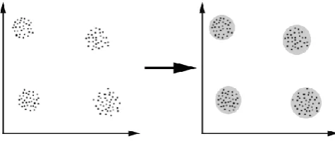
clusters. The grouping of pixels into clusters is based on [image:5.595.39.277.149.249.2]the principle of maximizing the intra class similarity and maximizing the interclass similarity.
Fig 1.4: Example for Clustering
K-Means Clustering Algorithm
A cluster is a collection of objects which are similar between them and are dissimilar to the objects belonging to other clusters. Clustering is an unsupervised learning method which deals with finding a structure in a collection of unlabelled data. A loose description of clustering could be the process of organizing objects into groups whose members are similar in some way. K-means clustering is an algorithm to group objects based on attributes/features into k number of groups where k is a positive integer. Fuzzy clustering algorithms include FCM (Fuzzy C means)algorithm, GK (Gustafson-Kessel), GMD (Gaussian Mixture Decomposition), FCV (Fuzzy C Varieties), AFC, FCS, FCSS,FCQS, FCRS algorithm and etc, among all the FCM is the most accepted method since it can preserve much more information than other approaches
.
Algorithm Implemantation
K-means Algorithm
Step 1: Decide on a value for K, the number of clusters.
Step 2: Initialize the K cluster (randomly, if necessary).
Step 3: Decide the class memberships of the N objects by
assigning them to the nearest cluster center.
Step 4: Re-estimate the K cluster centers, by assuming the
memberships found above are correct.
Step 5: Repeat 3 and 4 until none of the N objects changed
membership in the last iteration.
Modified Algorithm
Step 1:Initialize the closer center using this sub algorithm:
1) Compute the dmin and dmax from data set that we have.
2) For k = 0, 1, . . . , K −1, calculate new ck centers as:
k 1
Ck = dmin + K + 2K (dmax dmin)
Step 2:
Determine the membership matrix
1 if ||xj -Ci|| < ||xj – Ck||2 for each k = i
uij =
0 Otherwise
This means that xj belongs to group I if ci is the closest center among all centers. Since a given data point can belong to only one group. The membership matrix has the following properties:
c
j = ∑ uij=1.
Vj= 1,…….,n. i=1
Step 3:
Compute the cost function according to Eq. 1. Stop. If either, it is below a certain tolerance value or its improvement over previous iteration is below certain threshold.
Step 4:
Process Terminate
EVALUATION RESULT:
Analytical, MRI scan can also been used to assess the maturity of the central nervous system and diagnose malformations. The resonance is also crucial for imaging of vascular changes. Using this method of diagnostic, imaging allows obtaining information about aneurysms and accompanying symptoms. It is also helps in showing seditious changes of the central nervous system and gives accurate assessment of the degree of brain atrophy. The automatic classification of brain's MRI can thus be used to identify regions having various brain diseases like Cerebra-Vascular, Alzheimer, Brain Tumor, Inflammatory, etc
© 2015, IRJET.NET- All Rights Reserved
Page 733
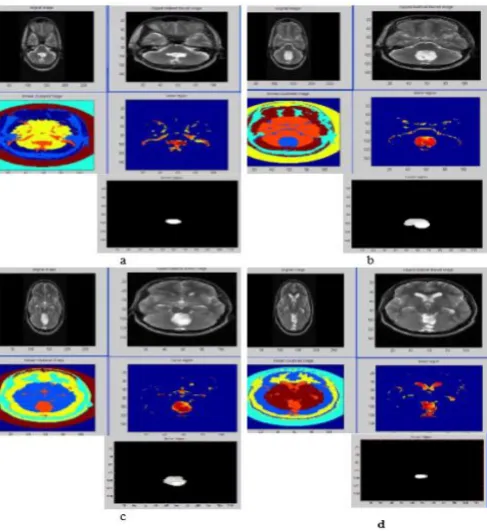
selected, using opening morphological operation with [image:6.595.306.560.97.376.2]structure element of shape disk (five pixels diameter), and the resulted image then convoluted with the original image to acquire the image of the tumor region.
Figure 1.5 Clustering Based on Intensity and Location 1st line, original image & original image after cutting background and smoothing; 2nd line, mat-to-gray of the background cutting and smoothed image & contrast adjusted image; 3rd line, extracted tumor image & contour of tumor region. a ,b , c & d for 5T2, 6T2, 7T2 & 8T2 images respectively.
Table 1.1 Illustrates the Values of the Surface Area of Tumor Regions for the Implemented Techniques.
[image:6.595.39.283.162.428.2]The tumor region area equals 165 pixels, using structure element of radius 4.
Figure 1.6 Modified K-Means Clustering
CONCLUSION
[image:6.595.38.289.555.680.2]© 2015, IRJET.NET- All Rights Reserved
Page 734
FUTURE WORK
Future research could focus on the performance differentials within the same level of technology integration. This would shed light on the reasons why healthcare providers selecting the same level of technology integration perform differently. Second, future research could focus on the role of supply chain readiness in the effective implementation of technologies by healthcare providers. Specifically, research could look into the role of three key participants administration, physicians, and support staff, - the moderating effects of the supply chain technology infrastructure. Third, from this study we found that there are unobservable factors that influence managerial decisions to select advanced information technologies. We can extend this research and empirically investigate the unobservable factors to understand the process of information technology selection by managers. Hence, another interesting topic of future inquiry can the study of the phenomenon of technology integration over time and investigate how performance is affected with the changes in the levels of technology integration over time. This proposed method is used for some other cancers like Breast Cancer etc.
REFERENCES:
1.
Limin Fu, “Neural Networks in Computer Intelligence”, McGraw Hill, 1994.2.
Adekunle M. Adesina, (2010), “Introduction and Overview of Brain Tumors”, [online], Available: http://link.springer.com/chapter/10.1007%2F978 -1-4419-1062-2_0.3.
SuchitaYadav, SachinMeshram, “Brain Tumor Detection using Clustering Method”, IJCER Journal, Vol.3, Issue 4.4.
A.K. Jain and R.C. Dubes, “Algorithms for Clustering Data”, Prentice Hall, Englewood Cliffs, NJ, 1988.5.
Ibrahim A. Almerhag, IdrisS.El-Feghi and Ali A.Dulla, “A Modified K-Means Clustering Algorithm for Gray Image Segmentation”.