Hybrid Capture 2 and Linear Array HPV DNA Tests
Julia C. Gage,aMark Sadorra,bBrandon J. LaMere,cRandi Kail,bCarrie Aldrich,dWalter Kinney,eBarbara Fetterman,fThomas Lorey,f
Mark Schiffman,aand Philip E. Castlegfor the PaP Cohort Study Group
Division of Cancer Epidemiology and Genetics, National Cancer Institute, National Institutes of Health, DHHS, Bethesda, Maryland, USAa; Roche Molecular Systems,
Alameda, California, USAb; Women’s Health Research Institute, Kaiser Permanente Medical Care Program, Oakland, California, USAc; Ventana Medical Systems Inc., Tucson,
Arizona, USAd; Regional Laboratory, Kaiser Permanente Northern California, Berkeley, California, USAe; Division of Gynecologic Oncology, Kaiser Permanente Medical Care
Program, Oakland, California, USAf; and American Society for Clinical Pathology, Washington, DC, USAg
The cobas human papillomavirus (HPV) test (cobas) was recently approved by the U.S. Food and Drug Administration (FDA)
and identifies HPV16 and HPV18 separately as well as detecting a pool of 11 HR-HPV genotypes (HPV31, -33, -35, -39, -45, -51,
-52, -56, -58, -59, -68) and also HPV66. We compared cobas, Linear Array (LA), and Hybrid Capture 2 (HC2) assays for detection
of carcinogenic HPV DNA, and cobas and LA for detection of HPV16 and HPV18 DNA, among the first 1,852 women enrolled in
the HPV Persistence and Progression Cohort (PaP Cohort) study. Specimens were tested by all 3 assays 1 year after an
HC2-positive result. In 1,824 specimens with cobas results, cobas had an 85.9% agreement with HC2 and 91.0% agreement with LA for
carcinogenic HPV detection. When results between cobas and HC2 disagreed, cobas tended to call more women HPV positive
(P
<
0.01). Categorizing cobas and LA results hierarchically according to cancer risk (HPV16, HPV18, other carcinogenic HPV
genotypes, or carcinogen negative), there was a 90% agreement for all categories of HPV (n
ⴝ
1,824). We found good agreement
between the two U.S. FDA-approved HPV tests, with discrepancies between the two assays due to specific characteristics of the
individual assays. Additional studies are needed to compare HC2 and cobas for detecting and predicting CIN3 to understand the
clinical implications of the discrepant test results between the two tests.
I
n the United States, testing for a pool of high-risk genotypes of
the human papillomavirus (HR-HPV) is currently used as an
adjunct to cervical cytology for general screening. There are data
supporting the individual detection of the two most carcinogenic
genotypes, HPV16 and HPV18, which might be clinically useful
for differentiating HPV-positive, cytology-negative women at
higher and lower cancer risk (3, 7). The cobas HPV test (cobas;
Roche Molecular Systems, Pleasanton, CA) was recently approved
by the U.S. Food and Drug Administration (FDA) and identifies
HPV16 and HPV18 separately as well as detecting a pool of 11
HR-HPV genotypes (HPV31, -33, -35, -39, -45, -51, -52, -56, -58,
-59, -68) and also HPV66. The test has been validated in some
initial studies (1, 13), and we sought to further add to the literature
by assessing the interassay agreement between cobas and two
other well-validated HPV DNA assays using samples collected in
specimen transport medium (STM) (Qiagen, Gaithersburg, MD).
Specifically, we compared cobas to (i) Linear Array (LA) (Roche
Molecular Systems), an HPV genotyping assay that, while not
ap-proved by the FDA, is widely used for research (4, 5, 11, 14) and is CE
marked for use in Europe, and (ii) the FDA-approved Hybrid
Cap-ture 2 assay (HC2) (Qiagen Corporation, Gaithersburg, MD), which
targets the 13 HR-HPV genotypes and cross-reacts with HPV66 (as
well as a few other possibly carcinogenic or low-risk types) (2).
MATERIALS AND METHODS
To enrich the study population for HPV positivity, this study was nested within the HPV Persistence and Progression Cohort (PaP Cohort) study (9). Kaiser Permanente Northern California (KPNC) routinely uses HC2 as an adjunct to cytology for cervical cancer screening in women 30 and older (“cotesting”) and as a triage prior to colposcopy for women with equivocal Pap results at all ages. After taking a specimen for making a conventional Pap smear, a cervical specimen for HPV testing is taken using a sampling kit composed of a collection brush and specimen
trans-port medium (STM; Qiagen, Gaithersburg, MD) for specimen storage and transportation after collection. STM specimens are sent to a central laboratory for routine HPV testing (1, 6).
To create the PaP cohort, we enrolled approximately 54,767 women aged 30 and older who underwent cotesting, 44,962 (82.1%) who tested HC2 positive, 9,778 (17.9%) who tested HC2 negative, and 27 (0.05%) with missing HC2 results. Women are followed prospectively as part of standard clinical guidelines for women 30 and older undergoing routine cotesting in the United States (15, 16). Selected women were mailed an opt-out letter to inform them of the study. If they did not want to partic-ipate, they could indicate their refusal by mailing back the opt-out letter using a prepaid envelope or calling a toll-free number. Women could opt out at any time; 6.7% of women who were selected elected not to partic-ipate. Women who did not opt out were considered enrolled into the study. Women were assigned a study ID that was linked to the enrollment and follow-up specimens, the latter of which were identified and flagged via the KPNC patient ID using KPNC’s tracking system. Test results were linked to clinical data (cytology and histology) for clinical management and then stripped of personal identifiers for research purposes. NCI and KPNC institutional review boards have approved the study.
Per KPNC protocol, specimens from follow-up visits were already tested in real time by HC2 according to the manufacturer’s instructions (9). For this study, specimens were neutralized within 14 h to minimize
DNA damage. Specifically, 0.5⫻volume of neutralization buffer was
added to the denatured samples (the exact amount of buffer depended on
Received5 October 2011Returned for modification10 October 2011
Accepted1 November 2011
Published ahead of print9 November 2011
Address correspondence to Julia C. Gage, [email protected].
Copyright © 2012, American Society for Microbiology. All Rights Reserved.
doi:10.1128/JCM.05989-11
on May 16, 2020 by guest
http://jcm.asm.org/
residual volume after HC2 testing), the sample was mixed using a vortex mixer, and the solution was examined for color to determine adequate neutralization (indicator turns yellow). Neutralization buffer was com-posed of 180 mM 2-morpholinoethanesulfonic acid and 100 mM acetic acid, available from HyClone, part number RR11093.01.
For this analysis, we selected a convenience sample of the first 1,852 women to test HC2 positive and return for a 1-year follow-up visit (me-dian, 12.8 months; range, 9.3 to 15.4). Baseline and 1-year follow-up visit specimens were tested by cobas and LA 2 to 3 years after the first (median, 34 months; range, 26 to 45) and follow-up (median, 22 months; range, 17 to 33) visits. To prepare DNA for both LA and cobas, automated sample
extraction was performed on the neutralized STM sample using the⫻480
sample extraction module of the cobas 4800 system. A 250-microliter
sample of cobas lysis buffer was added to 250l neutralized specimen in a
secondary tube (Falcon 5-ml polypropylene round-bottom tube, 12- by-75-mm style, nonpyrogenic, sterile). The sample was capped, vortexed,
uncapped, and placed on the⫻480 specimen rack. The⫻480 extraction
module of the cobas 4800 system then inputs 400l of this material into
the specimen preparation process.
The sample extraction for the cobas HPV test is based on lysis and digestion of cells followed by binding of nucleic acid to magnetic glass beads. The bead-bound DNA is then washed to remove impurities and
eluted from the beads in 120l of buffer at pH 8.7. The⫻480 sample
preparation module is also used to prepare and aliquot the master mix and to perform sample addition for the cobas HPV testing. Twenty-five
mi-croliters of sample is added to 25l of master mix in a 96-well PCR plate.
This plate is then manually transferred to the z480 real-time amplification and detection module of the cobas 4800.
HPV detection for the cobas HPV test is performed via real-time PCR on the z480 module as per the manufacturer’s recommended protocol (10). HPV genotypes 16 and 18 are identified and reported separately. This identification is accomplished by use of spectrally unique dyes to label TaqMan probes for HPV16, HPV18, and the other HR-HPV geno-types.
The HPV linear array test was carried out according to the manufac-turer’s protocol available within the package insert of the kit (11) with the
following exceptions. A 50-l portion of extracted sample from the⫻480
module of the cobas 4800 system was used as a target in the PCR. If extracted samples had to be stored before amplification, they were sealed tightly with foil film (USA Scientific TempPlate sealing foil, part number 2923-0100) and stored at 4°C until use (never frozen). Because the cobas
HPV extraction employs a different elution buffer than the recommended sample extraction for HPV LA, an addition to the master mix must be made to adapt it to this altered buffer. Ten microliters of 1 M Tris-HCl, 0.09% sodium azide, pH 7.4, buffer was added to the activated master mix. The solution was mixed by inverting a minimum of five times before dispensing into reaction tubes. Amplified samples were hybridized to the LA oligonucleotide probe strip and scored as per the manufacturer’s rec-ommendation. In addition, to reduce the chance of user read error, a research software program, HPV StripScan, was utilized to confirm HPV Linear Array genotypes. In the event of a discrepancy between the manual read and the StripScan result, a second, blinded manual read was per-formed of the 48-strip run; the consensus result (2 out of 3) was reported. Restricting our analysis to the first 1,852 1-year follow-up specimens, we compared all three assays at the level of test positivity/negativity for the pool of 14 HPV genotypes (13 HR-HPV and HPV66). Then, cobas and LA results were categorized hierarchically according to cancer risk: HPV16 positive (possibly positive for another HPV genotype), HPV18 positive (HPV16 negative and possibly positive for another HPV genotype), pos-itive for one or more other HR-HPV genotypes (-31, -33, -35, -39, -45, -51, -52, -56, -58, -59, and -68 as well as HPV66 but negative for both HPV16 and -18), and negative for all HR-HPV genotypes. We compared the agreement between cobas and Linear Array for this grouping. The paired results for cobas and LA were also stratified by concurrent HC2 and conventional Pap smear results as crude metrics of HPV viral load. Per-cent agreement, kappa, and weighted kappa values were calculated across the three tests. Statistical significance of differences of positive/negative agreement or multicategory agreement was determined using exact tests of McNemar’s chi-square tests (2 by 2) or exact tests of symmetry (4 by 4), respectively.
[image:2.585.39.548.78.246.2]To assess the genotypes present in nonconcordant cobas and HC2 tests, we further categorized LA HPV genotype results according to phy-logenetic hierarchy associated with cervical cancer risk (12): (i) HPV16 positive (possibly positive for another HPV genotype); (ii) positive for one or more high-risk HPV genotypes (HPV16 negative and positive for HPV18, -31, -33, -35, -39, -45, -51, -52, -56, -58, -59, or -68); (iii) positive for one or more non-high-risk HPV genotypes within the alpha 5, 6, 7, 9, or 11 species (negative for HPV16, -18, -31, -33, -35, -39, -45, -51, -52, -56, -58, -59, and -68 but positive for HPV26, -53, -66, -67, -69, -70, -73, or -82); (iv) positive for one or more non-high-risk HPV genotypes not in the alpha 5, 6, 7, 9, or 11 species (negative for HPV16, -18, -26, -31, -33, -35, -39, -45, -51, -52, -53, -56, -58, -59, -66, -67, -68, -69, -70, -73, and -82
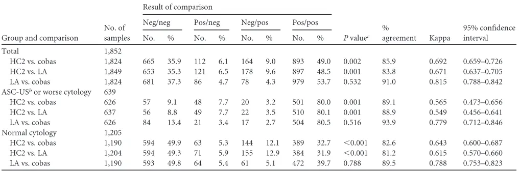
TABLE 1Comparison of HC2, cobas, and LA for detection of high-risk HPV genotypesa
Group and comparison
No. of samples
Result of comparison
Pvaluec
%
agreement Kappa
95% confidence interval
Neg/neg Pos/neg Neg/pos Pos/pos
No. % No. % No. % No. %
Total 1,852
HC2 vs. cobas 1,824 665 35.9 112 6.1 164 9.0 893 49.0 0.002 85.9 0.692 0.659–0.726
HC2 vs. LA 1,849 653 35.3 121 6.5 178 9.6 897 48.5 0.001 83.8 0.671 0.637–0.705
LA vs. cobas 1,824 681 37.3 86 4.7 78 4.3 979 53.7 0.532 91.0 0.815 0.788–0.842
ASC-USbor worse cytology 639
HC2 vs. cobas 626 57 9.1 48 7.7 20 3.2 501 80.0 0.001 89.1 0.565 0.473–0.656
HC2 vs. LA 637 56 8.8 49 7.7 22 3.5 510 80.1 0.001 88.9 0.549 0.456–0.641
LA vs. cobas 626 84 13.4 21 3.4 17 2.7 504 80.5 0.516 93.9 0.779 0.712–0.846
Normal cytology 1,205
HC2 vs. cobas 1,190 594 49.9 63 5.3 144 12.1 389 32.7 ⬍0.001 82.6 0.643 0.600–0.687
HC2 vs. LA 1,204 594 49.3 71 5.9 155 12.9 384 31.9 ⬍0.001 81.2 0.615 0.570–0.660
LA vs. cobas 1,190 593 49.8 64 5.4 61 5.1 472 39.7 0.788 89.5 0.788 0.753–0.823
aSamples were evaluated for the presence of 1 or more among 14 HPV genotypes (HPV16, -18, -31, -33, -35, -39, -45, -51, -52, -56, -58, -59, and -68, as well as HPV66) among
specimens taken at 1-year follow-up visit after a previous HC2-positive test result, stratified by cytologic result. Of 1,852 1-year follow-up specimens, cobas, LA, and cytology results were missing for 28, 3, and 8 samples, respectively.
bAtypical squamous cells of undetermined significance. cExact McNemar’s test.
on May 16, 2020 by guest
http://jcm.asm.org/
but positive for HPV6, -11, -40, -42, -52, -54, -55, -61, -62, -64, -71, -72, -81, -83, -84, or -89); and (v) negative for all HPV genotypes. Results were stratified by concurrent conventional Pap smear results classified by the Bethesda 2001 classification system. Differences in genotype distribution for cobas and HC2 concordant versus discordant results were compared using McNemar chi-square tests.
RESULTS
We compared overall positivity for 1 or more of 14 genotypes by
the 3 tests (Table 1). Positivity for one or more HPV genotypes was
similar for all three tests, as 55.1%, 57.9%, and 58.1% tested
pos-itive for HC2, cobas, and LA, respectively (chi-square
P
⫽
0.09).
However, in examining the data in a pairwise fashion, agreement
between cobas and LA was superior (91.0% agreement) to that
between cobas and HC2 (85.9% agreement). When results
be-tween cobas and HC2 disagreed, cobas tended to call more women
HPV positive (164 versus 112 women;
P
⬍
0.01).
After stratifying HC2, cobas, and LA results by cytologic result
(normal [
n
⫽
1,205] versus nonnormal [
n
⫽
639]) (Table 1), HPV
positivity was higher among women with nonnormal cytology
(87.8%, 83.2%, and 83.6% for HC2, cobas, and LA, respectively)
than women with a normal cytology (37.8%, 44.8%, and 44.8%
for HC2, cobas, and LA, respectively). Agreement was also higher
among women with a nonnormal cytology than among women
with normal cytology.
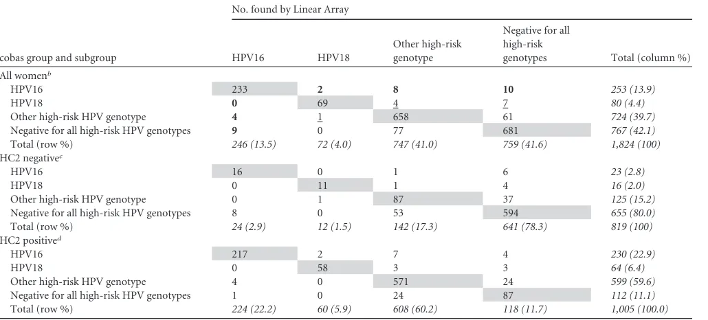
As shown in Table 2, using the hierarchical classification of
HPV genotypes according to cancer risk, the agreement and kappa
values between cobas and LA were also high: 90.0% agreement
with unweighted and weighted kappas of 0.844 and 0.871,
respec-tively. However, the exact test of symmetry showed that cobas
tended to classify women as higher risk (
P
⫽
0.03). For example,
when cobas and LA results disagreed, the cobas test labeled more
women as HPV16 positive (20 versus 13;
P
⫽
0.20; differences
highlighted in bold) and HPV18 positive (11 versus 1,
P
⬍
0.01;
differences underlined) compared to LA. Agreement was lower
among women testing HC2 negative (presumably because the
specimens had lower viral load) than among those testing HC2
positive (86.4% versus 92.8%, respectively, chi-square
P
⬍
0.01).
Our
post hoc
review of discordant HC2 and cobas test results
found that women who tested cobas positive and HC2 negative
were more likely to be called positive for HPV16 and carcinogenic
HPV genotypes detected by LA while cobas-negative and
HC2-positive women were more likely to be called LA HC2-positive for one
or more low-risk genotypes phylogenetically related to high-risk
genotypes (Fisher’s exact
P
⬍
0.01) (Table 3). Similar trends were
observed among women with abnormal cytology, although not
statistically significant (Fisher’s exact
P
⫽
0.09).
DISCUSSION
[image:3.585.41.556.78.311.2]Here we presented the largest study of the agreement for
carcino-genic HPV DNA detection for two of the three U.S.
FDA-approved tests, HC2 and cobas. We found that the agreement
between the two tests was modest, with cobas more likely to test
TABLE 2Hierarchical classification of HPV genotypes detected by cobas and Linear Arrayacobas group and subgroup
No. found by Linear Array
HPV16 HPV18
Other high-risk genotype
Negative for all high-risk
genotypes Total (column %)
All womenb
HPV16 233 2 8 10 253 (13.9)
HPV18 0 69 4 7 80 (4.4)
Other high-risk HPV genotype 4 1 658 61 724 (39.7)
Negative for all high-risk HPV genotypes 9 0 77 681 767 (42.1)
Total (row %) 246 (13.5) 72 (4.0) 747 (41.0) 759 (41.6) 1,824 (100)
HC2 negativec
HPV16 16 0 1 6 23 (2.8)
HPV18 0 11 1 4 16 (2.0)
Other high-risk HPV genotype 0 1 87 37 125 (15.2)
Negative for all high-risk HPV genotypes 8 0 53 594 655 (80.0)
Total (row %) 24 (2.9) 12 (1.5) 142 (17.3) 641 (78.3) 819 (100)
HC2 positived
HPV16 217 2 7 4 230 (22.9)
HPV18 0 58 3 3 64 (6.4)
Other high-risk HPV genotype 4 0 571 24 599 (59.6)
Negative for all high-risk HPV genotypes 1 0 24 87 112 (11.1)
Total (row %) 224 (22.2) 60 (5.9) 608 (60.2) 118 (11.7) 1,005 (100.0)
aClassification of specimens taken at 1-year follow-up visit after a previous HC2-positive test result, stratified by HC2 status at follow-up. LA and cobas results were categorized
hierarchically according to cancer risk: HPV16 positive (possibly positive for another high-risk HPV genotype), HPV18 positive (HPV16 negative, HPV18 positive, and possibly positive for another high-risk HPV genotype), positive for one or more other high-risk HPV genotypes (HPV31, -33, -35, -39, -45, -51, -52, -56, -58, -59, and -68 as well as HPV66 but negative for both HPV16 and -18), and negative for all high-risk HPV genotypes (negative for HPV16, -18, -31, -33, -35, -39, -45, -51, -52, -56, -58, -59, and -68 as well as HPV66). Bold type indicates values that indicate that cobas tended to classify more women as HPV 16 positive than LA when the tests disagreed; underlining indicates values that indicate cobas tended to classify more women as HPV18 positive than LA when the tests disagreed; italics indicate total; shading indicates agreement between cobas and LA. Of 37 specimens that were HPV66 positive by LA and negative for all 13 carcinogenic HPV genotypes, 26 (70.3%) were HC2 positive. Comparatively, 31 (86.1% of 36; one specimen was missing a cobas result) were cobas positive for any of the carcinogenic HPV genotypes.
bAgreement, 90.0%; overall kappa, 0.844 (95% CI, 0.822-0.866); weighted kappa, 0.871 (95% CI, 0.850-0.891); exactPvalue for symmetry, 0.029. cAgreement, 86.4%; overall kappa, 0.609 (95% CI, 0.543-0.675); weighted kappa, 0.629 (95% CI, 0.556-0.702); exactPvalue for symmetry, 0.229. dAgreement, 92.8%; overall kappa, 0.875 (95% CI, 0.848-0.903); weighted kappa, 0.901 (95% CI, 0.876-0.925); exactPvalue for symmetry, 0.101.
on May 16, 2020 by guest
http://jcm.asm.org/
positive. Agreement between the two tests was better among
women with abnormal cytology, probably because of the higher
HPV viral load (8). Discrepancies between the two assays had two
causes: cobas appeared to be more analytically sensitive for
carci-nogenic HPV than HC2, resulting in the overall increased
likeli-hood of testing positive (versus HC2), while HC2 was more likely
to cross-react with certain noncarcinogenic HPV genotypes, as
previously documented for HC2 (2), than cobas.
We found agreement between cobas and LA to be very good,
better than that between cobas and HC2, albeit lower than
previ-ously reported in a smaller study with similar HPV prevalence (1).
The reasons for differences in agreement between the two studies
are uncertain. One possibility is that the previous study used a
different specimen type, PreservCyt liquid-based cytology
me-dium, while this study used STM. Another difference is that the
previous study sampled more women with abnormal cytology,
consistent with the increased agreement between the two assays
among those with abnormal cytology shown here. For those
re-sults that differed, cobas tended to call more women positive for
HPV16 and especially HPV18, a finding similarly reported by
Cas-tle et al. (1). A recent analysis demonstrated that HPV16 and
HPV18 detection by cobas in conjunction with cytology is useful
in the management of HPV-positive women (3).
The cobas assay was recently U.S. FDA approved and the main
trial results published (13), again showing that carcinogenic HPV
DNA detection is more sensitive but less specific for identifying
women with precancerous lesions, specifically cervical
intraepi-thelial neoplasia grade 3 (CIN3). Here we showed that there is
good agreement between the two U.S. FDA-approved HPV tests,
with discrepancies between the two assays due to specific
charac-teristics of the individual assays. Additional studies are needed to
compare the HC2 and cobas for detecting and predicting CIN3 to
understand the clinical implications of the discrepant test results
between the two assays.
ACKNOWLEDGMENTS
Members of the PaP Cohort Study Group included Philip E. Castle (American Society for Clinical Pathology), Mark Schiffman and Sholom Wacholder (National Cancer Institute/Division of Cancer Epidemiology and Genetics) and Barbara Fetterman, Walter Kinney, Brandon J. LaMere, Thomas Lorey, Nancy Poitras, Ruth Shaber, Jen Shieh, and Mark Stanley (Kaiser Permanente Northern California).
REFERENCES
[image:4.585.40.547.78.353.2]1.Castle PE, et al.2009. Evaluation of a prototype real-time PCR assay for carcinogenic human papillomavirus (HPV) detection and simultaneous
TABLE 3Hierarchical classification of HPV genotypes according to phylogenicityadetected by LA given HC2 and cobas resultsc
LA group and subgroupsa
cobas positiveb cobas negative
Total
HC2 positive HC2 negative HC2 positive HC2 negative
No. Column % No. Column % No. Column % No. Column % No. Column %
Totald
1 221 24.7 16 9.8 1 0.9 8 1.2 246 13.4
2 616 69.0 95 57.9 24 21.4 48 7.3 783 42.9
3 32 3.6 10 6.1 42 37.5 46 7.0 130 7.1
4 6 0.7 12 7.3 10 8.9 96 14.7 124 6.8
5 18 2.0 31 18.9 35 31.3 457 69.8 541 29.7
Total (row %) 893 49.0 164 9.0 112 6.1 655 35.9 1,824 100.0
ASC-US or worse cytologyc,e
1 139 27.7 1 5.0 0 0.0 2 3.5 142 22.7
2 338 67.5 11 55.0 14 29.2 5 8.8 368 58.8
3 16 3.2 4 20.0 21 43.8 5 8.8 46 7.3
4 3 0.6 2 10.0 2 4.2 13 22.8 20 3.2
5 5 1.0 2 10.0 11 22.9 32 56.1 50 8.0
Total (row %) 501 80.0 20 3.2 48 7.7 57 9.1 626 100.0
Normal cytologyf
1 82 21.1 15 10.4 1 1.6 6 1.0 104 8.7
2 275 70.7 84 58.3 10 15.9 42 7.1 411 34.5
3 16 4.1 6 4.2 20 31.7 41 6.9 83 7.0
4 3 0.8 10 6.9 8 12.7 83 14.0 104 8.7
5 13 3.3 29 20.1 24 38.1 422 71.0 488 41.0
Total (row %) 389 32.7 144 12.1 63 5.3 594 49.9 1,190 100.0
aLA results were categorized hierarchically according to cancer risk: group 1, HPV16 positive (possibly positive for another HPV genotype); group 2, positive for one or more
high-risk HPV genotypes (HPV16-negative and positive for HPV18, -31, -33, -35, -39, -45, -51, -52, -56, -58, -59, or -68); group 3, positive for one or more non-high-high-risk HPV genotypes within the alpha 5, 6, 7, 9, or 11 species (negative for HPV16, -18, -31, -33, -35, -39, -45, -51, -52, -56, -58, -59, and -68 but positive for HPV26, -53, -66, -67, -69, -70, -73, or -82); group 4, positive for one or more non-high-risk HPV genotypes not in the alpha 5, 6, 7, 9, or 11 species (negative for HPV16, -18, -26, -31, -33, -35, -39, -45, -51, -52, -53, -56, -58, -59, -66, -67, -68, -69, -70, -73, and -82 but positive for HPV6, -11, -40, -42, -52, -54, -55, -61, -62, -64, -71, -72, -81, -83, -84, or -89); and group 5, negative for all HPV genotypes. Results are for specimens taken at 1-year follow-up visit after a previous HC2-positive test result. Italics indicate totals.
bcobas positive for one or more high-risk HPV genotypes (HPV16, -18, -31, -33, -35, -39, -45, -51, -52, -56, -58, -59, -68) as well as HPV66. cConcurrent cytology result was atypical squamous cells of undetermined significance or worse.
dFisher’s exactP⬍0.01 for difference between cobas-positive/HC2-negative and cobas-negative/HC2-positive results.
eFisher’s exactP⫽0.09 for difference between cobas-positive/HC2-negative and cobas-negative/HC2-positive results (for all high-risk genotypes combined). fFisher’s exactP⬍0.01 for difference between cobas-positive/HC2-negative and cobas-negative/HC2-positive results.
on May 16, 2020 by guest
http://jcm.asm.org/
HPV genotype 16 (HPV16) and HPV18 genotyping. J. Clin. Microbiol.
47:3344 –3347.
2.Castle PE, et al.2008. Human papillomavirus genotype specificity of
hybrid capture 2. J. Clin. Microbiol.46:2595–2604.
3.Castle PE, et al.2011. Performance of carcinogenic human papillomavi-rus (HPV) testing and HPV16 or HPV18 genotyping for cervical cancer screening of women aged 25 years and older: a subanalysis of the ATHENA
study. Lancet Oncol.12:880 – 890.
4.Froberg M, Johansson B, Hjerpe A, Andersson S.2008. Human papil-lomavirus ‘reflex’ testing as a screening method in cases of minor
cytolog-ical abnormalities. Br. J. Cancer.99:563–568.
5.Gravitt PE, Schiffman M, Solomon D, Wheeler CM, Castle PE.2008. A comparison of linear array and hybrid capture 2 for detection of carcino-genic human papillomavirus and cervical precancer in ASCUS-LSIL triage
study. Cancer Epidemiol. Biomarkers Prev.17:1248 –1254.
6.Katki HA, et al.2011. Cervical cancer risk for women undergoing concurrent testing for human papillomavirus and cervical cytology: a population-based
study in routine clinical practice. Lancet Oncol.12:663– 672.
7.Khan MJ, et al.2005. The elevated 10-year risk of cervical precancer and cancer in women with human papillomavirus (HPV) type 16 or 18 and the possible utility of type-specific HPV testing in clinical practice. J. Natl.
Cancer Inst.97:1072–1079.
8.Kovacic MB, et al.2006. Relationships of human papillomavirus type,
qualitative viral load, and age with cytologic abnormality. Cancer Res.
66:10112–10119.
9.LaMere BJ, et al.2007. Human papillomavirus genotyping after denatur-ation of specimens for Hybrid Capture 2 testing: feasibility study for the
HPV persistence and progression cohort. J. Virol. Methods146:80 – 85.
10. Roche Molecular Diagnostics.2008. AMPLICOR human papillomavirus test (package insert). Roche Molecular Diagnostics, Pleasanton, CA. 11. Sargent A, et al.2008. Prevalence of type-specific HPV infection by age
and grade of cervical cytology: data from the ARTISTIC trial. Br. J. Cancer
98:1704 –1709.
12. Schiffman M, et al.2005. The carcinogenicity of human papillomavirus
types reflects viral evolution. Virology337:76 – 84.
13.Stoler MH, et al. 2011. High-risk human papillomavirus testing in women with ASC-US cytology: results from the ATHENA HPV study.
Am. J. Clin. Pathol.135:468 – 475.
14. Szarewski A, et al.2008. Comparison of predictors for high-grade cervical intraepithelial neoplasia in women with abnormal smears. Cancer
Epide-miol. Biomarkers Prev.17:3033–3042.
15. Wright, TC, Jr, et al.2003. 2001 Consensus guidelines for the manage-ment of women with cervical intraepithelial neoplasia. Am. J. Obstet.
Gynecol.189:295–304.
16. Wright TC, Jr, et al.2007. 2006 consensus guidelines for the management of women with cervical intraepithelial neoplasia or adenocarcinoma in
situ. J. Low. Genit. Tract Dis.11:223–239.