0095-1137/08/$08.00⫹0 doi:10.1128/JCM.02171-07
Copyright © 2008, American Society for Microbiology. All Rights Reserved.
Method To Detect Only Live Bacteria during PCR Amplification
䌤
Takashi Soejima,
1,2* Ken-ichiro Iida,
1Tian Qin,
1Hiroaki Taniai,
1Masanori Seki,
1and Shin-ichi Yoshida
1Department of Bacteriology, Faculty of Medical Sciences, Kyushu University, Maidashi 3-1-1, Higashi-ku, Fukuoka 812-8582,
Japan,1and Biological Function Research Department, Food Science & Technology Institute,
Morinaga Milk Industry Co., Ltd., 5-1-83, Higashihara, Zama, Kanagawa 228-8583, Japan2
Received 9 November 2007/Returned for modification 23 December 2007/Accepted 19 April 2008
Ethidium monoazide (EMA) is a DNA cross-linking agent and eukaryotic topoisomerase II poison. We previously reported that the treatment of EMA with visible light irradiation (EMAⴙLight) directly cleaved chromosomal DNA of Escherichia coli (T. Soejima, K. Iida, T. Qin, H. Taniai, M. Seki, A. Takade, and S. Yoshida, Microbiol. Immunol. 51:763–775, 2007). Herein, we report that EMA ⴙ Light randomly cleaved chromosomal DNA of heat-treated, but not live,Listeria monocytogenescells within 10 min of treatment. When PCR amplified DNA that was 894 bp in size, PCR final products from 108heat-treatedL. monocytogeneswere completely suppressed by EMA ⴙ Light. When target DNA was short (113 bp), like the hly gene of L. monocytogenes, DNA amplification was not completely suppressed by EMAⴙLight only. Thus, we used DNA gyrase/topoisomerase IV and mammalian topoisomerase poisons (here abbreviated as T-poisons) together with EMAⴙLight. T-poisons could penetrate heat-treated, but not live,L. monocytogenescells within 30 min to cleave chromosomal DNA by poisoning activity. The PCR product of thehly gene from 108 heat-treatedL. monocytogenes cells was inhibited by a combination of EMA ⴙ Light and T-poisons (EMA ⴙ Light ⴙ T-poisons), but those from live bacteria were not suppressed. As a model for clinical application to bacteremia, we tried to discriminate live and antibiotic-treatedL. monocytogenescells present in human blood. EMA ⴙ LightⴙT-poisons completely suppressed the PCR product from 103to 107antibiotic-treatedL. monocytogenes cells but could detect 102live bacteria. Considering the prevention and control of food poisoning, this method was applied to discriminate live and heat-treatedL. monocytogenescells spiked into pasteurized milk. EMAⴙ LightⴙT-poisons inhibited the PCR product from 103to 107heat-treated cells but could detect 101liveL. monocytogenescells. Our method is useful in clinical as well as food hygiene tests.
PCR is widely used as an effective tool to detect bacteria in foods and clinical samples. The disadvantage of PCR is that it cannot discriminate dead from live bacteria. To overcome this disadvantage, reverse transcriptase PCR that targets mRNA has been used. The mRNA derived from high levels of dead bacteria (104 to 107/ml), however, cannot be removed from
samples, and subsequently the reverse transcriptase PCR be-comes positive (31, 35). Measuring the RNA/DNA molar ratio is not sensitive enough to detect low levels of live bacteria in samples containing high levels of dead bacteria.
To discriminate live and dead bacteria by PCR, cross-linking agents such as psoralen, a methylisopsoralen derivative (4⬘ -ami-nomethyl-4,5⬘-dimethylisopsoralen[4⬘-AMDMIP]), and ethidium monoazide (EMA) have been used (4, 5, 20, 23, 25, 27–29). They selectively permeate the cell walls of dead bacteria and irreversibly bind to chromosomal DNA by covalent attachment (20, 23, 27–29). It has been reported that EMA could cross-link to DNA at the rate of 1 agent per 10 to 80 bp in vitro (17). The PCR amplification of DNA from dead bacteria was inhibited by cross-linking action (23, 27–29), and the PCR signal from dead bacteria was reduced to 1/300 to 1/1,000 (23, 27, 29). It has been reported that pasteurized milk contains 105 to 107
cells/ml of injured/dead bacteria (1, 30). When these methods are applied to the pasteurized samples, the PCR products from injured/dead bacteria are amplified. It is very difficult to judge whether the PCR product is derived only from live bacteria in test samples.
DNase was added to cleave the chromosomal DNA of dead bacteria (21), and PCR signal intensity from dead bacteria decreased to 1/10. External DNase, however, could not com-pletely suppress PCR products from dead bacteria, because DNase could not penetrate the cell membranes of dead bac-teria due to its high molecular weight.
Developing rapid PCR methods to substitute for the culture method is a pressing matter in clinical and food hygiene tests. Most clinical samples are derived from patients administered antibiotics. Various foods have been pasteurized to kill bacte-ria while minimizing the denaturation of food components such as proteins. Therefore, the bacteria present in clinical samples and foods may be injured. Hyperthermophilic en-zymes are reported to be maintained in the bacteria of pas-teurized milk (9, 11, 36). The activities of bacterial DNA gyrase and topoisomerase IV (16) are likely to be maintained. Here, we focused on active bacterial DNA gyrase and topoisomerase IV retained in heat-treated bacteria. By utilizing the enzyme activity, we could completely suppress the PCR end products of heat-injured bacteria. There have been no reports of the inhibition of PCR products from heat-treated bacteria using DNA gyrase/topoisomerase IV poison (e.g., fluoroquinolones) and/or mammalian topoisomerase poisons (T-poisons). We
* Corresponding author. Mailing address: Biological Function Re-search Department, Food Science & Technology Institute, Morinaga Milk Industry Co., Ltd., 5-1-83, Higashihara, Zama, Kanagawa 228-8583, Japan. Phone: 81-46-252-3047. Fax: 81-46-252-3055. E-mail: t_soezim @morinagamilk.co.jp.
䌤Published ahead of print on 30 April 2008.
2305
on May 16, 2020 by guest
http://jcm.asm.org/
cytogenesstrain, because this bacterium is important in both clinical and food hygiene applications.
MATERIALS AND METHODS
Reagents.EMA (Sigma, St. Louis, MO) was used for DNA cross-linking and the DNA cleavage of bacteria. CIN, purchased from Fluka Chemie GmbH (Buchs, Germany), was dissolved in physiological saline. Ampicillin (AMP) and gentamicin (GEN) were from Sigma. Camptothecin (CAM), etoposide (ETP), ellipticine (ELP), mitoxantrone (MIT), and amsacrine (m-AMSA) were pur-chased from Sigma and were dissolved in dimethyl sulfoxide (DMSO).
Bacteria and culture.Listeria monocytogenesJCM 2873 was cultured at 30°C in brain heart infusion (BHI) broth (Eiken Kagaku, Tokyo, Japan). To prepare live bacterial suspensions, bacteria in the logarithmic growth phase were sus-pended in physiological saline. The number of live bacteria was counted by plating the bacterial suspension on Luria (L) agar after the appropriate dilution. Preparation of heat-treatedL. monocytogenes.The live bacterial suspension (1 ml) was transferred to a 1.5-ml microtube (Eppendorf, Tokyo, Japan), and the tube was soaked in a boiling water bath for 50 s. Thereafter, it was immediately chilled in an ice-water bath. This treatment simulated high-temperature, short-time pasteurization and avoided denaturing the DNA gyrase/topoisomerase IV of bacteria. The temperature of the contents was measured by a thermal sensor (TX 10; Yokogawa M C Corp., Musashino, Japan).
Preparation of antibiotic-treatedL. monocytogenes.L. monocytogenes(3.0⫻ 106cells/ml) was treated in L broth with AMP and GEN (the final concentrations were 500 and 200g/ml, respectively). The suspension was incubated at 30°C for 3 weeks to bringL. monocytogenescompletely to the injured/dead state and/or to examine whether DNA gyrase/topoisomerase IV is retained during long-term administration. The cell counts for the antibiotic-treatedL. monocytogeneswere done by a standard curve made from live bacterial counts and its optical density at 600 nm (OD600).
EMA treatment and visible light irradiation (EMA ⴙLight). EMA was dissolved in sterile water at the concentration of 1 mg/ml and filtrated through a 0.20-m microfilter (Minisart-plus; Sartorius AG, Gottingen, Germany). After EMA was added at the concentration of 10g/ml to each heat- or antibiotic-treated and live bacterial suspension, it was kept at 4°C for 5 min in the dark. The suspension was then set in an ice-water bath and irradiated for 5 min with visible light (Flood PRF; 100 V, 500 W; Iwasaki Electric Co., Ltd., Tokyo, Japan) set 20 cm from the solution. The concentration of EMA was set at 10g/ml, because ⬎10g/ml of EMA could penetrate liveL. monocytogenesas well.
Use of T-poisons together with EMAⴙLight.After EMA⫹Light treatments, bacteria were washed by centrifugation. T-poisons were used to make DNA more degraded by interfering with the breakage reunion function of DNA gyrase/ topoisomerase IV that is retained in the heat-treated cells. T-poisons were added to 1 ml of bacterial suspensions at volumes and concentrations (in parentheses) as follows: 8l of CIN (0.5 mg/ml), 10l of CAM (1 mg/ml), 10l of ETP (1 mg/ml), 5l of ELP (0.1 mg/ml), 10l of MIT (0.1 mg/ml), and 10l of m-AMSA (1 mg/ml). The bacterial suspensions were then incubated at 30°C for 30 min.
CIN treatment to confirm DNA gyrase and topoisomerase IV activities re-tained in heat-treatedL. monocytogenes.Live and heat-treatedL. monocytogenes
cells were suspended in fresh BHI broth, and CIN was added at a final concen-tration of 20g/ml. They were then incubated at 30°C for 1.5, 3.5, 5, and 72 h. Simultaneously, the heat-treated bacterial suspension not treated with CIN was prepared as a control to examine the influence of DNase retained in heat-treated
L. monocytogeneson chromosomal DNA. It was incubated for 72 h.
Treatment ofL. monocytogenes-added human blood.Heparinized blood from a healthy human was cooled beforehand at 4°C. Live and antibiotic-treatedL. monocytogeneswas inoculated to the heparinized blood at concentrations of 1.8⫻ 100
to 1.8⫻107
cells/ml. After the sample was diluted twofold with physiological saline, 1 ml was slowly overlaid on 1.0 ml of Ficoll-Paque Plus (GE Healthcare Bio-Sciences AB, Uppsala, Sweden) in a sterilized microtube (2 ml of volume). The layers were then subjected to centrifugation at 100⫻gfor 5 min at 4°C, and the blood plasma containing microorganism was collected. EMA⫹Light
treat-beforehand by culture and noL. monocytogeneswas detected by PCR without EMA as described below (detection limit for liveL. monocytogenesin milk, 2.2⫻101 cells/ml). T-poisons initially were added to 1 ml of milk inoculated withL. monocytogenesand then incubated at 30°C for 3 h. One milliliter of 1% Triton X-100–2 mM EDTA solution (pH 8.0) was added and centrifuged at 3,000⫻g
for 5 min at room temperature. After the lipid and supernatant were removed, the washing step (at 15,000⫻gfor 10 min at room temperature) was done with 2 ml of physiological saline, and then 1 ml of physiological saline was added. EMA⫹Light treatment and the washing of bacteria were performed as men-tioned above. As a control, 0.5% DMSO and sterile water were added instead of T-poisons and EMA, respectively.
DNA extraction from bacteria.After 0.5 ml of 5 mM EDTA was added to bacterial pellets in a microtube (2 ml), 20l of achromopeptidase (Wako Pure Chemical Industries, Ltd., Osaka, Japan), dissolved at 5 mg/ml in 10 mM NaCl, was added and incubated at 50°C for 30 min. At this point, 0.5 ml of 10 mM Tris-HCl (pH 8.0), 20l of 1,250-U/ml proteinase K (Sigma, St. Louis, MO), and 400l of 10% (wt/vol) lauryl sulfate sodium salt solution were added one after another. The solution was incubated at 50°C overnight. The chromosomal DNA was purified by the usual phenol-chloroform extraction and ethanol precipita-tion. TE buffer (150l; 10 mM Tris-HCl, 1 mM disodium EDTA) was added to the purified DNA. The concentration of DNA was calculated from the OD260, and purity was evaluated from the ratio of OD260/OD280. Furthermore, the usual RNase treatment was carried out successively for PCR.
Gel electrophoresis of chromosomal DNA.Seakem GTG agarose (FMC Bio-Products, Rockland, ME) was dissolved in 0.8% Tris-acetate-EDTA (TAE) buffer. After 1g of purified DNA was applied to wells, electrophoresis was performed at 100 V. The-EcoT14 I digest and/or a 100-bp DNA ladder (Takara-Bio, Ohtsu, Japan) were used as DNA markers. After the gel was stained with 1g/ml ethidium bromide, the result was visualized with a UV transilluminator at 254 nm of UV light and recorded on Polaroid film (type 667; Nippon Polaroid, Tokyo, Japan).
Real-time PCR.Reactions were performed in the real-time PCR system (iCycler iQ; Bio-Rad, Hercules, CA). The fluorescence threshold was set at a value of 10⫻the standard deviation calculated from the fluorescence values, from 0 to 10 cycles. The first cycle for which the signals of real-time PCR amplification were above the threshold fluorescence value was set as the threshold cycle (CT)
value.
Targeting genes and primers used for PCR.The 23S rRNA gene primers to precisely discriminate live and heat-treatedL. monocytogeneswere 23S-MF (5⬘ -ACCAGGATTTTGGCTTAGAAG-3⬘) and 23S-MR (5⬘-CACTTACCCCGAC AAGGAAT-3⬘) (12). The length of the PCR product was 894 bp.
The listeriolysin O gene (hly) also was targeted to discriminate liveL. mono-cytogenesfrom heat- and antibiotic-treatedL. monocytogenes. Thehlyprimers werehly-F (5⬘-TGCAAGTCCTAAGACGCCA-3⬘) andhly-R (5⬘-CACTGCAT CTCCGTGGTATACTAA-3⬘) (22). The length of the PCR product was 113 bp. Amplification of 23S rRNA gene andhlyby real-time PCR.Fifty microliters of PCR master mix was prepared and contained 150 ng of template DNA, 5l of 10⫻Ex-Taqbuffer (Takara-Bio), 200M each deoxynucleoside triphosphate (Takara-Bio), 0.25M of 23S rRNA gene orhlygene primers (Takara-Bio), 0.4⫻SYBR green (BMA, Rockland, ME), and 1.25 U of Ex-Taqpolymerase (Takara-Bio).
The PCR protocol for the 23S rRNA gene ofL. monocytogeneswas 1 cycle at 4°C for 3 min, 1 cycle at 94°C for 30 s, and 40 cycles at 94°C for 20 s, 46°C for 30 s, and 72°C for 1 min. After PCR, theTm(melting-point measurement) pattern
analysis of PCR product was carried out with 1 cycle at 95°C for 3 min, followed by being cooled at 60°C and heated to 95°C at the rate of 0.75°C per min.
The PCR protocol forhlywas 1 cycle at 4°C for 3 min, 1 cycle at 94°C for 30 s, and 40 cycles at 95°C for 20 s, followed by 60°C for 1 min. After PCR, theTm
pattern analysis of the PCR product was performed with the same procedures as that for the 23S rRNA gene.
In an experimental procedure applied to blood and milk, a direct PCR cocktail (G & g Science, Fukushima, Japan) was added to the bacterial pellet suspended in sterilized water (10l) after treatment with a combination of EMA⫹Light and T-poison (EMA⫹Light⫹T-poison) and successive washing, taking into consideration the simplification of DNA extraction. That is, 5l of bacterial
on May 16, 2020 by guest
http://jcm.asm.org/
suspension treated by EMA⫹Light⫹T-poison was added to 50.5l of PCR cocktail. The thermal cycle profile, which was the same as that of the PCR protocol forhly, was utilized.
Electrophoresis of PCR final products amplified by real-time PCR.A 0.8 or 3% agarose gel was made from the Seakem GTG agarose and TAE buffer for PCR final products from 23S rRNA gene andhly, respectively. The-EcoT14 I digest and 100-bp DNA ladder (Takara-Bio) were used as DNA markers. After 10l of PCR product was applied to the wells, it was separated at 100 V.
RESULTS
Time course of temperature and number of CFU of bacteria when bacterial suspension was inserted in boiling bath.The relationship between the time of insertion into a boiling bath and the temperature of the contents was the following: 0 s, 25.0⫾0.15°C; 27 s, 65.0⫾0.20°C; 34 s, 70.0⫾0.10°C; 47 s, 80.0⫾ 0.15°C; 70 s, 90.0⫾0.25°C; 90 s, 93.8 ⫾0.60°C; and 120 s, 99.0⫾0.45°C (n ⫽3). The relation of the immersion time and the viable cell counts (in log10CFU/milliliter) ofL.
monocytogenes JCM 2873 was the following: 0 s, 8.1⫾ 0.20
counts; 10 s, 7.5⫾0.10 counts; 20 s, 6.1⫾0.10 counts; 30 s, 4.7⫾0.15 counts; 40 s, 2.4⫾0.10 counts; and 50 s, no counts (n⫽3). The detection limit was 5 CFU/ml. Insertion for 50 s offered a condition similar to that of high-temperature, short-time pasteurization; that is, 72 to 75°C for 15 to 16 s.
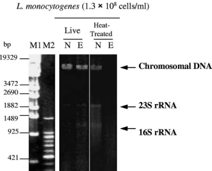
Effects of EMA on the cleavage of chromosomal DNA of heat-treated and liveL. monocytogenes.Live and heat-treated cells ofL. monocytogeneswere treated with EMA⫹Light (4°C for 5 min in the dark; irradiation time, 5 min), and chromo-somal DNA was purified. The gel electrophoresis patterns are shown in Fig. 1. When EMA⫹Light was performed on live bacteria, long fragments near 19,329 bp (derived from chro-mosomal DNA, because the band is fuzzy on the lower site, i.e., the anode side of the gel) were detected. As for the heat-treatedL. monocytogenes, the long fragments did not ap-pear, but smear bands were detectable at a range of less than 1,489 bp when EMA⫹Light treatment was done.
Discrimination by PCR of live and heat-treatedL. monocy-togenesafter EMAⴙLight using the 23S rRNA gene.PCR was performed to target the 23S rRNA gene ofL. monocytogenes
after being treated with EMA⫹Light. The PCR band from
liveL. monocytogeneswas observed after EMA⫹Light
treat-ment, but that from heat-treated cells was not observed by EMA⫹ Light (Fig. 2). Evidently, the discrimination of live from heat-treatedL. monocytogenescould be done.
Discrimination of live and heat-treatedL. monocytogenesby targeting short DNA.When PCR targets pathogenic bacteria, short genes specific for the organism often are amplified. Our method tested whether thehlygene (113 bp) can be used for discrimination between live and heat-treated L.
monocyto-genes. The PCR final product from heat-treated cells was not
suppressed after processing by EMA⫹ Light; thus, discrimi-nation between live and heat-treatedL. monocytogeneswas not successful (Fig. 3, lanes N and E). Therefore, combination methods (EMA⫹Light⫹T-poison) were performed (Fig. 3). When ETP, MIT, and m-AMSA were treated at 30°C for 30 min after EMA ⫹ Light treatment, the PCR final products from heat-treated cells were greatly inhibited, although those from live cells were not.
[image:3.585.55.271.68.242.2]Involvement of DNA gyrase/topoisomerase IV in DNA cleav-age.T-poisons impair DNA activity by accelerating the forward rate (breakage) and inhibiting the reunion of the breakage reunion activity of topoisomerases. CIN is one T-poison. The effect of CIN on the cleavage of chromosomal DNA was ex-amined using live and heat-treatedL. monocytogenes. The re-sults for live and heat-treatedL. monocytogenesare shown in Fig. 4 in CIN (⫹) lanes. When live L. monocytogenes was treated with CIN, the level of long fragments decreased time dependently during 0 to 3.5 h of incubation. The intensity of long fragments increased at 5 h but decreased to near the detection limit at 72 h. Heat-treatedL. monocytogenesnext was incubated at 30°C for 72 h with and without CIN. With CIN, the band intensity of long fragments decreased in a time-dependent manner and was near the detection limit at 72 h. Without CIN, the band intensity of long fragments (near 19,329 bp) did not decrease. These results show that DNA gyrase/topoisomerase IV is active in heat-treatedL.
monocy-togenes, because T-poisons work only when topoisomerase is
active. The results also imply that the influence of DNase
FIG. 1. Gel electrophoresis patterns of chromosomal DNA puri-fied from live and heat-treated L. monocytogenesbefore and after EMA⫹Light treatment. N, no EMA⫹Light treatment; E, EMA⫹ Light treatment (at 4°C in the dark for 5 min; irradiation for 5 min); M1,-EcoT14 I digest; and M2, 100-bp DNA ladder. The experiments for DNA extraction were performed in duplicate, and the electro-phoresis patterns were the same.
FIG. 2. Amplification results of 23S rRNA gene PCR after no treatment or EMA⫹Light treatment using live and heat-treatedL. monocytogenes. The amplification results for the 23S rRNA gene (894 bp) ofL. monocytogenesare represented. N, no EMA⫹Light treat-ment; E, EMA⫹ Light treatment (at 4°C in the dark for 5 min; irradiation for 5 min); M1,-EcoT14 I digest; and M2, 100-bp DNA ladder. The PCR experiments were done in three replicates, and the results were the same.
on May 16, 2020 by guest
http://jcm.asm.org/
retained in heat-treatedL. monocytogeneson DNA cleavage is minimal.
Discrimination of live and heat-treatedL. monocytogenesby CIN using real-time PCR that targeted the 23S rRNA gene.In a comparison of the thickness of bands in gels (Fig. 3), we cannot see the additive effect of CIN on EMA⫹Light treat-ment. To make the slight difference in discrimination power more distinct, the effect of CIN on the degree of PCR sup-pression was evaluated by theCTvalue of real-time PCR. The higher the CT value, the greater the PCR inhibition. ⌬CT
means the degree of PCR suppression, which is represented as
CT(at treatment time) minus CT(at 0 h). For live cells, the ⌬CT(at 0 to 72 h) was 0.0⫾0.00 (at 0 h), 0.0⫾0.00 (at 1.5 h), 1.6⫾0.10 (at 3.5 h), 1.0⫾0.08 (at 5 h), and 2.9⫾0.10 (at 72 h) (means⫾standard deviation;n⫽3). As for heat-treated
L. monocytogenes, the⌬CT(at 0 to 72 h) was 0.0⫾ 0.00 (at
0 h), 0.0⫾0.00 (at 1.5 h), 1.0⫾0.10 (at 3.5 h), 1.4⫾0.10 (at 5 h), and 6.1⫾0.15 (at 72 h).
The⌬CTvalue of 2.9⫾0.01 after 72 h of CIN treatment of live organisms was significantly different from the⌬CTvalue (6.1 ⫾ 0.15) of heat-treated cells (P ⬍ 0.05 by the t test). Hence, the degree of PCR suppression was greater for heat-treated cells than for liveL. monocytogenesby CIN treatment for 72 h.
Detection limit of liveL. monocytogenesin human blood with PCR that targeted thehlygene.Figure 5A shows the results of
detection for live L. monocytogenes inoculated into healthy human blood using PCR that targeted thehlygene. The PCR bands stemming from 1.8⫻ 102 to 1.8 ⫻ 107 cells/ml ofL.
monocytogenes in blood were clear but were not detectable
fromL. monocytogenesat concentrations of 1.8⫻100to 1.8⫻
101cells/ml.
PCR after EMAⴙLightⴙT-poison on live and antibiotic-treatedL. monocytogenesinoculated into healthy human blood.
LiveL. monocytogeneswas treated with 500g/ml of AMP plus
200g/ml of GEN. Live and antibiotic-treatedL.
monocyto-genescells were inoculated into healthy human blood at
con-centrations of 1.8 ⫻ 107, 2.9 ⫻ 104, and 2.9⫻ 103 cells/ml.
Figure 5B to D presents the amplifiedhlygene by PCR after EMA⫹Light⫹T-poisons (CAM, ETP, ELP, and m-AMSA) treatment. In the case of no treatment and EMA⫹Light (1.8⫻ 107cells/ml) (Fig. 5B), the bands of live and antibiotic-treated
L. monocytogeneswere clearly detected. However, the intensity
of the bands of antibiotic-treatedL. monocytogenesdecreased to under or near the detection limit after adding CAM, ETP, or m-AMSA. The effect of ELP treatment was weak.
When 2.9⫻104cells/ml ofL. monocytogeneswere mixed in
blood (Fig. 5C), the PCR bands of live L. monocytogenes
[image:4.585.136.451.68.190.2]clearly appeared without treatment (lane N), after EMA ⫹ Light treatment (lane E), and after EMA⫹Light⫹T-poison treatment. In the case of antibiotic-treated cells, the effects of
[image:4.585.139.451.587.676.2]FIG. 3. Amplification results of PCR that targeted thehly gene after the combination treatment of EMA⫹Light and CIN/mammalian topoisomerase (I and II) poisons on live and heat-treatedL. monocytogenes. The targetedhlygene was 113 bp. T-poison treatments were carried out for 30 min after EMA⫹Light treatment. N, no EMA⫹Light treatment; E, EMA⫹Light treatment (at 4°C in the dark for 5 min; irradiation for 5 min); M, 100-bp DNA ladder; CIN, 4g/ml CIN; CAM, 10g/ml CAM; ETP, 10g/ml ETP; ELP, 0.5g/ml ELP; MIT, 1g/ml MIT; and m-AMSA, 10 g/ml m-AMSA. The concentrations represent final concentrations. The experiments were carried out in triplicate, and the electrophoresis images were the same.
FIG. 4. Effects of CIN on the retaining activities of DNA gyrase and topoisomerase IV in heat-treatedL. monocytogenes. The influence of CIN on the chromosomal DNA of live and heat-treatedL. monocytogenesand the effect of DNase retained in heat-treated bacteria on the chromosomal DNA of heat-treatedL. monocytogenesis shown. N, no treatment; CIN (⫹), CIN treatment. CIN (⫺) represents that heat-treatedL. monocytogenes
cells were incubated at 30°C for 24, 48, or 72 h without CIN. The evaluations were done in duplicate, and the same electrophoresis patterns were obtained.
on May 16, 2020 by guest
http://jcm.asm.org/
T-poisons were clear; that is, PCR bands did not appear after EMA⫹Light⫹T-poison treatments.
Even when theL. monocytogenesdose was lowered to 2.9⫻ 103cells/ml in blood (Fig. 5D), the effects of T-poisons were
almost the same as those shown in Fig. 5C.
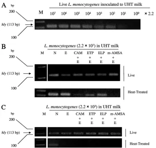
Detection limit of liveL. monocytogenes-spiked commercially available milk with PCR that targeted thehlygene.Figure 6A shows the results of the detection of live L. monocytogenes
inoculated into pasteurized commercial milk using PCR that targeted thehlygene. The PCR bands derived from 2.2⫻101
to 2.2⫻107cells/ml ofL. monocytogenesin milk were
detect-able. No band ofL. monocytogenesfor 2.2⫻100cells/ml was
detected, which means that the detection limit is between 2.2⫻ 101and 2.2⫻100cells/ml.
Amplifications of PCR targetinghlygene after EMAⴙLightⴙ T-poison to live and heat-treatedL. monocytogenes-spiked com-mercial milk.L. monocytogenes was inoculated into milk at concentrations of 2.2⫻107(Fig. 6B) and 2.2⫻103cells/ml
[image:5.585.119.464.63.519.2](Fig. 6C). Figures 6B and C indicate the final products (hly) amplified by PCR, which was performed after EMA⫹Light⫹
FIG. 5. Detection limit ofL. monocytogenesand the discrimination of the live or antibiotic-treatedL. monocytogenescells in human blood by PCR that targeted thehlygene. (A) LiveL. monocytogenes(1.8⫻100to 1.8⫻107cells/ml) inoculated into heparinized healthy human blood was
harvested, and thehly(listeriolysin O) gene (113 bp; short DNA) was targeted by PCR. (B to D) Live or antibiotic-treatedL. monocytogenescells were mixed in human blood, and then treatments with EMA⫹Light⫹T-poisons and PCR methods were carried out. M, 100-bp DNA ladder;
hly, listeriolysin O gene (113 bp; short DNA) ofL. monocytogenes; N, no treatment, as a control; E, EMA⫹Light (at 4°C in the dark for 5 min; irradiation for 5 min); CAM, 25g/ml CAM; ETP, 25g/ml ETP; ELP, 2.5g/ml ELP; and m-AMSA, 25g/ml m-AMSA. The concentrations represent final concentrations. Each examination was performed in duplicate, and the same results were observed.
on May 16, 2020 by guest
http://jcm.asm.org/
T-poison (CAM, ETP, ELP, and m-AMSA) treatment on live and heat-treatedL. monocytogenes. In the case of 2.2⫻ 107
cells/ml (Fig. 6B), bands of live and heat-treatedL.
monocyto-genesapparently were detected both without treatment (lane
N) and after EMA⫹Light (lane E) treatment. As for CAM⫹ EMA⫹Light and m-AMSA⫹EMA⫹Light, the bands of heat-treatedL. monocytogeneswere near or under the detec-tion limit, although the bands of live cells were clearly detected and the intensity was near that of nontreated live cells. In ETP or ELP⫹ EMA⫹ Light treatment, PCR bands of live and heat-treated bacteria appeared; thus, the effectiveness of the discrimination of ETP and ELP was weak when milk was used. For 2.2⫻103cells/ml ofL. monocytogenesin milk (Fig. 6C),
the PCR bands of liveL. monocytogenesobviously appeared in no treatment, EMA⫹Light treatment, and EMA⫹Light⫹ T-poison (four kinds of agents) treatment. However, PCR bands did not appear after EMA⫹Light and EMA⫹Light⫹ T-poisons (four agents).
DISCUSSION
Rapid PCR methods that detect only viable and culturable bacteria are required in food hygiene and clinical tests in place of the culture method. Rudi et al. (27, 29) applied EMA as a
cross-linking agent to discriminate between live and dead pathogens and targeted 85-bp DNA forCampylobacter jejuni
and the 113-bp DNA of thehlygene ofL. monocytogenes. PCR signals from dead, but not live, bacteria were suppressed to 1/1,000 in dead cell counts, which means that theCTvalue of dead bacteria would increase by 10 cycles compared to values from no use of cross-linking EMA. Rudi et al., however, could not suppress real-time PCR final products from dead patho-gens. In the case of food or clinical samples, in which the concentrations of live and dead pathogens are unknown, it is very difficult to judge whetherCTvalues of test samples are derived from live or dead bacteria. No end product of PCR from dead cells is necessary in the factory or bedside, and the FDA requests the complete inhibition of PCR product from dead bacteria.
We have completely suppressed PCR signals from 107to 108
cells of heat- and antibiotic-treated bacteria that were consid-ered background signals in many food and clinical samples. Furthermore, PCR products derived from 102 cells/ml (in
blood) and 101 cells/ml (in pasteurized milk) of live bacteria
[image:6.585.133.453.65.368.2]were detected. It is a very important consequence in clinical diagnostics and food testing that a high level of heat- and antibiotic-treated bacteria was not detected, but a low level of
FIG. 6. Detection limit ofL. monocytogenesand discrimination of live or heat-treated cells spiked into pasteurized milk with PCR that targeted thehlygene. (A) LiveL. monocytogenesspiked into pasteurized (125°C for 2 s) commercial milk (2.2⫻100to 2.2⫻107cells/ml) was recovered
as bacterial pellets and supplied to a direct PCR (hly) cocktail. Thehly(listeriolysin O) gene (113 bp; short DNA) was targeted by PCR. (B and C) Live or heat-treatedL. monocytogeneswas spiked in milk and then treated by EMA⫹Light⫹T-poisons and PCR methods. M, 100-bp DNA ladder;hly, listeriolysin O gene (113 bp; short DNA) ofL. monocytogenes; N, no treatment, as a control; E, EMA⫹Light (at 4°C in the dark for 5 min; irradiation for 5 min); CAM, 25g/ml CAM; ETP, 25g/ml ETP; ELP, 2.5g/ml ELP; and m-AMSA, 25 g/ml m-AMSA. The concentrations represent final concentrations. The experiments were carried out in duplicate, and the results were reproducible. UHT, ultra-high-temperature pasteurized.
on May 16, 2020 by guest
http://jcm.asm.org/
live bacteria was detectable by our PCR method. Although gel electrophoresis was used as the detection step for PCR-ampli-fied genes in the present study, the automated Tm analysis (using a real-time PCR apparatus) for PCR products described in Materials and Methods may be used, considering the rapid-ity, simplicrapid-ity, and sensitivity to the dose of antibiotic used of live bacteria during the detection steps. If EMA⫹ Light or EMA⫹Light⫹T-poison was combined withTmanalysis after real-time PCR, 102and 101cells/ml of live bacteria in blood or
milk, respectively, could be detected within 3 to 6 h.
We recently demonstrated that 1 to 10g/ml of EMA has the direct DNA cleavage function without mediating enzymes in bacteria, and that 50 to 100 ng/ml of EMA has the function of single-stranded breaks (32). For heat-treatedL.
monocyto-genes, PCR that targeted long DNA (894 bp in the 23S rRNA
gene) was suppressed by EMA⫹Light more than that of short DNA (113 bp inhly) (Fig. 2 and 3). This may be because there are more cleavage sites in long DNA. It was reported that the nanograms-per-milliliter level of EMA as a topoisomerase II poison could be cross-linked to DNA at the rate of 1 agent per 10 to 80 bp in vitro (17). In the present study, which used 10
g/ml of EMA, direct DNA cleavage was not induced in thehly
region of heat-treatedL. monocytogenes(Fig. 3). We suppose that Rudi et al. (27) did not notice that PCR inhibition by EMA was not satisfactory when short DNA had been targeted. As seen above, the main cause of the failure to suppress the PCR product is that the PCR target gene was short. Even if PCR was targeted to short DNA such as that ofhly, liveL.
monocytogenes could clearly be discriminated from
heat-treatedL. monocytogenes(108cells/ml) by combining EMA⫹
Light together with T-poisons (Fig. 3).
The mechanism of our method for discriminating live from dead bacteria is shown in Fig. 7. After EMA (dark red bar) penetrates heat- and antibiotic-treated cells and intercalates to the chromosomal DNA (23, 27–29), the cleavage of DNA is greatly induced by the irradiation of visible light (32). The cleavage sites are seen in double-stranded DNA. When the targeted gene is as short as hly, DNA cleavage sites are not likely to be contained in every bacterial cell. The cell mem-branes of heat- and antibiotic-treated L. monocytogenes are physiologically injured. Therefore, when a T-poison such as m-AMSA is added, m-AMSA randomly cross-links to
chromo-FIG. 7. (Left) Scheme for PCR suppression by EMA, psoralen, and a methylisopsoralen derivative (4⬘-AMDMIP) as a DNA cross-linking agent (current scheme). (Right) Scheme for PCR suppression through DNA cleavage by a new function of EMA and T-poisons containing fluoroquin-olone (new scheme). Dark red bar, EMA, psoralen, or 4⬘-AMDMIP; yellow bar, T-poison (m-AMSA, etc.); Topo IV, bacterial topoisomerase IV. The cleavage points are represented in double-stranded DNA.
on May 16, 2020 by guest
http://jcm.asm.org/
[image:7.585.44.540.68.427.2]The effectiveness of EMA ⫹ Light ⫹ T-poison (mainly CAM, ETP, and m-AMSA) was demonstrated in a model of bacteremia (Fig. 5). In adult bacteremia patients, the numbers of microorganisms present in blood are fewer than 10 cells/ml, and 30 ml of blood is used for culture to maximize microbial recovery. A 30-ml volume of blood could be concentrated to approximately 1 ml for PCR testing. Thus, the concentration of live bacteria would be approximately 3⫻102cells/ml (26). In
the case of bacteremia in infants, the number of bacteria ex-isting in blood often is more than 1.0⫻103cells/ml, but only
1 to 4.5 ml of blood should be cultured, taking into consider-ation the weight of the infant (13). Bacteria injured or killed by antibiotics are supposed to exist in blood together with live bacteria. In the present study, therefore, the live and antibiotic-treatedL. monocytogenescells were spiked into healthy human blood at the concentrations of 2.9⫻104(Fig. 5C) and 2.9⫻103
cells/ml (Fig. 5D). On the other hand, live and antibiotic-treatedL. monocytogenescells were inoculated into blood at a concentration of 1.8⫻107cells/ml, considering the presence of
high levels of injured/dead bacteria in urine from urinary tract infection and sputa of tuberculosis patients given anti-tuber-culosis agents (6). EMA⫹Light⫹T-poison may be effective to rapidly discriminate live from injured/dead pathogen.
Pasteurized milk contains 105to 107cells/ml of injured/dead
bacteria, and approximately half of the bacteria are gram pos-itive (1, 30). IfL. monocytogeneswas estimated to be the major contaminant, only liveL. monocytogenesshould be detected by PCR in a high level of background injured/deadL.
monocyto-genes. The effectiveness of EMA⫹Light and EMA⫹Light⫹
T-poison (mainly m-AMSA and CAM) was tested in food hygiene tests of dairy products. Hence, as shown in Fig. 6B, live and heat-treatedL. monocytogenes (2.2 ⫻ 107 cells/ml) cells
were inoculated into pasteurized milk. It is conceivable that the discriminating power of EMA⫹Light⫹T-poisons was infe-rior in milk compared to that in blood (Fig. 5B), except for the case of m-AMSA⫹EMA⫹Light. It has been reported that 2.4 to 7.5% of raw milk is contaminated by liveL.
monocyto-genes(10), and liveL. monocytogenesexists at a concentration
of 2.0 ⫻102 cells/g in raw milk cheese (2). Hence, live and
heat-treated L. monocytogenes(2.2 ⫻ 103 cells/ml) also was
inoculated into pasteurized milk (Fig. 6C). WhenL.
monocy-togenesin milk is of low concentration (live and heat-treated
cells, 2.2 ⫻ 103 cells/ml), EMA ⫹ Light without T-poisons
could discriminate live from heat-treatedL. monocytogenesas well (Fig. 6C).
The verification of active DNA gyrase/topoisomerase IV retained in heat-treated bacteria is speculated as follows. As shown in Fig. 4, when live and heat-treatedL. monocytogenes
cells are treated with CIN for 72 h, the levels of long fragments derived from chromosomal DNA (close to 19,329 bp) obvi-ously decrease. Fluoroquinolones, such as CIN, cause the in-hibition of DNA synthesis and trigger cell killing by interfering with breakage reunion that is mediated by DNA gyrase (33).
peratures and for longer periods, the foods contain mainly dead bacteria in which no activity of DNA gyrase/topoisomer-ase IV is retained. In such cgyrase/topoisomer-ases, at least CAM and m-AMSA, among the T-poisons, would cross-link to chromosomal DNA and might specifically suppress PCR final products from dead bacteria (7). In this case, however, the PCR suppression is due to the cross-linking effect but not poisoning activity (7). EMA could function as a random and direct cleavage agent of chro-mosomal DNA with the irradiation of visible light, even if DNA gyrase and/or topoisomerase IV are completely dena-tured in dead cells (32).
ACKNOWLEDGMENT
We are grateful to Hiroaki Nakayama for expertise and advice regarding the mechanism of DNA gyrase poison and topoisomerase IV poisons.
REFERENCES
1.Adesiyun, A. A.1994. Bacteriological quality and associated public health risk of pre-processed bovine milk in Trinidad. Int. J. Food Microbiol.21: 253–261.
2.Almeida, G., A. Figueiredo, M. Roˆla, R. M. Barros, P. Gibbs, T. Hogg, and P. Teixeira.2007. Microbiolgical characterization of randomly selected Por-tuguese raw milk cheeses with reference to food safety. J. Food Prot.70: 1710–1716.
3.Burden, D. A., P. S. Kingma, S. J. Froelich-Ammon, M.-A. Bjornsti, M. W. Patchan, R. B. Thompson, and N. Osheroff.1996. Topoisomerase II䡠etoposide interactions direct the formation of drug-induced enzyme-DNA cleavage com-plexes. J. Biol. Chem.271:29238–29244.
4.Cimino, G. D., K. C. Metchette, J. W. Tessman, J. E. Hearst, and S. T. Isaacs.1991. Post-PCR sterilization: a method to control carryover contam-ination for the polymerase chain reaction. Nucleic Acids Res.19:99–107. 5.Dall’Acqua, F., D. Vedaldi, S. Caffieri, A. Guiotto, P. Rodighiero, F.
Bac-cichetti, F. Carlassare, and F. Bordin.1981. New monofunctional reagents for DNA as possible agents for the photochemotherapy of psoriasis: deriv-atives of 4,5⬘-dimethylangelicin. J. Med. Chem.24:178–184.
6.Desjardin, L. E., Y. Chen, M. D. Perkins, L. Teixeira, M. D. Cave, and K. D. Eisenach.1998. Comparison of the ABI 7700 system (TaqMan) and com-petitive PCR for quantification of IS6110DNA in sputum during treatment of tuberculosis. J. Clin. Microbiol.36:1964–1968.
7.Froelich-Ammon, S. J., and N. Osheroff.1995. Topoisomerase poisons: har-nessing the dark side of enzyme mechanism. J. Biol. Chem. 270:21429– 21432.
8.Gaunt, P. N., and B. E. Lambert.1988. Single dose ciprofloxacin for the eradication of pharyngeal carriage ofNeisseria meningitidis. J. Antimicrob. Chemother.21:489–496.
9.Guipaud, O., B. Labedan, and P. Forterre.1996. A gyrB-like gene from the hyperthermophilic bacterionThermotoga maritima. Gene174:121–128. 10.Hamdi, T. M., M. Naı¨m, P. Martin, and C. Jacquet.2007. Identification and
molecular characterization ofListeria monocytogenesisolated in raw milk in the region of Algiers (Algeria). Int. J. Food Microbiol.116:190–193. 11.Hethke, C., A. Bergerat, W. Hausner, P. Forterre, and M. Thomm.1999.
Cell-free transcription at 95 degrees: thermostability of transcriptional components and DNA topology requirements ofPyrococcus transcrip-tion. Genetics152:1325–1333.
12.Hong, B.-X., L.-F. Jiang, Y.-S. Hu, D.-Y. Fang, and H.-Y. Guo.2004. Appli-cation of oligonucleotide array technology for the rapid detection of patho-genic bacteria of foodborne infections. J. Microbiol. Methods58:403–411. 13.Kaditis, A. G., A. S. O’Marcaigh, K. H. Rhodes, A. L. Weaver, and N. K.
Henry.1996. Yield of positive blood cultures in pediatric oncology patients by a new method of blood culture collection. Pediatr. Infect. Dis. J.15:615– 620.
14.Kjeldsen, E., B. J. Bonven, T. Andoh, K. Ishii, K. Okada, L. Bolund, and O. Westergaard. 1988. Characterization of a camptothesin-resistant human DNA topoisomerase I⬘. J. Biol. Chem.263:3912–3916.
15.Licitra, C. M., R. G. Brooks, and B. E. Sieger.1987. Clinical efficiency and
on May 16, 2020 by guest
http://jcm.asm.org/
levels of ciprofloxacin in tissue in patients with soft tissue infection. Antimi-crob. Agents Chemother.31:805–807.
16.Luttinger, A.1995. The twisted ‘life’ of DNA in the cell: bacterial topoisom-erases. Mol. Microbiol.15:601–606.
17.Marx, G., H. Zhou, D. E. Graves, and N. Osheroff.1997. Covalent attach-ment of ethidium to DNA results in enhanced topoisomerase II-mediated DNA cleavage. Biochemistry36:15884–15891.
18.Mistry, A. R., C. A. Felix, R. J. Whitmarsh, A. Mason, A. Reiter, B. Cassinat, A. Parry, C. Walz, J. L. Wiemels, M. R. Segal, L. Ades, I. A. Blair, N. Osheroff, A. J. Peniket, M. Lafage-Pochitaloff, N. C. Cross, C. Chomienne, E. Solomon, P. Fenaux, and D. Grimwade.2005. DNA topoisomerase II in therapy-related acute promyelocytic leukemia. N. Engl. J. Med.352:1529– 1538.
19.Nelson, E. M., K. M. Tewey, and L. F. Liu.1984. Mechanism of antitumor drug action: poisoning of mammalian DNA topoisomerase II on DNA by 4⬘-(9-acridinylamino)-methanesulfon-m-anisidide. Proc. Natl. Acad. Sci. USA81:1361–1365.
20.Nocker, A., and A. K. Camper.2006. Selective removal of DNA from dead cells of mixed bacterial communities by use of ethidium monoazide. Appl. Environ. Microbiol.72:1997–2004.
21.Nogva, H. K., A. Bergh, A. Holck, and K. Rudi.2000. Application of the 5⬘-nuclease PCR assay in evaluation and development of methods for quan-titative detection of Campylobacter jejuni. Appl. Environ. Microbiol. 66: 4029–4036.
22.Nogva, H. K., K. Rudi, K. Naterstad, A. Holck, and D. Lillehaug.2000. Application of 5⬘-nuclease PCR for quantitative detection ofListeria mono-cytogenesin pure cultures, water, skim milk, and unpasteurized whole milk. Appl. Environ. Microbiol.66:4266–4271.
23.Nogva, H. K., S. M. Dromtorp, H. Nissen, and K. Rudi.2003. Ethidium monoazide for DNA-based differentiation of viable and dead bacteria by 5⬘-nuclease PCR. BioTechniques34:804–813.
24.Pan, X.-S., J. Ambler, S. Mehtar, and L. M. Fisher.1996. Involvement of topoisomerase IV and DNA gyrase as ciprofloxacin targets inStreptococcus pneumoniae. Antimicrob. Agents Chemother.40:2321–2326.
25.Piette, J. G., and J. E. Hearst.1983. Termination sites of the in vitro nick-translation reaction on DNA that had photoreacted with psoralen. Proc. Natl. Acad. Sci. USA80:5540–5544.
26.Reimer, L. G., M. L. Wilson, and M. P. Weinstein.1997. Update on detection of bacteremia and fungemia. Clin. Microbiol. Rev.10:444–465.
27.Rudi, K., B. Moen, S. M. Dromtorp, and A. L. Holck.2005. Use of ethidium monoazide and PCR in combination for quantification of viable and dead cells in complex samples. Appl. Environ. Microbiol.71:1018–1024. 28.Rudi, K., H. K. Nogva, B. Moen, H. Nissen, S. Bredholt, T. Moretro, K.
Naterstad, and A. Holck.2002. Development and application of new nucleic acid-based technologies for microbial community analysis in foods. Int. J. Food Microbiol.78:171–180.
29.Rudi, K., K. Naterstad, S. M. Dromtorp, and H. Holo.2005. Detection of viable and deadListeria monocytogeneson gouda-like cheeses by real-time PCR. Lett. Appl. Microbiol.40:301–306.
30.Santos, E. C., C. Genigeorgis, and T. B. Farver.1981. Prevalence of Staph-ylococcus aureusin raw and pasteurized milk used for commercial manufac-turing of Brazilian minas cheese. J. Food Prot.44:172–176.
31.Sheridan, G. E. C., C. I. Masters, J. A. Shallcross, and B. M. Mackey.1998. Detection of mRNA by reverse transcription-PCR as an indicator of viability inEscherichia colicells. Appl. Environ. Microbiol.64:1313–1318. 32.Soejima, T., K. Iida, T. Qin, H. Taniai, M. Seki, A. Takade, and S. Yoshida.
2007. Photoactivated ethidium monoazide directly cleaves bacterial DNA and is applied to PCR for discrimination of live and dead bacteria. Micro-biol. Immunol.51:763–775.
33.Tanaka, M., K. Sato, Y. Kimura, I. Hayakawa, Y. Osada, and T. Nishino. 1991. Inhibition by quinolones of DNA gyrase fromStaphylococcus aureus. Antimicrob. Agents Chemother.35:1489–1491.
34.Tewey, K. M., G. L. Chen, E. M. Nelson, and L. F. Liu.1984. Intercalative antitumor drugs interfere with the breakage-reunion reaction of mammalian DNA topoisomerase II. J. Biol. Chem.259:9182–9187.
35.Vaitilingom, M., F. Gendre, and P. Brignon.1998. Direct detection of viable bacteria, molds and yeasts by reverse transcriptase PCR in contaminated milk samples after heat treatment. Appl. Environ. Microbiol.64:1157–1160. 36.Viard, T., R. Cossard, M. Duguet, and C. B. de La Tour.2004.Thermotoga maritima-Escherichia colichimeric topoisomerases. J. Biol. Chem.279:30073– 30080.
37.Wolfson, J. S., and D. C. Hooper. 1989. Fluoroquinolone antimicrobial agents. Clin. Microbiol. Rev.2:378–424.