J
OURNAL OFC
LINICALM
ICROBIOLOGY, Apr. 2006, p. 1405–1412
Vol. 44, No. 4
0095-1137/06/$08.00
⫹
0
doi:10.1128/JCM.44.4.1405–1412.2006
Use of TaqMan Real-Time Reverse Transcription-PCR for Rapid
Detection, Quantification, and Typing of Norovirus
A. Angelica Trujillo,
1,2* Karen A. McCaustland,
1Du-Ping Zheng,
1Leslie A. Hadley,
1George Vaughn,
3Susan M. Adams,
1,2Tamie Ando,
1Roger I. Glass,
1and Stephan S. Monroe
1National Center for Infectious Diseases, Centers for Disease Control and Prevention, Atlanta, Georgia
1; Atlanta Research and
Education Foundation, Decatur, Georgia
2; and National Center for Environmental Health,
Centers for Disease Control and Prevention, Atlanta, Georgia
3Received 28 October 2005/Returned for modification 27 December 2005/Accepted 15 February 2006
Noroviruses (NoVs) are the most commonly identified cause of outbreaks and sporadic cases of acute
gastroenteritis. We evaluated and optimized NoV-specific TaqMan real-time reverse transcription (RT)-PCR
assays for the rapid detection and typing of NoV strains belonging to genogroups GI and GII and adapted them
to the LightCycler platform. We expanded the detection ability of the assays by developing an assay that detects
the GIV NoV strain. The assays were validated with 92 clinical samples and 33 water samples from confirmed
NoV outbreaks and suspected NoV contamination cases. The assays detected NoV RNA in all of the clinical
specimens previously confirmed positive by conventional RT-PCR and sequencing. Additionally, the TaqMan
assays successfully detected NoV RNA in water samples containing low viral concentrations and inhibitors of
RT and/or PCR, whereas the conventional method with region B primers required dilution of the inhibitors.
By means of serially diluted NoV T7 RNA transcripts, a potential detection limit of <10 transcript copies per
reaction mixture was observed with the GII assay and a potential detection limit of <100 transcript copies per
reaction mixture was observed with the GI assay. These results and the ability to detect virus in water that was
negative by RT-PCR demonstrate the higher sensitivity of the TaqMan assay compared with that of a
conventional RT-PCR assay. The TaqMan methods dramatically decrease the turnaround time by eliminating
post-PCR processing. These assays have proven useful in assisting scientists in public health and diagnostic
laboratories report findings quickly to outbreak management teams.
Noroviruses (NoVs) are a group of noncultivable,
genet-ically diverse single-stranded RNA viruses belonging to the
family
Caliciviridae
. These viruses are responsible for the
majority of outbreaks of acute gastroenteritis in
industrial-ized countries (11, 12, 15, 20, 23, 28). In the United States
alone, an estimated 23 million cases of NoV illnesses are
reported each year, and the economic consequences of these
cases are likely to be substantial (21, 23, 24). Outbreaks of
NoV have been caused by contaminated food and/or
drink-ing water, person-to-person virus transmission, and airborne
droplets of infected vomitus (14, 21, 22, 23). Contaminated
water poses an especially serious health risk since results
from human volunteer studies indicate that the minimum
infectious dose of NoV may be as low as 10 to 100 PCR
units (C. Moe, Emory University, personal
communica-tion). Waterborne outbreaks have been caused by
contam-inated surface water, ground water, drinking water, and
mineral water (1, 6, 18, 19, 35). NoV outbreaks are difficult
to control and present a major public health challenge; thus,
rapid diagnosis can be critical for the control of outbreaks.
NoVs are separated into five genogroups on the basis of
sequence comparison of the RNA polymerase and capsid
re-gion of the genome. Genogroups I, II, and IV are associated
with infections in humans. To date, 29 genetic clusters (8 from
GI; 17 from GII; 2 from GIII; and 1 each from GIV and GV)
have been identified, demonstrating a high degree of genomic
diversity among NoVs (2, 13, 41, 42). NoV traditionally has
been detected by conventional reverse transcription
(RT)-PCR, a method that requires confirmation by probe or
se-quence analysis (3, 9). Human NoVs cannot be cultivated, and
immunoassays developed to date have not been adequately
sensitive for detecting sporadic cases. Additionally, because
NoVs are so diverse, designing one set of primers to detect all
strains with equal efficiency is difficult. More-sensitive
tech-niques are required to detect NoV in food and water samples,
in which viral loads are typically much lower than those found
in clinical samples. To date, several methods have been
devel-oped to increase the detection rate of NoV, including PCR and
probe hybridization with multiple genotype-specific
oligonu-cleotides and SYBR green analysis (3, 4, 9, 25, 26, 29, 31, 40).
However, these assays have limitations. Conventional RT-PCR
is time-consuming, and SYBR green analysis uses an
interca-lating dye that binds to all double-stranded DNA, including
primers-dimers and nonspecific products. Additionally,
detec-tion of NoV in water requires methods for concentrating the
virus from a large volume of water in order to increase
sensi-tivity, a process that can coconcentrate RT-PCR inhibitors
and/or erroneous targets (32). Unlike SYBR green-based
quantitative methods, probe-based quantitative RT-PCR uses
a fluorescently labeled, target-specific probe that results in
increased specificity and sensitivity. In addition to their utility
with clinical samples, probe-based quantitative methods can be
especially useful in the detection of virus in samples with low
viral concentrations.
To improve the detection of NoV, we developed, expanded,
* Corresponding author. Mailing address: 1600 Clifton Road N.E.,
Mail Stop G-04, Atlanta, GA 30333. Phone: (404) 639-4256. Fax: (404)
639-3645. E-mail: [email protected].
1405
on May 16, 2020 by guest
http://jcm.asm.org/
and evaluated real-time RT-PCR methods based on TaqMan
probe hydrolysis technology, which was previously described
(17). These TaqMan assays provide sensitive, specific, and
quantitative results in NoV diagnostic assays and have been
successfully applied to clinical and environmental samples
con-taining NoVs in low copy numbers. These assays rapidly detect
and identify NoVs belonging to genogroups I, II, and IV and
do not require any post-PCR processing. This straightforward,
sensitive, and specific method will assist scientists in public
health and diagnostic laboratories reduce the time needed for
routine diagnosis of NoV infection and will aid in the
manage-ment of outbreaks.
MATERIALS AND METHODS
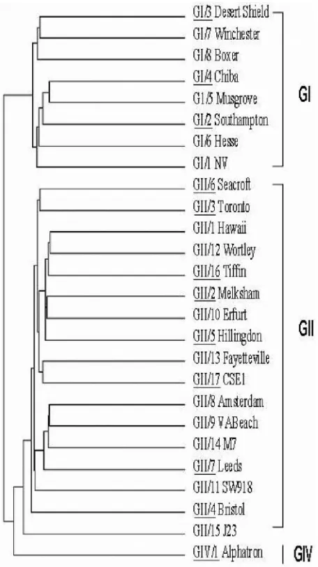
Clinical specimens tested.The TaqMan assay was optimized for the Light-Cycler platform and validated by using an archived panel of 65 NoV-positive stool samples collected as part of the investigation of sporadic cases and outbreaks of acute gastroenteritis that occurred in the United States between June 1999 and June 2004. Because NoVs are genetically diverse, specimens were chosen to include a good representation of NoV clusters that have been reported thus far (2, 43; Fig. 1). In addition, we tested 20 clinical samples from NoV outbreaks that were confirmed negative when tested with primers for region A (the RdRp gene
located in open reading frame 1 [ORF1]), region B (the 3⬘end of ORF1), region
C (a short stretch close to the 5⬘end of ORF2), and RNA polymerase region 5
(primer pair 289 and 290) (2, 16). Finally, seven stool samples from
NoV-negative but rotavirus (n⫽4)-, astrovirus (n⫽2)-, and sapovirus (n⫽
1)-positive outbreaks were also tested.
Specimens were determined to be positive for NoV by conventional duplex RT-PCR with individual primer sets GI, GII, and GIV, and they were confirmed positive and classified into genogroups on the basis of analysis of the nucleotide sequences of the amplified products. Samples were drawn from two to four outbreaks each from three clusters of GI strains, eight clusters of GII strains, and one GIV strain.
Stool sample preparation and extraction of total nucleic acids.Fecal samples were prepared as previously described, with some modifications (38). In brief, 0.1 g of formed stool or 0.1 ml of watery stool was suspended in 1 ml of diethyl pyrocarbonate-treated water (Ambion, Austin, TX), yielding a 10% suspension. To clarify the stool suspension, 1 ml of 2,3-dihydrodecafluoropentane (Vertrel, New Britain, CT) was added and the mixture was vortexed for 1 min and clarified
by centrifugation at 2,060⫻gfor 10 min at 4°C. The suspension (0.2 ml) was
mixed with NucliSens lysis buffer (0.9 ml; BioMe´rieux, Durham, NC), and total
nucleic acid was extracted by the method of Boom et al. (8), with the NucliSens
extractor (BioMe´rieux, Durham, NC), as directed for small sample volumes.
RNA samples were stored at⫺70°C until ready for use.
NoV detection and genetic characterization by region B conventional duplex RT-PCR.Partial NoV sequences for region B were amplified by conventional RT-PCR with primers Mon 431 and Mon 433 for genogroup I and primers Mon 432 and Mon 434 for genogroup II (Table 1). These primers amplify a
small region within the 3⬘ end of the ORF1 portion of the genome. The
product length is 213 bp, with a unique sequence length of 172 bp. In cases where region B primers produced a negative or indeterminate result, primers for region C that are specific only for amplifying GII strains were used (7).
The region B RT-PCR mixture consisted of 1l of RNA and a final volume
of 49l of a reaction mixture containing 19.3l of diethyl
pyrocarbonate-treated water; 2.5l of 20 mM dithiothreitol (Invitrogen); 0.1l of Triton
X-100; 0.07l of 1.44 M-mercaptoethanol; 0.3M each primers Mon 431,
Mon 432, Mon 433, and Mon 434; 25l of Master-Amp G 2X PCR Pre-Mix
(Epicenter); 0.5l of 40 U/l Protector RNase inhibitor (Roche Inc.,
Indi-anapolis, IN); 0.09l of 20 U/l Super Reverse Transcriptase (Molecular
Genetic Resources, Tampa, FL); and 0.25l of 5 U/l AmpliTaq DNA
polymerase (Applied Biosystems, Foster City, CA). The capsid region
RT-PCR mixture consisted of 1l of RNA and a final volume of 49l of a
reaction mixture containing 0.6M each capsid primer (Mon 381, Mon 383),
33.8l of H2O from the QIAGEN One Step RT-PCR kit (QIAGEN Inc.,
Valencia, CA), 10l of QIAGEN One Step RT-PCR 5⫻buffer, 2l of a
deoxynucleoside triphosphate mixture (10 mM each), and 2l of One Step
enzyme mix (QIAGEN Inc., Valencia, CA). The thermocycling program for the one-step conventional region B RT-PCR consisted of RT for 10 min at 42°C; denaturation for 3 min at 94°C; 40 PCR cycles consisting of 94°C for
30 s, 50°C for 90 s, and 60°C for 30 s; and then 72°C for 7 min. The thermocycling conditions for the capsid L region RT-PCR consisted of
lin-earizing the RNA by heating 1l of RNA with 0.6M each capsid primer at
94°C for 3 min, followed by addition of the cocktail. The RNA was then reverse transcribed for 60 min at 42°C and then subjected to activation for 15 min at 95°C; 40 PCR cycles consisting of 94°C for 60 s, 50°C for 90 s, and 60°C for 120 s; and 72°C for 7 min.
PCR products were purified with the QIAquick PCR purification kit (QIAGEN Inc., Valencia, CA). Fluorescent dideoxy-chain terminators (Applied Biosystems, Foster City, CA) were used to sequence both strands with an auto-mated sequencer (model 3100; Applied Biosystems). All strains tested were previously found to be NoV positive, and this was confirmed through sequencing.
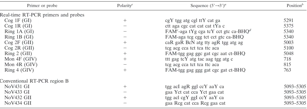
[image:2.585.308.536.69.475.2]Primers and probes for real-time RT-PCR.For detecting NoV GI and GII, we used primers and TaqMan probes previously described (17; Table 1). These primers and probes target NoV sequences at the ORF1-ORF2 junction, a highly conserved region of the NoV genome. The primers and probes for GI were chosen from the corresponding Norwalk/68 virus (GenBank accession no. M87661), and those for GII were chosen from the corresponding Camberwell virus (GenBank accession no. AF145896). Primers and TaqMan probes for GIV NoV detection were developed at the Centers for Disease Control and Prevention (CDC) and used the corresponding
FIG. 1. Phylogenetic dendrogram of strains belonging to clusters of
human NoVs. Underlined clusters denote GI, GII, and GIV strains
tested by real-time RT-PCR (43).
on May 16, 2020 by guest
http://jcm.asm.org/
Saint Cloud virus (GenBank accession no. AF414427) (Table 1). These primers target the ORF1-ORF2 junction of the genome.
Preparation of in vitro RNA transcripts.The 3-kb RT-PCR products obtained in a prior study were used as the source of DNA for preparation of in vitro RNA transcripts (4). The amplification products were cloned into either the ClaI/NotI
or the ClaI/XbaI sites of the pBluescript II SK (⫹) plasmid vector (Stratagene,
La Jolla, CA) after reamplification with the original primers modified to contain the restriction enzyme sites. After the cloned plasmid DNA was purified by use of the QIAfilter plasmid Mega kit (QIAGEN Inc., Valencia, CA), the DNA insert was cut out with the restriction enzymes, gel purified, and used as the template for in vitro transcription with T7 RNA polymerase with the MEGA script kit (Ambion Inc., Austin, TX). After digestion of the template DNA with RNase-free DNase I (Ambion Inc.) at 37°C for 30 min and phenol-chloroform extraction, followed by ethanol precipitation, the RNA transcripts were dissolved in RNase-free water and the concentrations were calculated after measuring the absorbance at 260 nm. The RNA transcript solutions were diluted with RNA
Storage Solution (Ambion Inc., Austin, TX), aliquoted, and kept at⫺70°C for
long-term storage. The genetic groups (2) (GenBank accession numbers) of the 3-kb products that served as the templates for the RNA transcripts were GI/3 (Honolulu 219/1992/US, S4 [accession no. AF414403]) and GII/4 (Burwash Landing 331/1995/ US, S24 [accession no. AF414425]). The 3-kb products were reamplified with three
primers, SR48ad (forward primer for GI; 5⬘-CCA TCG ATA CTA GTG AAC AGC
ATA AA-3⬘), SR46ad (forward primer for GII; 5⬘-C CAT CGA TAC TAG TCC
ATC GCC CAC TGG-3⬘), and VNad (reverse primer for both GI and GII; 5⬘-A
AGG ATC CGC GGC CGC TCT AGA TTT TTT-3⬘).
TaqMan real-time RT-PCR.We performed quantitative real-time RT-PCR assays with the LightCycler RNA amplification kit hybridization probes (Roche
Diagnostics, Alameda, CA). The final reaction mixture (20l) consisted of 1l
of RNA, 4l of the LightCycler RT-PCR mixture, 0.4l of the LightCycler
RT-PCR enzyme mixture, and 5 mM MgCl2. In the final optimized format, the
concentrations of the primers and probes were as follows: for the GI assay, 0.2
M each GI probe with 0.4M each primer; for the GII assay, 0.4M probe and
0.4M each primer; and for the GIV assay, 0.4M probe and 0.3M each
primer. The thermal cycling conditions consisted of RT for 30 min at 55°C, followed by denaturation at 95°C for 30 s, amplification for 45 cycles, followed by denaturation at 95°C for 0 min, and annealing-extension at 60°C for 60 s.
Fluorescence measurements were taken, and the crossing point (cycle number [CN]) for each sample was calculated by the fit points method. The noise band was set to a minimum value of 3 standard deviations above the background fluorescence (34). The algorithm to minimize error for the LightCycler platform was used to set the crossing point for analysis. The threshold for a positive value was set at greater than three times the background fluorescence. A test result was considered positive
if the genomic target showed positive results (CN) at less than 40 cycles and all positive and negative control reactions gave expected values.
NoV RNA concentration from water samples.In the fall of 2003, the CDC Vessel Sanitation Program collected 12 water samples from cruise ship A, which had repeated outbreaks of NoV gastroenteritis suspected to be linked to its water supply, although the exact source had not been determined. Potable-water samples (10 to 20 liters) were collected from various sites aboard the ship. Prefilters (AP 2504700; Millipore, Bedford, MA) were used to prevent clogging by large-particulate matter.
The samples were filtered through a positively charged membrane (0.45-m pore
size; Zetapore; AMF-Cuno, Meriden, CT). After samples were collected, the pre-filter and pre-filters were packaged by a sterile technique, placed at 4°C, and transported to the CDC, where they were processed within 24 h. The prefilter was discarded, and the membrane was transferred to a 200-ml beaker. Sterile 0.5 M lysine (15 ml), pH 8.5, was added, and the filter was shaken for 20 min at room temperature. Fifteen milliliters of sterile 0.5 M arginine, pH 8.5, was added to the beaker and vigorously shaken for an additional 20 min. A sufficient volume of 1 M HCl was added to the supernatant to decrease the pH to 7.3. The solution was vigorously shaken for an additional 20 min at room temperature. The virus was concentrated by adding 15 ml of sterile 30% polyethylene glycol and 0.9 M NaCl and shaken vigorously for an additional 20 min at room temperature. The mixture was transferred into a 50-ml
conical tube and centrifuged at 10,000⫻gfor 30 min at 4°C. The supernatant was
discarded, and the pellet was resuspended in 1 ml of H2O and mixed by inverting the
tube. The mixture was washed by adding 0.5 ml of a 30% polyethylene glycol–0.9 M
NaCl solution and then centrifuged at 10,000⫻gfor 30 min at 4°C. This process was
repeated twice. The retained material was adjusted to 250l with sterile H2O. Total
nucleic acid was extracted with the automated Nuclisens Extractor (BioMe´rieux,
Durham, NC) as directed for small sample volumes. RNA samples were stored at
⫺70°C until ready for use. Five microliters of RNA, representing 10% of the water
concentrate, was used for NoV detection.
In a separate case of outbreaks of NoV among river rafters, 21 water samples were collected by filtration as described above. Since the river was suspected as the source of NoV contamination, water samples from different locations on the river were collected. Due to the remote location and the lack of refrigeration, the samples were stored at room temperature for 1 week. Upon arrival at the CDC, the samples were refrigerated and processed within 12 h. RNA samples were
stored at⫺70°C until ready for use.
RESULTS
We selected 12 NoV strains that each represented a different
genetic cluster to evaluate and validate the three
genogroup-TABLE 1. Primer and probe oligonucleotides used for real-time quantitative RT-PCR for genogroups I, II, and IV and primers for region B
Primer or probe Polaritye Sequence (5⬘33⬘)a Positionb
Real-time RT-PCR primers and probes
Cog 1F (GI)
⫹
cgY tgg atg cgl ttY cat ga
5291
Cog 1R (GI)
⫺
ctt aga cgc cat cat cat tYa c
5375
Ring 1A (GI)
⫺
FAM
c-aga tYg cga tcY cct gtc ca-BHQ
d5340
Ring 1B (GI)
⫺
FAM-aga tcg cgg tct cct gtc ca-BHQ
5340
Cog 2F (GII)
⫹
caR gaR BcN atg tty agR tgg atg ag
5003
Cog 2R (GII)
⫺
tcg acg cca tct tca ttc aca
5100
Ring 2 (GII)
⫹
FAM-tgg gag ggc gat cgc aat ct-BHQ
5048
Mon 4F (GIV)
⫹
ttt gag tcY atg tac aag tgg atg c
718
Mon 4R (GIV)
⫺
tcg acg cca tct tca ttc aca
815
Ring 4 (GIV)
⫹
FAM-tgg gag ggg gat cgc gat ct-BHQ
763
Conventional RT-PCR region B
NoV431 GI
⫹
tgg acI agR ggl ccY aaY ca
5093–5305
NoV433 GI
⫺
gaa Yct cat cca Yct gaa cat
5093–5305
NoV432 GII
⫹
tgg acl cgY ggI ccY aaY ca
5093–5305
NoV434 GII
⫺
gaa Rcg cat cca Rcg gaa cat
5093–5305
a
R⫽A or G, Y⫽C or T, N⫽any.
b
Nucleotide positions for conventional RT-PCR were taken from reference NoV strains in genogroups GI (Norwalk virus 68 [Genbank accession no. M87661]), GII (Hawaii virus [GenBank accession no. U07611]), and GIV (Saint Cloud virus [GenBank accession no. AF414426]). Nucleotide positions for real-time RT-PCR were taken from reference NoV strains in genogroups GI (Norwalk virus 68 [Genbank accession no. M87661]) and GII (Camberwell [Genbank accession no. AF145896]).
c
FAM, 6-carboxyfluorescein reporter dye.
d
BHQ, black hole quencher dye.
e⫹
, virus sense;⫺, antivirus sense.
V
OL. 44, 2006
QUANTITATIVE RT-PCR FOR NOROVIRUS GI, GII, AND GIV
1407
on May 16, 2020 by guest
http://jcm.asm.org/
[image:3.585.44.542.79.257.2]specific real-time TaqMan RT-PCR assays. These strains
be-longed to GI (GI/3, GI/4, and GI/2), eight bebe-longed to GII (GII/2,
GII/3, GII/4, GII/5, GII/6, GII/7, GII/16, and GII/17), and we
developed primers and probes to test the single GIV strain (Fig.
1; Table 1). A total of 92 clinical specimens were tested, 7 of
which were negative controls for NoV and 65 of which were
positive for NoV (Fig. 2B). Additionally, we tested 20 samples
confirmed to be NoV negative in regions A, B, C, and 5. These
samples were also negative for rotavirus, astrovirus, and
sapovi-rus. All of the negative samples were negative by the TaqMan
assays (data not shown). The NoV-positive clinical samples
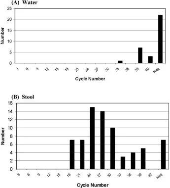
rep-resented 12 clusters from GI, GII, and GIV (Fig. 1 and 2B). All
65 stool samples (100%) that tested positive for NoV by means of
conventional duplex RT-PCR also tested positive by the TaqMan
real-time RT-PCR assay (Fig. 2B). The specificity of the assays
was tested against seven NoV-negative specimens that were
pos-itive for rotavirus (
n
⫽
4), astrovirus (
n
⫽
2), and sapovirus (
n
⫽
1); no cross-reactivity was observed (Fig. 2B).
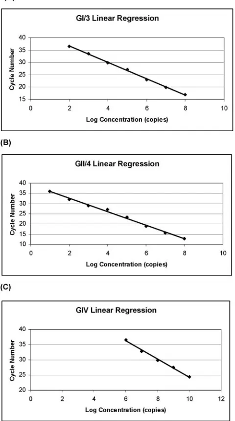
T7 RNA transcripts representing genogroups I and II were
selected as standards to determine the copy detection limits and
dynamic range of the optimized TaqMan real-time RT-PCR
as-say (Fig. 3A and B). No GIV transcript was available at the time
of this study; therefore, a stool standard was used for genogroup
GIV (Fig. 3C). Standards were diluted in a 10-fold series ranging
from undiluted to 10
0, and the intensity of fluorescence was
plot-ted against the CN. RNA standards demonstraplot-ted excellent
neg-ative linearity with a wide dynamic range (Fig. 3A and B). The
GI/3 transcript standard had a detection limit of
⬍
100 copies per
reaction mixture (Fig. 3A). The slope was
⫺
3.370, the
y
intercept
was 43.97, and the
R
2value was 1.00. A detection limit of
⬍
10
copies per reaction mixture was observed in the GII T7 RNA
transcript standards. The slope was
⫺
3.324, the
y
intercept was
39.20, and the
R
2value was 1.00 (Fig. 3B). We examined the
specificity of the GIV primers and probe with six archived
sam-ples collected more than 5 years previously that had tested
posi-tive for genogroup IV by conventional RT-PCR. Five of the six
specimens previously found positive for GIV were found to be
positive by use of the TaqMan assay. The sensitivity of the GIV
assay was tested with a serial dilution from undiluted to 10
⫺9to
determine the detection limit. The dynamic range of the GIV
stool standard was narrower, with positive detection from
undi-luted to 10
5. The slope and
y
intercept were
⫺
3.310 and 58.96,
respectively, and the
R
2value was 1.00 (Fig. 3C).
[image:4.585.120.465.52.432.2]These positive results and the high sensitivity of this method
FIG. 2. CNs detected from archived specimens of water and stool. A comparison of water (A) (
n
⫽
33) and stool (B) (
n
⫽
72) samples is shown.
Specimens with CNs of
⬎
40 or not detected were considered negative (Neg).
1408
on May 16, 2020 by guest
encouraged us to use it to detect NoV in water samples presumed
to contain low viral loads. For testing, we chose potable-water
samples from a cruise ship (cruise ship A) that had previously
reported numerous gastroenteritis outbreaks that were suspected
to be linked to the potable-water supply. Twelve samples were
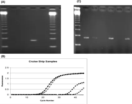
tested twice by conventional RT-PCR and found negative for
NoV with region B primers (Fig. 4A; Table 1). The samples were
then retested for NoV RNA with the TaqMan assay, and two
were positive for low concentrations of NoV, with CNs of 39.63
and 33.42 for samples 1 and 2, respectively (Fig. 4B). The samples
were diluted 100-fold and retested with region B primers (Table
1) to rule out the possible presence of RT and/or PCR inhibitors.
Faint bands were detected by ethidium bromide staining. Of the
two samples, no. 2 was successfully sequenced, confirming it to be
a true positive (Fig. 4C).
In addition to the 92 archived stool specimens, we tested 33
water samples from environmental sources suspected to be
contaminated with NoVs. Of these, eight were positive for
low-level NoV contamination, with a mean CN of 37.911 (Fig.
2A). The TaqMan assay easily and rapidly detected previously
identified clusters of GI, GII, and GIV.
DISCUSSION
Newly developed, commercially available nucleic acid
am-plification systems designed to decrease assay time by
moni-toring amplification of target sequences in real time by
fluo-rescence resonance energy transfer analysis are widely used for
absolute and relative quantification and are a critical tool for
basic research, diagnostics, and biotechnology. In this report,
we describe the expansion and validation of broadly reactive
and semiquantitative TaqMan real-time RT-PCR assays to
detect the three NoV genogroups (GI, GII, and GIV) that
target the ORF1-ORF2 junction, a highly conserved region of
the NoV genome (17, 27). A wide range of NoV strains
rep-resenting numerous clusters archived at the CDC were tested
with the TaqMan assays. The assays detected 64/65 NoV
sam-ples that had been previously identified by duplex conventional
RT-PCR assay. Viral instability and/or RNA degradation of
one specimen may account for the negative result since the
samples had been stored at
⫺
70°C for more than 6 years and
retesting with a conventional RT-PCR assay also yielded a
negative result.
NoVs are especially diverse; therefore, designing one set of
primers to detect all strains with equal efficiency is difficult.
Since NoVs circulating in the community are frequently
chang-ing and new clusters are recognized regularly, we entertained
the possibility that the primers and probes, over time, would
not continue to provide broadly reactive detection of GI, GII,
and GIV NoVs. To investigate this possibility, we tested two
recently identified NoV strains, GII/16 (accession no. AY502006)
and GII/17 (accession no. AY502009) (43). Robust signals were
observed with the GII assay, demonstrating continued detection
albeit strain evolution (data not shown). The primer and probe
sets for GI and GII NoVs designed by Kageyama et al. and our
GIV primers and probe set maximize detection over time by using
the ORF1-ORF2 region, which contains the highest nucleotide
homology (17).
[image:5.585.44.284.79.508.2]RT-PCR was the first molecular diagnostic method used for
the diagnosis of NoV infection in clinical and environmental
samples (2, 28, 36). Although this method is powerful, it lacks
the sensitivity and specificity needed for samples with low viral
loads or samples containing RT-PCR inhibitors. As observed
in our study, samples containing low viral concentrations and
FIG. 3. Relationship between known copy numbers of T7 RNA
(GI and GII) or viral RNA from stool extract (GIV) and the detection
threshold. T7 RNA transcripts were made for representative GI and
GII strains, and a fecal specimen was used for GIV. (A) Serial dilution
of a GI/3 NoV RNA transcript demonstrating a wide dynamic range.
The detection limit was 9.836
⫻
10
1copies/
l, the slope was
⫺
3.370,
the
y
intercept was 43.97, and the
R
2value was
⫺
1.00. (B) GII/4 serial
dilution of a NoV RNA transcript showing a wide dynamic range. The
detection limit was 9.05 copies/
l, the slope was
⫺
3.324, the
y
intercept
was 39.20, and the
R
2value was
⫺
1.00. The mean squared error was
0.195. (C) (GIV) NoV RNA stool standard serial dilutions. The slope
was
⫺
3.310, the
y
intercept was 58.96, and the
R
2value was
⫺
1.00. The
mean squared error was 0.0763. The detection limit of the GIV stool
standard curve was 8.644
⫻
10
5copies/
l. The slope,
y
intercept, and
mean squared error were 3.392, 59.27, and 0.0764, respectively, and the
R
2value was
⫺
1.00.
V
OL. 44, 2006
QUANTITATIVE RT-PCR FOR NOROVIRUS GI, GII, AND GIV
1409
on May 16, 2020 by guest
http://jcm.asm.org/
RT-PCR inhibitors may test falsely negative by conventional
RT-PCR methods. However, with the TaqMan assay,
sensitiv-ity greater than that of the conventional method was
repeat-edly observed with samples containing low NoV
concentra-tions or RT-PCR inhibitors. Additionally, the TaqMan assay
has the increased specificity of the TaqMan probe, as opposed
to previously described SYBR green real-time RT-PCR assays
(29). This TaqMan real-time RT-PCR method detects NoV in
a genogroup-specific manner, making it possible to determine
the genogroup directly without resorting to conventional
RT-PCR methods that use degenerate primers and require
se-quencing for confirmation. By means of serially diluted NoV
transcripts, potential detection limits of
⬍
10 transcript copies
per reaction mixture and
⬍
100 transcript copies per reaction
mixture were achieved with the GII and GI transcripts,
respec-tively. The assays demonstrated a large dynamic range of at
least 6 logs for quantification. The GIV stool had a lower
detection limit of
⬍
10,000 transcript copies per reaction
mix-ture. As opposed to T7 RNA transcripts, the presence of
nucleases and other RT and/or PCR inhibitors in stool may
account for the decreased sensitivity of the GIV assay.
How-ever, much like the GII transcript standard, a detection limit of
⬍
10 copies per reaction mixture was also observed with the
GII stool standard (data not shown). The sensitivity difference
among the GI, GII, and GIV assays can also be due to many
other factors, such as primer design and differences in PCR
conditions.
Real-time RT-PCR offers obvious advantages over more
traditional RT-PCR formats, but some caution is required
when interpreting results. The efficiency of a real-time assay
can be estimated by analyzing the exponential phase of the
amplification curve (5, 33, 39). Quantitative RT-PCR
meth-ods presume that the target and the sample are amplified
with similar efficiencies. However, small variations in
effi-ciency reflecting a decline in DNA polymerase activity
be-tween standards and unknowns can negatively impact true
quantification.
[image:6.585.81.505.73.408.2]The risk with external standards is that some of the unknown
FIG. 4. Detection of NoV in water samples from cruise ship A by conventional (A) versus real-time (B) RT-PCR assay. (A) A
conventional RT-PCR assay failed to detect NoV RNA from potable water (lanes 2 and 3) compared with a positive control (lane 4, GII
strain) and a negative control (lane 5, water). Lanes 1 and 6 were 123-bp ladders in a 3% agarose gel with 0.5
g of ethidium bromide run
at 120 V for 60 min. (B) A real-time RT-PCR assay identified 2 positive water samples (
Œ
,
〫
) among 12 samples tested. Controls included
a GII/4 strain T7 RNA transcript (
■
), a GII/4 fecal specimen (
E
), and a no-template negative control water sample (
⫺
). (C) Conventional
RT-PCR detected NoV RNA in potable water after samples were diluted 1:100. Lane 1 was a 123-bp ladder in a 3% agarose gel with 0.5
g of ethidium bromide run at 120 V for 60 min. Lanes 2 and 5 were positive controls (GII strain); lanes 3 and 7 were water samples 1 and
2, respectively; and lanes 4 and 6 were negative controls.
on May 16, 2020 by guest
http://jcm.asm.org/
samples may contain substances that significantly reduce the
efficiency of the PCR. When inhibitors are suspected, sample
dilution is often effective and the inhibitory factors can be
diluted out if the target nucleic acid is present in sufficient
quantities. The presence of inhibitors may affect the ability to
determine the absolute or true starting copy number in these
samples, but for detection and subtyping, semiquantitation or
approximation of the concentration is sufficient. Various other
approaches have been developed to detect RT and PCR
in-hibitors in clinical and environmental samples containing NoV.
Housekeeping genes such as that for RNase P have been used
with fecal samples but have yielded inconsistent results (S.
Monroe, personal communication; 10). Internal standard
con-trols of synthetic NoVs can be generated by transcribing NoV
plasmids, but this significantly increases the cost of testing (5,
36, 37).
The TaqMan assay is ideal for laboratories handling a large
volume of clinical and environmental specimens and provides
quantification and immediate typing of GI, GII, and GIV NoV
strains. A multiplex real-time RT-PCR assay for the detection
of GI and GII NoVs has recently been described (30), but
further development to include the GIV primer set is needed.
After an initial rapid NoV diagnosis has been made with the
TaqMan assays, strain comparison with capsid VP1 (region D)
primers can be done since phylogenetic analysis of sequences
from this region are consistent with those based on entire
capsid sequences (42).
This assay has proven useful for routine diagnostic assays in
clinical settings because it eliminates postamplification product
processing and thus shortens the turnaround time for reporting
results, which can prove critical in an outbreak setting. Finally,
the TaqMan assay can assist in determining if NoV
contami-nation is present in samples containing very low viral
concen-trations.
ACKNOWLEDGMENTS
We gratefully acknowledge Charles E. Rose for assistance with
statistical analysis and Claudia C. Chesley and Robert M. Wohlhueter
for editorial assistance. We also thank Jaret T. Ames and Jon Schnoor
from Vessel Sanitation and the Biotechnology Core Facility at the
CDC for help with water collection and the preparation of
oligonu-cleotides, respectively.
This research was supported in part by the Atlanta Research and
Education Foundation (AREF) and the CDC.
The findings and conclusions in this report are ours and do not
necessarily represent the views of the funding agency.
REFERENCES
1.Abbaszadegan, M., Lechevallier, and C. P. Gerba. 2003. Occurrence of
viruses in US ground water. J. Am. Water Works Assoc.95:107–120.
2.Ando, T., J. S. Noel, and R. L. Fankhauser.2000. Genetic classification of
Norwalk-like viruses. J. Infect. Dis.181:S336–S348.
3.Ando, T., S. S. Monroe, J. R. Gentsch, Q. Jun, D. C. Lewis, and R. I. Glass.
1995. Detection and differentiation of antigenically distinct small round-structured viruses (Norwalk-like viruses) by reverse transcription-PCR and
Southern hybridization. J. Clin. Microbiol.33:64–71.
4.Ando, T., S. S. Monroe, J. S. Noel, and R. I. Glass.1997. A one-tube method of reverse transcription-PCR to efficiently amplify a 3-kilobase region from RNA polymerase gene to the poly(A) tail of small round-structured viruses
(Norwalk-like viruses). J. Clin. Microbiol.35:570–577.
5.Atmar, R. L., and M. K. Estes.2001. Diagnosis of noncultivable
gastroen-teritis viruses, the human caliciviruses. Clin. Microbiol. Rev.14:15–37.
6.Beuret, C., D. Kohler, A. Baumgartner, and T. M. Luthi.2002. Norwalk-like virus sequences in mineral waters: one-year monitoring of three brands.
Appl. Environ. Microbiol.68:1925–1931.
7.Blanton, L. H., S. M. Adams, R. S. Beard, G. Wei, S. N. Bulens, M. A. Widdowson, R. I. Glass, and S. S. Monroe.2006. Molecular and epidemio-logic trends of caliciviruses associated with outbreaks of acute gastroenteritis
in the United States, 2000–2004. J. Infect. Dis.193:413–421.
8.Boom, R., C. J. Sol, M. M. Saliman, C. L. Jansen, P. M. Wertheim-van Dillen, and J. Van der Noordaa.1990. Rapid and simple method for
puri-fication of nucleic acids. J. Clin. Microbiol.28:495–503.
9.Burton-MacLeod, J. A., E. M. Kane, R. S. Beard, L. A. Hadley, and R. I. Glass.2004. Evaluation and comparison of two commercial enzyme-linked immunosorbent assay kits for detection of antigenically diverse human
noro-viruses in stool samples. J. Clin. Microbiol.42:2587–2595.
10.Emery, S. L., D. D. Erdman, M. D. Bowen, B. R. Newton, J. M. Winchell, R. F. Meyer, S. Tong, B. T. Cook, B. P. Holloway, K. A. McCaustland, P. A. Rota, B. Bankamp, L. E. Lowe, T. G. Ksiazek, W. J. Bellini, and L. J. Anderson.2004. Real-time reverse transcription-polymerase chain reaction
assay for SARS-associated coronavirus. Emerg. Infect. Dis.10:311–316.
11.Frankhauser, R. L., S. S. Monroe, J. S. Noel, C. D. Humphrey, J. S. Bresee, U. D. Parashar, T. Ando, and R. I. Glass.2002. Epidemiologic and molecular trends of Norwalk-like viruses associated with outbreaks of gastroenteritis in
the United States. J. Infect. Dis.186:1–7.
12.Glass, R. I., J. Noel, T. Ando, R. Frankhauser, G. Belliot, A. Mounts, U. D. Parashar, J. S. Bresee, and S. S. Monroe.2000. The epidemiology of enteric caliciviruses from humans; a reassessment using new diagnostics. J. Infect.
Dis.181:S254–261.
13.Green, K. Y., T. Ando, M. S. Balayan, T. Berke, I. N. Clarke, M. K. Estes, D. O. Matson, S. Nakata, J. D. Neill, M. J. Studdert, and H. J. Thiel.2000.
Taxonomy of the caliciviruses. J. Infect. Dis.181(Suppl. 2):S322–S330.
14.Ho, M. S., R. I. Glass, S. S. Monroe, H. P. Madore, S. Stine, P. F. Pinsky, D. Cubitt, C. Ashley, and E. O. Caul.1989. Viral gastroenteritis aboard a cruise
ship. Lancetii:961–965.
15.Inouye, S., K. Yamashita, S. Yamadera, M. Yoshikawa, N. Kato, and N. Okabe.2000. Surveillance of viral gastroenteritis in Japan: pediatric cases
and outbreak incidents. J. Infect. Dis.181:S270–274.
16.Jiang, X., P. W. Huang, W. M. Zhong, T. Farkas, D. W. Cubitt, and D. O. Matson.1999. Design and evaluation of a primer pair that detects both
Norwalk and Sapporo-like caliciviruses by RT-PCR. J. Virol. Methods83:
145–154.
17.Kageyama, T., S. Kojima, M. Shinoharra, K. Uchida, S. Fukushi, F. B. Hoshino, N. Takeda, and K. Katayama.2003. A broadly reactive and highly sensitive assay for Norwalk-like viruses on real-time quantitative RT-PCR.
J. Clin. Microbiol.41:1548–1557.
18.Kukkula, M., P. Arstila, M. L. Klossner, L. Maunula, C. V. Bonsdorff, and P. Jaatinen.1997. Waterborne outbreak of viral gastroenteritis. Scand. J.
In-fect. Dis.29:415–418.
19.Laverick, M. A., A. P. Wyn-Jones, and M. J. Carter.2004. Quantitative RT-PCR for the enumeration of noroviruses (Norwalk-like viruses) in water
and sewage. Lett. Appl. Microbiol.39:127–136.
20.Lopman, B. A., M. H. Reacher, Y. Van Duijnhoven, F. X. Hanon, D. Brown, and M. Koopmans.2003. Viral gastroenteritis outbreaks in Europe, 1995–
2000. Emerg. Infect. Dis.9:90–96.
21.Marks, P. J., I. B. Vipond, F. M. Regan, K. Wedgwood, R. E. Fey, and E. O. Caul.2003. A school outbreak of Norwalk-like virus: evidence for airborne
transmission. Epidemiol. Infect.131:727–736.
22.McIver, C. J., G. Hansman, P. White, J. C. Doultree, M. Catton, and W. D. Rawlinson.2001. Diagnosis of enteric pathogens in children with
gastroen-teritis. Pathology33:353–358.
23.Mead, P. S., L. Slutsker, V. Dietz, L. F. McCaig, J. S. Bresee, C. Shapiro, P. M. Griffin, and R. V. Tauxe.1999. Food-related illness and death in the
United States. Emerg. Infect. Dis.5:607–625.
24.Meschke, J. S., M. Sobsey, and G. Britton.2001. Human caliciviruses: basic
virology. Encycl. Environ. Microbiol.3:1586–1598.
25.Miller, I., R. Gunson, and W. F. Carman.2002. Norwalk like virus by light
cycler PCR. J. Clin. Virol.25:231–232.
26.Moore, C., E. M. Clark, C. I. Gallimore, S. A. Corden, J. J. Gray, and D. Westmoreland.2004. Evaluation of a broadly reactive nucleic acid sequence based amplification assay for the detection of noroviruses in faecal material.
J. Clin. Virol.29:290–296.
27.Nishida, T., H. Kimura, M. Saitoh, M. Shinohora, M. Kato, S. Fukuda, T. Munemura, T. Mikami, A. Kawamoto, M. Akiyama, Y. Kato, K. Nishi, K. Kozawa, and O. Nishio.2003. Detection, quantitation, and phylogenetic
analysis of noroviruses in Japanese oysters. Appl. Environ. Microbiol.69:
5782–5786.
28.Noel, J. S., R. L. Fankhauser, T. Ando, S. S. Monroe, and R. I. Glass.1999. Identification of a distinct common strain of “Norwalk-like viruses” having a
global distribution. J. Infect. Dis.179:1334–1344.
29.Pang, X., B. Lee, L. Chui, J. K. Preiksaitis, and S. S. Monroe.2004. Eval-uation and validation of real-time reverse transcription-PCR assay using the LightCycler system for detection and quantitation of norovirus. J. Clin.
Microbiol.42:4679–4685.
30.Pang, X., J. K. Preiksaitis, and B. Lee.2005. Multiplex real time RT-PCR for
V
OL. 44, 2006
QUANTITATIVE RT-PCR FOR NOROVIRUS GI, GII, AND GIV
1411
on May 16, 2020 by guest
http://jcm.asm.org/
the detection and quantitation of norovirus genogroups I and II in patients
with acute gastroenteritis. J. Clin. Virol.33:168–171.
31.Peirson, S. N., J. N. Butler, and R. G. Foster.2003. Experimental validation of novel and conventional approaches to quantitative real-time PCR data
analysis. Nucleic Acids Res.31:e73.
32.Queiroz, A. P., F. M. Santos, A. Sassaroli, C. M. Harsi, T. A. Monezi, and D. U. Mehnert.2001. Electropositive filter membrane as an alternative for the elimination of PCR inhibitors from sewage and water samples. Appl.
Environ. Microbiol.67:4614–4618.
33.Ramakers, C., J. M. Ruijter, R. H. Deprez, and A. F. Moorman.2003. Assumption-free analysis of quantitative real-time polymerase chain reaction
(PCR) data. Neurosci. Lett.339:62–66.
34.Roche Molecular Biochemicals.2000. LightCycler operator’s manual—ver-sion 3.5. Roche Molecular Biochemicals, Indianapolis, Ind.
35.Schaub, S. A., and R. K. Oshiro.2000. Public health concerns about
calici-viruses as waterborne contaminants. J. Infect. Dis.181(Suppl. 2):374–380.
36.Schwab, K. J., M. K. Estes, F. H. Neill, and R. L. Atmar.1997. Use of heat release and an internal RNA standard control in reverse transcription-PCR
detection of Norwalk virus from stool samples. J. Clin. Microbiol.35:511–
514.
37.Schwab, K. J., F. H. Neill, F. L. Guyader, M. K. Estes, and R. L. Atmar.2001. Development of a reverse transcription-PCR-DNA enzyme immunoassay
for detection of “Norwalk-like” viruses and hepatitis A virus in stool and
shellfish. Appl. Environ. Microbiol.67:742–749.
38.Tai, J. H., M. S. Ewert, G. Belliot, R. I. Glass, and S. S. Monroe.2003. Development of a rapid method using nucleic acid sequence-based
amplifi-cation for the detection of astrovirus. J. Virol. Methods110:119–127.
39.Tichopad, A., M. Dilger, G. Schwartz, and M. W. Pfaffl.2003. Standardized determination of real-time PCR efficiency from a single reaction set-up.
Nucleic Acids Res.31:e122.
40.Vinje´, J., and M. P. Koopmans.2000. Simultaneous detection and genotyp-ing of “Norwalk-like viruses” by oligonucleotide array in a reverse line blot
hybridization format. J. Clin. Microbiol.38:2595–2601.
41.Vinje, J., J. Green, D. C. Lewis, C. I. Gallimore, D. W. Brown, and M. P. Koopmans.2000. Genetic polymorphism across regions of the three open
reading frames of “Norwalk-like viruses.” Arch. Virol.145:223–241.
42.Vinje, J., R. A. Hamidjaja, and M. D. Sobsey. 2004. Development and application of a capsid VP1 (region D) based reverse transcription PCR assay for genotyping of genogroup I and II noroviruses. J. Virol. Methods
116:109–117.
43.Zheng, D. P., T. Ando, R. L. Fankhauser, R. S. Beard, R. I. Glass, and S. S. Monroe.2005. Norovirus classification and proposed strain nomenclature.
Virology346:312–323.