Copyright © 2004, American Society for Microbiology. All Rights Reserved.
Highly Sensitive Assay for Detection of Enterovirus in Clinical
Specimens by Reverse Transcription-PCR with an
Armored RNA Internal Control
Marcel Beld,
1* Rene´ Minnaar,
1Jan Weel,
1Cees Sol,
1Marjolein Damen,
1Harry van der Avoort,
2Pauline Wertheim-van Dillen,
1Alex van Breda,
1and Rene´ Boom
1Laboratory of Clinical Virology, Department of Medical Microbiology, Academic Medical Center, University of Amsterdam,
Amsterdam,
1and National Institute of Public Health and the Environment, Bilthoven,
2The Netherlands
Received 10 July 2002/Returned for modification 7 October 2002/Accepted 7 March 2004
The objective of the present study was the development of a diagnostic reverse transcription (RT)-PCR for
the specific detection of enterovirus (EV) RNA in clinical specimens controlled by an internal control (IC)
RNA. The IC RNA contains the same primer binding sites as EV RNA but has a different probe region. The
IC RNA was packaged into an MS2 phage core particle (armored) and was added to the clinical sample to allow
monitoring of both extraction efficiency and RT-PCR efficiency. Serial dilutions of the IC RNA were made, and
the detection limit of the RT-PCR was tested in a background of EV RNA-negative cerebrospinal fluid. The
sensitivity and specificity of the RT-PCR assay were tested by using all 64 known EV serotypes, several non-EV
serotypes, and two Quality Control for Molecular Diagnostics (QCMD) Program EV proficiency panels from
2001 and 2002. In total, 322 clinical specimens were tested by RT-PCR, and to establish the clinical utility of
the RT-PCR, a comparison of the results of viral culture and RT-PCR was done with 87 clinical specimens. The
lower limit of sensitivity was reached at about 150 copies of IC RNA/ml. All 64 EV serotypes were positive, while
all non-EV serotypes were negative. All culture-positive samples of the 2001 QCMD proficiency panel
(accord-ing to the 50% tissue culture infective doses per milliliter) were positive by RT-PCR. Invalid results, i.e.,
negativity for both EV RNA and IC RNA, due to inhibition of RT-PCR were observed for 33.3% of the members
of the 2002 QCMD proficiency panel and 3.1% of the clinical specimens. Inhibition of RT-PCR could be
relieved by the addition of 400 ng of bovine
␣
-casein per
l to both the RT reaction mixture and the PCR
mixture. With this optimized protocol, the results for all samples of the 2002 QCMD proficiency panel and all
clinical specimens except one fecal sample (0.3%) were valid. Evaluation of the clinical samples demonstrated
that EV infection could be detected in 12 of 87 samples (13.8%) by RT-PCR, while viral culture was negative.
Our data show that the RT-PCR with armored IC RNA offers a very reliable and rapid diagnostic tool for the
detection of EV in clinical specimens and that the addition of bovine
␣
-casein relieved inhibition of the RT-PCR
for 99.7% of clinical specimens.
The human enteroviruses (EVs) are members of the family
Picornaviridae
, are ubiquitous, and are mainly enterically
trans-mitted. EVs have traditionally been identified by
serotype-spe-cific antisera in a virus-neutralizing test, and 66 EV types are
known to infect humans (19). The 66 EV serotypes were
initial-ly recognized and divided into five major groups: polioviruses
(PV; types 1 to 3), coxsackieviruses A (CVAs; types 1 to 22 and
24), coxsackieviruses B (CVBs; types 1 to 6), echoviruses (types
1 to 7, 9, 11 to 27, and 29 to 33), and EV types 68 to 71 (17).
Recent molecular analyses have proved that echovirus types 22
and 23 are genetically distinct from the members of the genus
Enterovirus
and have been reclassified in a separate genus,
Parechovirus
, in the family
Picornaviridae
(13, 20, 23).
Infections with EVs cause a wide range of clinical outcomes,
such as asymptomatic infections, aseptic meningitis (meningeal
inflammation in the absence of a bacterial pathogen),
enceph-alitis, paralytic poliomyelitis, and myocarditis. Although the
majority of EV infections do not cause significant disease,
infection can cause serious illness, especially in infants and
immune-compromised patients. EV infections are the most
common cause of aseptic meningitis and account for 80 to 90%
of all cases of central nervous system infections for which a
possible causative agent is identified (24). In the neonate,
asep-tic meningitis-induced complications and poor outcomes of
EV infections generally occur within the first 2 days of life (1,
2). Aseptic meningitis in immune-competent adults is
charac-terized by sudden onset of fever, but neurological
abnormali-ties are rare, and both short-term and long-term outcomes are
generally good. Encephalitis caused by EV infections is a less
common but a more severe disease than aseptic meningitis (18,
29, 30). Immune-compromised children and adults who are
infected with EV may develop chronic meningitis and
enceph-alitis, which may last for years before becoming fatal (16).
The early clinical symptoms of meningitis caused by viruses,
bacteria, and fungi are quite similar and are difficult to
distin-guish, but the diagnosis, therapy, and outcome of disease
caused by these pathogens vary considerably. A reliable means
of laboratory detection of EV is needed, since the specific and
rapid diagnosis of EV meningitis has a significant impact on
* Corresponding author. Mailing address: Laboratory of Clinical
Virology, Department of Medical Microbiology, Academic Medical
Center, University of Amsterdam, Meibergdreef 15, 1105 AZ,
Amster-dam, The Netherlands. Phone: 31 20 5665472. Fax: 31 20 6974005.
E-mail: [email protected].
3059
on May 15, 2020 by guest
http://jcm.asm.org/
patient management (10). Although reverse transcription
(RT)-PCR may become the method of choice for the diagnosis of EV
infections of the central nervous system, in many laboratories
the diagnosis of EV infections still relies on cell culture
tech-niques. Stool specimens or rectal swab, throat swab, and
cere-brospinal fluid (CSF) specimens are used for virus culture.
Early in the acute phase of the illness, EV is frequently isolated
from the throat, whereas isolation of the virus from CSF
pro-vides the most direct link to disease but is usually less
success-ful. A cytopathic effect (CPE) is not recognizable within a few
days, and some EVs do not grow in cell culture and therefore
do not cause a CPE at all (15, 25). In recent years, several EV
RT-PCR assays which are sensitive and rapid have been
de-veloped (8, 9, 14, 25, 26). In those previous studies, EV RNA
was purified without an internal control (IC) RNA that
mon-itored both RNA extraction efficiency and RT-PCR efficiency.
Like the wild-type target, these IC RNAs should have the same
length, contain the same primer binding sites, and have the
same G
⫹
C content for identical extraction and amplification
efficiency but should contain a different probe binding site for
the differential detection of IC RNA. In molecular diagnostics,
the use of an IC RNA is crucial for a reliable interpretation of
results because it enables the verification of the sensitivity of
the assay and avoids false-negative results. RNA transcribed in
vitro can be used as IC RNA but is prone to degradation by
RNases. Packaging of IC RNA into a phage protects the RNA
from enzymatic degradation (22). These armored RNAs can
be spiked into clinical specimens without degradation, thus
enabling simultaneous monitoring of the complete nucleic acid
extraction and amplification process for each specimen.
MATERIALS AND METHODS
Chemicals and enzymes.Lysis buffer, wash buffers, and silica suspensions were prepared as described previously (3). SuperScript II was obtained from Life Technologies (Gaithersburg, Md.). RNase inhibitor (RNAsin) was obtained from Promega (Madison, Wis.). Bovine serum albumin (BSA) was obtained from Roche Diagnostics (Almere, The Netherlands). Deoxynucleotides (dATP, dCTP, dGTP, dTTP, and dUTP),TaqDNA polymerase (Amplitaq Gold), and uracil-N-glycosylase (Amperase) were from Applied Biosystems (Nieuwerkerk a/d IJssel, The Netherlands). Streptavidin-coated magnetic beads (Dynabeads M-280) were from Dynal (Hamburg, Germany). Tris, KCl, MgCl2, calf thymus DNA, and bovine␣-casein (lot no. 17H9551) were obtained from Sigma (Zwi-jndrecht, The Netherlands). Bovine␣-casein was dissolved in 1⫻PCRII buffer (Applied Biosystems) at 20 mg/ml, and the mixture was stored at⫺20°C. Triton X-100 was obtained from Packard Instrument Corporation (Downers Grove, Ill.). Serum and plasma samples were obtained in Vacutainer tubes (Becton Dickinson Systems, Meylan, France).
Clinical specimens.In total, 322 clinical specimens, including 281 CSF sam-ples, 18 fecal samsam-ples, 10 throat swab samsam-ples, 3 vesicle fluid samsam-ples, 3 pleural fluid samples, 2 broncheoalveolar lavage fluid samples, 2 amniotic fluid samples, 1 urine sample, and 2 brain biopsy specimens, were obtained from patients suspected to be infected with EV and were tested by the RT-PCR.
CPE for EV infection.Viral culture was done by cocultivation of CSF with human diploid fibroblasts, tertiary monkey kidney cells, or Vero cells. These viral cultures were examined twice weekly for the appearance of EV-specific CPEs. Preliminary identification of isolates was performed according to either the unstained CPE or the CPE obtained by incubation with a specific monoclonal antibody (DAKO-Enterovirus monoclonal antibody 5-D8/1; DAKO, Glostrup, Denmark).
Serotyping of isolates.Typing of EVs was performed at the National Institute of Public Health and the Environment (Bilthoven, The Netherlands) by neutral-ization tests with antiserum pools. These pools are prepared in combinations designed so that an isolate can be screened for identity by using eight pools of 42 antisera in a single test.
Viral strains.EV serotypes CVA (types 1 to 6, 18 to 22, 24), CVB (types 1 to 6), echovirus (types 1 to 9, 11 to 22, 24 to 27, 29 to 33), EV (types 68 to 71), PV vaccine types (types 1 to 3), and parechovirus types 1 and 2 were kindly provided by the National Institute of Public Health and the Environment. The rhinovirus serotypes (types 1A, 1B, 3, 8, 11, 13, 14, 15, 16, and 88) were kindly provided by the Department of Virology of the Utrecht Medical Center (Utrecht, The Neth-erlands). The hepatitis A virus (HAV) serotype (type HM175) was kindly pro-vided by the Municipal Health Service, Amsterdam.
Primers.Random hexamers (Roche Diagnostics), which were diluted to 1.5
g/l in TE buffer (10 mM Tris-HCl, 1 mM EDTA [pH 8.0]), were used for RT. PCR primers were designed by computer-assisted analysis (OMIGA; Oxford Molecular, Oxford, England) of all available 5⬘noncoding regions and the full genomes of the EV serotypes. High-pressure liquid chromatography-purified PCR primers were from Applied Biosystems and were diluted to 100 ng/l in TE buffer. The sequences of the primers used for amplification of both wild-type EV RNA and IC RNA are located in the conserved 5⬘noncoding region of the EV sequence. The primer pair used for amplification consisted of entero-1 (5⬘-CCC TGA ATG CGG CTA AT-3⬘; nucleotide positions [nt] 452 to 468) and Bio-entero-2 (5⬘-ATT GTC ACC ATA AGC AGC C-3⬘; 5⬘biotinylated; nt 579 to 597). Nucleotide numbering was that for the Sabin PV type 2 strain, as described by Toyoda et al. (28).
Armored EV RNA control.The armored EV RNA control, which contained part of the 5⬘ noncoding region (nt 428 to 691), was obtained from RNA Diagnostics, Ambion, Inc. (Austin, Tex.), and was constructed by the armored RNA technique (22). The method is based on the packaging of recombinant RNA into MS2-like particles, which are produced inEscherichia coli. The par-ticles are isolated through a series of conventional protein purification proce-dures. According to Ambion, the approximate factor used for the conversion of 1 mg of armored RNA to the numbers of RNA copies is about 2⫻1014. The armored EV RNA stock solution used in the present study (lot no. 040D49012A) contained approximately 6.5⫻1014copies of EV RNA/ml.
Construction of armored IC RNA control.We designed two oligonucleotides for the construction of IC RNA, and these were synthesized by Applied Biosys-tems: Ent-hyb-3 (5⬘-CCC TGA ATG CGG CTA ATC CTA ACC ACG GAA CAG GCG GTC GCG AAC CAG TGA CTG GTC TGT CGT AAC GCG CAA GTC TGT GCT TGA GAC GTG-3⬘) and Ent-hyb-4 (5⬘-ATT GTC ACC ATA
AGC AGC CAT GAT AAA AAT AAC AGG AAA CAC GGA CGG TTA CCA
CGC ACG TCT CAA GCA CAG ACT T-3⬘). These two oligonucleotides, which together represent the same part of the 5⬘noncoding region (nt 428 to 691) present in the armored EV RNA control, overlapped over a stretch of 20 nt (underlined) and contained the same primer binding sites as the armored EV RNA control (italics), but with a different probe region (boldface). This IC probe region allowed discrimination between EV and IC RNA amplimers after hybrid-ization and detection. The IC RNA control was constructed by hybridhybrid-ization and elongation of 1 ng of oligonucleotide Ent-hyb-3 and 1 ng of oligonucleotide Ent-hyb-4 in a mixture of 2.5 U of Amplitaq Gold; 5g of BSA; 1⫻PCRII buffer (10 mM Tris-HCl [pH 8.3], 50 mM KCl) dATP, dCTP, dGTP, and dTTP at a concentration of 200M each; and 3 mM MgCl2. The mixture was incubated for 10 min at 95°C, 5 min at 55°C, and 10 min at 72°C. The resulting hybrid was subsequently amplified with primer pair entero-1 and non-Bio-entero-2 in the same mixture described above and was incubated for 10 min at 95°C, followed by 35 cycles, with each cycle consisting of 20 s at 95°C, 20 s at 55°C, and 1 min at 72°C, followed by 5 min at 72°C. The concentration of the resulting amplimer was estimated by measuring the UV absorption at 260 nm, and subsequently, 2 ng was cloned into a plasmid vector (PCRII; Promega), resulting in plasmid pEntIC 2. The IC RNA sequence was confirmed by dideoxynucleotide sequencing (Vis-ible Genetics Inc., Toronto, Ontario, Canada). Packaging of the IC RNA control into MS2-like particles was performed with plasmid pEntIC 2 as the template and was custom made by Ambion. According to Ambion, the approximate factor for the conversion of 1 mg of armored RNA to the numbers of copies of RNA is about 2⫻1014. The armored IC RNA stock solution used in the present study (lot no. 021D49013A) contained approximately 5.4⫻1014copies of IC RNA/ml.
Dilution buffer for armored RNA controls.The armored RNA controls were diluted in TSM dilution buffer, which contained 20 ng of calf thymus DNA per
l, 10 mM Tris (pH 7.5), 100 mM NaCl, 1 mM MgCl2, and 0.1% gelatin (catalog no. G-9382; Sigma), and stored at⫺20°C. The final solution of armored EV RNA contained 500 copies/l, and the final solution of armored IC RNA con-tained 100 copies/l.
RNA purification.EV RNA was purified from clinical specimens as described earlier (4), with the following modifications: 20l (50l for fecal samples [7]) of size-fractionated silica particles was used in combination with 900l of lysis buffer L6. The clinical specimen and 500 armored IC RNA copies were added to this silica-lysis buffer mixture. RNA was eluted in 100l of TE buffer.
on May 15, 2020 by guest
http://jcm.asm.org/
RT-PCR.Forty microliters of the 100l (2/5) of RNA eluate was used for RT. The final RT mixture (50l) contained 1,500 ng of hexamers, 1⫻CMB1 buffer (10 mM Tris-HCl [pH 8.3], 50 mM KCl, 0.1% Triton X-100), 0.4 U of Super-Script II perl, 120M (each) deoxynucleoside triphosphate, 0.08 U of RNAsin perl, and 5 mM MgCl2. The mixture was incubated for 30 min at 42°C, and 25
l (corresponding to 40l [1/5] of CSF) was subsequently used as input in the PCR. The PCR was performed in a 50-l volume containing 200 ng of primer entero-1 (5⬘-CCC TGA ATG CGG CTA AT-3⬘; nt 452 to 468) and 200 ng of primer Bio-entero-2 (5⬘-ATT GTC ACC ATA AGC AGC C-3⬘; 5⬘biotinylated; nt 579 to 597); 0.05 U of Amplitaq Gold DNA polymerase perl; 0.01 U of Amperase perl; 0.1g of BSA perl; 1⫻PCRII buffer (10 mM Tris-HCl [pH 8.3], 50 mM KCl); 400 ng of bovine␣-casein perl; dATP, dCTP, and dGTP at a concentration of 200M each; and 400M dUTP. The final MgCl2 concen-tration in the PCR mixture was 2.5 mM. PCRs were performed in an Applied Biosystems 9600 thermocycler, as follows: 2 min at 50°C and 10 min at 95°C,
followed by 45 cycles each consisting of 20 s at 95°C, 20 s at 55°C, and 1 min at 72°C, followed by 5 min at 72°C.
Optimized RT-PCR.Bovine␣-casein (400 ng/l) was added to the RT mix-ture, and the mixture was processed as described above.
Hybridization and measurement by ECL.After RT-PCR, excess primers were removed as described earlier (5), and amplicons were hybridized with Tris (2,2⬘ -bipyridine) ruthenium [II] chelate (TBR)-labeled probes specific for EV RNA and IC RNA, as described recently (5). The probes (nt 531 to 551) were labeled with TBR at the 5⬘end and were as follows: TBR-entero-1 (EV-specific probe; 5⬘-GCG GAA CCG ACT ACT TTG GGT-3⬘) and TBR-entero-2 (IC-specific probe; 5⬘-CTT GAG ACG TGC GTG GTA ACC-3⬘). Hybrids were captured with streptavidin-coated magnetic beads; and the electrochemiluminescence (ECL) signal, expressed in luminosity units (LU), was measured with the M8 system (IGEN, Oxford, England). A 96-well plate is used with this device, and unhy-bridized TBR-labeled probes are automatically removed by washing. The amount of labeled hybrids is determined after excitation by applying an electric field.
Criteria for diagnostic RT-PCR.A signal of more than 500 LU (2.5 times the mean background signal for either probe) was considered a positive result. A clinical specimen was considered positive for EV RNA if more than 500 LU was measured with the EV-specific probe, regardless of the result obtained with the IC-specific probe. A clinical specimen was considered negative for EV RNA if less than 500 LU was measured with the EV-specific probe and IC RNA was detected at more than 500 LU. In the diagnostic RT-PCR, four controls, two positive controls and two negative controls, were included in the RNA extraction procedure. The high-positive control contained 12,500 armored EV RNA copies/ extraction, and the low-positive control contained 2,500 armored EV RNA copies/extraction, both together with 500 armored IC RNA copies. The first negative control contained 500 armored IC RNA copies and served as a control for the entire procedure and should be negative for the EV-specific probe but positive for the IC-specific probe. The second negative control contained no armored EV RNA copies or armored IC RNA copies and should be negative for both probes with a mean of 200 LU.
Parechovirus-specific RT-PCR.The conditions of the RT-PCR for the detec-tion of parechoviruses were similar to those for the protocol described above, except for the primers used in the PCR. For the specific detection of parecho-viruses, we used the primer pair described by Oberste et al. (21).
RESULTS
[image:3.603.44.284.87.294.2]Determination of the lower limit of detection of the EV
RT-PCR assay.
To determine the lower limit of detection of
the EV RT-PCR assay, we spiked decreasing amounts of both
armored EV RNA and armored IC RNA diluted in TSM
buffer into 200
l of EV-negative CSF before RNA extraction
by the method of Boom et al. (4). RNA was eluted in 100
l,
and 40
l of extracted RNA (2/5 of the extracted RNA) was
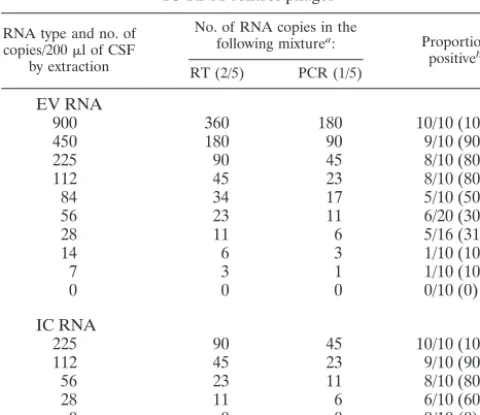
TABLE 1. PCR sensitivity for armored EV RNA and
IC RNA control phages
RNA type and no. of copies/200l of CSF
by extraction
No. of RNA copies in the
following mixturea: Proportion
positiveb
RT (2/5) PCR (1/5)
EV RNA
900
360
180
10/10 (100)
450
180
90
9/10 (90)
225
90
45
8/10 (80)
112
45
23
8/10 (80)
84
34
17
5/10 (50)
56
23
11
6/20 (30)
28
11
6
5/16 (31)
14
6
3
1/10 (10)
7
3
1
1/10 (10)
0
0
0
0/10 (0)
IC RNA
225
90
45
10/10 (100)
112
45
23
9/10 (90)
56
23
11
8/10 (80)
28
11
6
6/10 (60)
0
0
0
0/10 (0)
aThe following assumptions were made for the copy numbers presented:
(i) the armored RNA stocks contain the specified number of phage particles, and all contain EV RNA or IC RNA; and (ii) the efficiencies of extraction, RT, and PCR were 100% each.
bThe data represent number of samples positive/number of samples tested
(percent).
TABLE 2. Specificity of EV RT-PCR for prototype strains of EVs and controls
Virus (virus serotype or titer) Source Result Mean (range) LU of EV RNA
CVA (2–4, 6–18, 20, 21, 24
a)
Cell culture
Positive
87,000 (57,000–117,000)
CVA (1, 5, 19, 22)
Suckling mice
Positive
87,000 (73,000–100,000)
CVB (1–6)
Cell culture
Positive
91,000 (75,000–107,000)
Echovirus (1–9, 11–16, 17–21, 24–27, 29–33
b)
Cell culture
Positive
98,000 (86,000–111,000)
Echovirus (22, 23
c)
Cell culture
Negative
218 (209–227)
Enterovirus (68–71)
Cell culture
Positive
77,000 (68,000–86,000)
Poliovirus (1, 2, 3)
Cell culture
Positive
104,000 (79,000–129,000)
Rhinovirus (1A, 1B, 3, 8, 11, 13, 14, 15, 16, 88)
Cell culture
Negative
228 (205–277)
HAV
Cell culture
Negative
237
Negative control
Negative
226 (217–235)
HPC
d(25,000 EV RNA copies/ml)
Positive
85,000 (62,000–106,000)
LPC
e(5,000 EV RNA copies/ml)
Positive
58,000 (46,000–70,000)
IC (2,500 IC RNA copies/ml)
Positive
51,000 (40,000–63,000)
aEchovirus type 34 is a variant of CVA type 24.
bEchovirus type 8 is a variant of echovirus type 1, CVA type 23 is a variant of echovirus type 9, echovirus type 10 was reclassified as reovirus type 1, and echovirus
type 28 was reclassified as human rhinovirus type 1A.
cEchovirus types 22 and 23 were reclassified as parechovirus types 1 and 2, respectively. dHPC, high-positive control.
eLPC, low-positive control.
on May 15, 2020 by guest
http://jcm.asm.org/
[image:3.603.40.542.536.675.2]used for RT; subsequently, 50% of the cDNA was used for
PCR (1/5 of the extracted RNA). Limiting dilutions of the
armored EV RNA revealed a detection limit of 84 EV RNA
copies in the extraction procedure with a 50% hit rate (5 of 10
runs), resulting in a detection limit of 17 copies of EV DNA by
PCR (1/5). Limiting dilutions of the armored IC RNA revealed
a detection limit of 28 IC RNA copies in the extraction
pro-cedure with a 60% hit rate (6 of 10 runs), resulting in a
detection limit of 6 copies of IC DNA by PCR (1/5) (Table 1).
Identical results were found after the direct release of EV
RNA and IC RNA from its phage (without CSF as a
back-ground) after incubation at 70°C for 5 min (results not shown).
Testing of EV serotypes.
All known EV serotypes (
n
⫽
64)
were tested by the EV RT-PCR described here. The extraction
of every single EV serotype and non-EV serotype was done
alternately with negative extraction controls that contained IC
RNA. All EV serotypes were positive, whereas the non-EVs,
including echovirus types 22 and 23, rhinoviruses, HAV, and
all negative controls, were negative. The negative results were
truly negative, since the ECL signals for coextracted armored
IC RNA were all positive (Table 2).
Testing of 2001 and 2002 QCMD proficiency panels.
The
Quality Control for Molecular Diagnostics (QCMD) Program
EV proficiency panels from 2001 and 2002 consisted of coded
freeze-dried samples, including an echovirus type 11 and CVA
type 9 dilution series, other EV serotypes representing
differ-ent genetic clusters of human EVs, and negative controls. The
freeze-dried samples were reconstituted with 1 ml of sterile
water by the QCMD Program protocol, and 200
l of the
reconstituted sample was used for extraction and amplification,
as described in Materials and Methods. All samples of the 2001
QCMD proficiency panel were positive, regardless of the
se-rotype, and all negative controls included tested truly
nega-tive, since the ECL signals for coextracted armored IC RNA
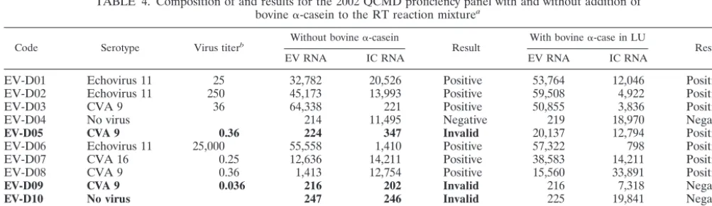
were all positive (Table 3). However, when we tested the 2002
QCMD proficiency panel, four invalid results (33%), i.e., no
result for either EV or IC, were found, which meant that no
conclusive result could be reported (Table 4). These four
in-valid results were probably caused by inhibition of the RT
reaction, and we therefore decided to add 400 ng of bovine
␣
-casein per
l to the RT reaction mixture as well as to the
PCR mixture. After the addition of 400 ng of bovine
␣
-casein
per
l to the RT reaction mixture, the results for all panel
members were valid. All samples expected to be positive were
found to be positive except for one sample with CVA type 9
with the lowest virus titer, which was negative. All negative
results were truly negative, since the ECL signals for
coex-tracted armored IC RNA were all positive (Table 4).
[image:4.603.44.285.88.225.2]Testing of clinical specimens.
A total of 322 clinical
speci-mens were obtained from patients suspected to be infected
with EV and were tested by the RT-PCR. Forty-five positive
results (14%), 267 negative results (82.9%), and 10 invalid
results (3.1%) were found. These invalid results were found for
7 of 281 CSF samples (2.5%), 2 of 18 fecal samples (11.1%),
and 1 of 10 throat swab samples (10%). Repeat tests by the
optimized RT-PCR with these 10 samples with invalid results
revealed that all invalid results apart from those for one fecal
TABLE 3. Composition of and results for the 2001
QCMD proficiency panel
Code Serotype Virus titera LU Result
EV RNA IC RNA
EV-C01 CVA 9
0.036
31,104
65,482
Positive
EV-C02 CVA 9
0.36
87,325
81,793
Positive
EV-C03 CVA 9
3.6
90,570
71,435
Positive
EV-C04 No virus
214
89,860
Negative
EV-C06 Echovirus 11
25.2
96,460
85,396
Positive
EV-C11 Echovirus 11
252
99,930
43,751
Positive
EV-C09 Echovirus 11 25,200
87,380
860
Positive
EV-C07 CVB 5
317
83,867
32,092
Positive
EV-C08 EV 71
56.4
98,508
31,411
Positive
EV-C10 No virus
226
74,234
Negative
EV-C05 Echovirus 6
20,000
82,529
27,549
Positive
aThe titers of the original virus stocks before inactivation and freeze-drying
were as follows: CVA type 9, 3.6 ⫻10650% tissue culture infective doses (TCID50s) per ml; echovirus type 6, 2.0⫻108TCID50s/ml; echovirus type 11, 2.5⫻107TCID50s/ml; CVB type 5, 3.2⫻107TCID50s/ml; EV type 71, 5.6⫻106 TCID50s/ml.
TABLE 4. Composition of and results for the 2002 QCMD proficiency panel with and without addition of
bovine
␣
-casein to the RT reaction mixture
aCode Serotype Virus titerb Without bovine␣-casein Result With bovine␣-case in LU Result
EV RNA IC RNA EV RNA IC RNA
EV-D01
Echovirus 11
25
32,782
20,526
Positive
53,764
12,046
Positive
EV-D02
Echovirus 11
250
45,173
13,993
Positive
59,508
4,922
Positive
EV-D03
CVA 9
36
64,338
221
Positive
50,855
3,836
Positive
EV-D04
No virus
214
11,495
Negative
219
18,970
Negative
EV-D05
CVA 9
0.36
224
347
Invalid
20,137
12,794
Positive
EV-D06
Echovirus 11
25,000
55,558
1,410
Positive
57,322
798
Positive
EV-D07
CVA 16
0.25
12,636
14,211
Positive
38,583
14,211
Positive
EV-D08
CVA 9
0.36
1,413
12,754
Positive
15,560
33,891
Positive
EV-D09
CVA 9
0.036
216
202
Invalid
216
7,318
Negative
EV-D10
No virus
247
246
Invalid
225
19,841
Negative
EV-D11
CVB 5
32
225
237
Invalid
32,701
8,544
Positive
EV-D12
CVA 9
3.6
197
8,156
Positive
38,271
15,007
Positive
aBovine␣-casein was used at a concentration of 400 ng/l. Invalid results are in boldface.
bThe titers of the original virus stocks before inactivation and freeze-drying were as follows: echovirus type 11, 2.5⫻10750% tissue culture infective doses (TCID50s) per ml; CVA type 9, 3.6⫻106TCID50s/ml; CVA type 16, 2.5⫻105TCID50s/ml; CVB type 5, 3.2⫻107TCID50s/ml.
on May 15, 2020 by guest
http://jcm.asm.org/
[image:4.603.42.544.554.699.2]sample were valid and negative for EV and could be reported
to the clinician (Table 5).
Comparison of virus culture with RT-PCR.
Eighty-seven
clinical specimens suspected to be infected with EV were
avail-able to be tested by virus culture and RT-PCR. Agreement of
the results between virus culture and RT-PCR was found for
72 of 87 clinical specimens (82.8%). A negative result by both
virus culture and RT-PCR was found for 54 clinical specimens,
whereas positive results by virus culture and RT-PCR were
found for 18 clinical specimens. Discordant results were found
for 15 clinical specimens. For three of these the result of virus
culture was positive for EV RNA, whereas the result of
RT-PCR was truly negative for EV RNA, since the results for IC
RNA were positive. Two of these RT-PCR-negative samples
were subjected to a parechovirus-specific RT-PCR and were
found to be positive (results not shown). For the other
RT-PCR-negative sample, a CPE was found only after 4 weeks.
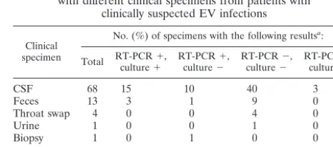
The remaining samples with discordant results consisted of 12
clinical specimens that were RT-PCR positive and virus culture
negative (Table 6).
DISCUSSION
Isolation of EV in cell culture is still regarded as the
diag-nostic “gold standard.” There are, however, many
disadvan-tages to culture, such as its labor-intensiveness, the delay of
days to weeks to obtain a positive result, and its false negativity
rate of approximately 25 to 35% because of failures of antibody
neutralization and the inability of certain CVA serotypes to
grow in cell culture (15, 25).
Many of the disadvantages of cell culture can be overcome
by RT-PCR. The primer and probe sets most frequently used
for the detection of EV are those described by Chapman et al.
(9) and Rotbart et al. (25). These primers and probes have
been proved to be reactive with all known EVs and fail to
am-plify echovirus types 22 and 23, which have been reported to be
members of a genetically distinct genus in the family
Picorna-viridae
(13, 20, 23). We designed a primer and probe set with
minor modifications in comparison to those first described by
Chapman et al. (9) and Rotbart et al. (25) not only to ensure that
all known EV serotypes would be detected but also to avoid
cross-reactivity between EV serotypes and rhinoviruses. This
cross-reactivity with rhinoviruses could have an important
im-pact if nasal or pharyngeal swab specimens are used for testing.
We have described an RT-PCR assay in a nonnested format
for the detection of EV RNA in clinical specimens in which
2,500 copies of armored IC RNA mimicking the EV target per
ml were included in the RNA extraction step and all
subse-quent steps of the procedure. This armored IC RNA can be
spiked directly into clinical specimens without degradation of
the RNA and enables monitoring of the complete nucleic acid
extraction and amplification processes for each specimen.
The sensitivity of the RT-PCR was evaluated with limiting
dilutions of both armored EV RNA and armored IC RNA. We
found a detection limit of 6 copies of IC DNA in 60% of the
samples by PCR. Poisson statistics predict that 63% of the
reactions will be positive by PCR with a single copy of DNA
(12). Thus, if RNA is extracted and reverse transcribed with
efficiencies of 100%, the 60% detection rate by PCR should
represent 1 copy of IC DNA. Since we found comparable
results after the direct release of RNA from the armored RNA
by incubation at 70°C for 5 min and after extraction of the
RNA from the armored RNA by the method of Boom et al.
(4), our results might suggest a less efficient RT step or the
presence of a certain percentage of empty armored RNA
phages. In addition, the approximately threefold difference in
detection between EV RNA and IC RNA that we observed
might be explained by inaccuracies in the determination of the
concentration and the steps used for dilution of the stock
solutions. The lower limit of sensitivity of the RT-PCR was
based on the lower detection rate of IC RNA and was about
150 copies of IC RNA/ml. The presence of 500 copies of IC
RNA during extraction from 200
l of clinical specimens
al-lowed us to draw the conclusion that a specimen found to be
negative for EV RNA but positive for IC RNA would contain
less than 2,500 copies of EV RNA/ml.
[image:5.603.44.284.82.197.2]The use of the armored IC RNA was critical in the detection
of false-negative reactions or invalid results. Recently, it was
shown (6) that an inhibitor(s) of DNA-processing enzymes
could be introduced by the extraction method of Boom et al.
(4) itself and after DNA extraction from clinical specimens,
such as CSF and urine. This inhibition could be relieved by a
novel buffer containing bovine
␣
-casein. Similar results were
found in a recent study by Boom et al. (7), in which inhibitors
were relieved after extraction of DNA from fecal specimens by
the addition of bovine
␣
-casein directly to the PCR mixture.
Despite the presence of bovine
␣
-casein in the PCR mixture,
strong inhibition of IC RNA was still found for 4 of 12 (33.3%)
samples of the 2002 QCMD proficiency panel and 10 of 322
(3.1%) clinical specimens. Thus, the invalid results were
TABLE 5. Summary of results after testing of clinical specimens
Specimen
No. (%) of specimens with the following results:
Total Positive Negative
Invalid
Without bovine
␣-casein
With bovine
␣-casein at 400 ng/l
CSF 281 36 238 7 (2.5) 0
Feces 18 6 10 2 (11.1) 1 (50) Throat swab 10 0 9 1 (10) 0
Other 13 3 10 0 0
Total 322 45 (14) 267 (82.9) 10 (3.1) 1 (0.3)
TABLE 6. Comparison of results of RT-PCR and virus culture
with different clinical specimens from patients with
clinically suspected EV infections
Clinical specimen
No. (%) of specimens with the following resultsa:
Total RT-PCRculture⫹⫹, RT-PCRculture⫺⫹, RT-PCRculture⫺⫺, RT-PCRculture⫹⫺,
CSF 68 15 10 40 3
Feces 13 3 1 9 0
Throat swap 4 0 0 4 0
Urine 1 0 0 1 0
Biopsy 1 0 1 0 0
Total 87 18 (20.7) 12 (13.8) 54 (62.1) 3 (3.4)b a⫹, positive result;⫺, negative result.
bTwo isolates were identified by culture as parechovirus and not EV, and one
isolate was positive by culture only after 4 weeks, indicating a very low virus titer.
on May 15, 2020 by guest
http://jcm.asm.org/
[image:5.603.301.542.595.701.2]caused not only by inhibition of the PCR but also by inhibition
of the RT reaction. The optimized RT-PCR protocol with the
addition of bovine
␣
-casein in the RT reaction relieved the
in-hibition of the RT-PCR, resulting in valid results for all panel
members of the 2002 QCMD proficiency panel and for all
clinical specimens except one fecal specimen. We found similar
results by the extraction of MS2 RNA in a background of fecal
specimens and a subsequent RT reaction as a model system
(unpublished data). The mechanism of relief of inhibition is not
known, but it has been speculated (6) that stretches of
phosphor-ylated serine residues present in bovine
␣
-casein may be involved.
These phosphorylated regions give bovine
␣
-casein a high
chelat-ing force toward positively charged metal ions (3, 11, 27). This
chelating power may be involved in the enhancing properties of
bovine
␣
-casein by capturing coextracted metal ions which might
otherwise act as inhibitors of both RT and PCR.
At present, viral culture is still the gold standard for
detec-tion of EV infecdetec-tions. Validadetec-tion of an RT-PCR assay for the
diagnosis of EV infections is difficult because cases are defined
clinically and viral culture has a poor sensitivity (approximately
70%), partly due to the inability of certain CVA serotypes to
grow in cell culture (15, 25). We found an overall good
agree-ment of 82.8% between the results of virus culture and those of
RT-PCR. CSF provides the most direct link to disease but is
usually less successful in virus culture. We found high rates of
discordant results (17.2%) between virus culture and RT-PCR,
mainly with CSF samples. Delayed processing and improper
handling of the clinical specimens may have contributed to the
false-negative results of virus culture, and our results showed
the added value of the RT-PCR assay described here for the
diagnosis of EV infections.
In conclusion, viral culture for EV will still be of use for the
clinical diagnosis of EV infections. However, the EV RT-PCR
assay described here will be of benefit as a sensitive, highly
specific, and faster assay for the detection of EV in clinical
specimens. The use of armored IC RNA strongly reduces the
rates of false-negative results, and the addition of bovine
␣
-ca-sein relieves inhibition of the RT-PCR in 99.7% of cases.
ACKNOWLEDGMENTS
We thank Tim Schuurman for technical assistance with the
construc-tion of the IC and Gerrit Koen and Hetty van Eijk for outstanding
technical assistance with viral culture and EV detection by
immuno-fluorescence.
REFERENCES
1. Abzug, M. J., M. J. Levin, and H. A. Rotbart.1993. Profile of enterovirus disease in the first two weeks of life. Pediatr. Infect. Dis. J.12:820–824. 2. Berlin, L. E., M. L. Rorabaugh, F. Heldrich, K. Roberts, T. Doran, and J. F.
Modlin.1993. Aseptic meningitis in infants⬍2 years of age: diagnosis and etiology. J. Infect. Dis.168:888–892.
3. Bernos, E., J. M. Girardet, G. Humbert, and G. Linden.1997. Role of the
O-phosphoserine clusters in the interaction of the bovine milk alpha-, beta-, kappa-caseins and the PP3 component with immobilized iron (III) ions. Biochim. Biophys. Acta1337:149–159.
4. Boom, R., C. J. Sol, M. M. Salimans, C. L. Jansen, P. M. Wertheim-van Dillen, and J. van der Noorda.1990. Rapid and simple method for purifi-cation of nucleic acids. J. Clin. Microbiol.28:495–503.
5. Boom, R., C. Sol, J. Weel, Y. Gerrits, M. de Boer, and P. Wertheim-van Dillen. 1999. A highly sensitive assay for detection and quantitation of human cytomegalovirus DNA in serum and plasma by PCR and electro-chemiluminescence. J. Clin. Microbiol.37:1489–1497.
6. Boom, R., C. Sol, M. Beld, J. Weel, J. Goudsmit, and P. Wertheim-van
Dillen.1999. Improved silica-guanidiniumthiocyanate DNA isolation proce-dure based on selective binding of bovine alpha-casein to silica particles. J. Clin. Microbiol.37:615–619.
7. Boom, R., C. Sol, J. Weel, K. Lettinga, Y. Gerrits, A. van Breda, and P. Wertheim-van Dillen.2000. Detection and quantitation of human cytomeg-alovirus DNA in faeces. J. Virol. Methods84:1–14.
8. Casas, I., P. E. Klapper, G. M. Cleator, J. E. Echevarria, A. Tenorio, and J. M. Echevarria.1995. Two different PCR assays to detect enteroviral RNA in CSF samples from patients with acute aseptic meningitis. J. Med. Virol.
47:378–385.
9. Chapman, N. M., S. Tracy, C. J. Gauntt, and U. Fortmueller.1990. Molec-ular detection and identification of enteroviruses using enzymatic amplifica-tion and nucleic acid hybridizaamplifica-tion. J. Clin. Microbiol.28:843–850. 10. Chonmaitree, T., C. D. Baldwin, and H. L. Lucia.1989. Role of the virology
laboratory in diagnosis and management of patients with central nervous system disease. Clin. Microbiol. Rev.2:1–14.
11. Chruscinska, E., M. Dyba, G. Micera, W. Ambroziak, J. Olczak, J. Zabrocki, and H. Kozlowski.1997. Binding ability of Cu2⫹ions by opiate-like
frag-ments of bovine casein. Inorg. Biochem.66:19–22.
12. Diaco, R.1995. Practical considerations for the design of quantitative PCR assays, p. 84–108.InM. A. Innis, D. H. Gelfand, and J. J. Sninsky (ed.), PCR strategies. Academic Press, Inc., New York, N.Y.
13. Hyypia, T., T. Hovi, N. J. Knowles, and G. Stanway.1997. Classification of enteroviruses based on molecular and biological properties. J. Gen. Virol.
78(Pt 1):1–11.
14. Kessler, H. H., B. Santner, H. Rabenau, A. Berger, A. Vince, C. Lewinski, B. Weber, K. Pierer, D. Stuenzner, E. Marth, and H. W. Doerr.1997. Rapid diagnosis of enterovirus infection by a new one-step reverse transcription-PCR assay. J. Clin. Microbiol.35:976–977.
15. Lipson, S. M., R. Walderman, P. Costello, and K. Szabo.1988. Sensitivity of rhabdomyosarcoma and guinea pig embryo cell cultures to field isolates of difficult-to-cultivate group A coxsackieviruses. J. Clin. Microbiol.26:1298–1303. 16. McKinney, R. E., Jr., S. L. Katz, and C. M. Wilfert.1987. Chronic enteroviral meningoencephalitis in agammaglobulinemic patients. Rev. Infect. Dis.
9:334–356.
17. Melnick, J. L.1996. Enteroviruses: polioviruses, coxsackieviruses, echovi-ruses, and newer enteroviechovi-ruses, p. 655–712.InD. M. Knipe and P. M. Howley (ed.), Fields virology, 3rd ed. Lippincott-Raven, Philadelphia, Pa. 18. Modlin, J. F., R. Dagan, L. E. Berlin, D. M. Virshup, R. H. Yolken, and M.
Menegus.1991. Focal encephalitis with enterovirus infections. Pediatrics
88:841–845.
19. Muir, P., U. Kammerer, K. Korn, M. N. Mulders, T. Poyry, B. Weissbrich, R. Kandolf, G. M. Cleator, A. M. van Loon, and The European Union Concerted Action on Virus Meningitis and Encephalitis.1998. Molecular typing of enteroviruses: current status and future requirements. Clin. Micro-biol. Rev.11:202–227.
20. Oberste, M. S., K. Maher, and M. A. Pallansch.1998. Complete sequence of echovirus 23 and its relationship to echovirus 22 and other human entero-viruses. Virus Res.56:217–223.
21. Oberste, M. S., K. Maher, and M. A. Pallansch.1999. Specific detection of echoviruses 22 and 23 in cell culture supernatants by RT-PCR. J. Med. Virol.
58:178–181.
22. Pasloske, B. L., C. R. Walkerpeach, R. D. Obermoeller, M. Winkler, and D. B. DuBois.1998. Armored RNA technology for production of ribonucle-ase-resistant viral RNA controls and standards. J. Clin. Microbiol.36:3590– 3594.
23. Poyry, T., L. Kinnunen, T. Hyypia, B. Brown, C. Horsnell, T. Hovi, and G. Stanway.1996. Genetic and phylogenetic clustering of enteroviruses. J. Gen. Virol.77(Pt 8):1699–1717.
24. Rotbart, H. A., M. H. Sawyer, S. Fast, C. Lewinski, N. Murphy, E. F. Keyser, J. Spadoro, S. Y. Kao, and M. Loeffelholz.1994. Diagnosis of enteroviral meningitis by using PCR with a colorimetric microwell detection assay. J. Clin. Microbiol.32:2590–2592.
25. Rotbart, H. A.1995. Enteroviral infections of the central nervous system. Clin. Infect. Dis.20:971–981.
26. Stellrecht, K. A., I. Harding, F. M. Hussain, N. G. Mishrik, R. T. Czap, M. L. Lepow, and R. A. Venezia.2000. A one-step RT-PCR assay using an enzyme-linked detection system for the diagnosis of enterovirus meningitis. J. Clin. Virol.17:143–149.
27. Swaisgood, H. E.1992. Chemistry of the caseins, p. 63–110.InP. F. Fox (ed.), Advanced dairy chemistry, vol. 1. Elsevier Applied Science, Barking, United Kingdom.
28. Toyoda, H., M. Kohara, Y. Kataoka, T. Suganuma, T. Omata, N. Imura, and A. Nomoto.1984. Complete nucleotide sequences of all three poliovirus serotype genomes. Implication for genetic relationship, gene function and antigenic determinants. J. Mol. Biol.174:561–585.
29. Whitley, R. J.1990. Viral encephalitis. N. Engl. J. Med.323:242–250. 30. Whitley, R. J., and J. W. Gnann.2002. Viral encephalitis: familiar infections
and emerging pathogens. Lancet359:507–513.