Original Article
Pathologic features of small hepatocellular carcinoma:
analysis of 61 cases
Hyeong Chan Shin1, Seok Ju Park1, Mi Jin Gu1, Dong Shik Lee2, Sung Soo Yun2, Hong Jin Kim2, Jae Woon Kim3, Joon Hyuk Choi1
1Department of Pathology, Yeungnam University College of Medicine, Daegu, Korea; 2Department of Surgery,
Yeungnam University College of Medicine, Daegu, Korea; 3Department of Radiology, Yeungnam University College
of Medicine, Daegu, Korea
Received January 25, 2016; Accepted April 23, 2016; Epub July 1, 2016; Published July 15, 2016
Abstract: Small hepatocellular carcinoma (HCC) is defined as hepatocellular carcinoma measuring less than 2 cm in diameter. Sixty-one cases of small HCC were analyzed to investigate the pathologic features of small HCC. Sixty-one cases of small HCC were selected from HCC cases which underwent surgical resection at Yeungnam University Hospital. The pathologic findings were analyzed. There were 48 male patients and 13 female patients, with ages ranging from 37 to 75 years (median, 57 years). Hepatitis B virus infection was present in 75.4% (46/61), hepatitis C virus infection in 11.4% (7/61), and alcoholic liver disease in 6.6% (4/61). Cirrhosis was present in 77.0% (47/61). Grossly, vaguely nodular type was present in 16.4% (10/61) and distinctly nodular type in 83.6% (51/61). Histologically, well differentiated type was present in 62.3% (38/61), moderately differentiated type in 36.1% (22/61), and poorly differentiated type in 1.6% (1/61). Vascular invasion was present in 8.2% (5/61). In the non-neoplastic liver, high grade dysplastic nodule was present in 16.4% (10/61). In conclusion, a large percentage of small HCCs are well differentiated. The majority of small HCCs arise in cirrhotic liver. Hepatitis B virus infection is the most common cause of small HCCs in Korea.
Keywords: Small hepatocellular carcinoma, hepatitis B, liver cirrhosis, pathology
Introduction
Hepatocellular carcinoma (HCC) is a common malignant tumor and many people in Korea as well as the rest of the world suffer from this tumor. Advances in imaging techniques and establishment of surveillance protocols for high-risk populations have led to the early detection of small hepatic nodules in patients with chronic liver diseases [1]. The increase in the number of resected liver specimens has provided pathologists more opportunity to examine earlier HCC lesions [2]. The Inter- national Working Party (IWP) for nodular hepa-tocellular lesions defined small HCC as a tumor measuring less than 2 cm [3]. Small HCC is classified according to two major types: the vaguely nodular and distinctly nodular type [4].
Vaguely nodular small HCC corresponds to early HCC [5]. To date few studies providing pathologic data on small HCC have been report-ed [6-9].
In the current study, we studied 61 surgically resected small HCCs in order to clarify the pathologic features of small HCC.
Materials and methods
Patients and specimen
nodular type with extranodular growth, multi-nodular confluent type, and 3) infiltrative type [10]. Histological grading of HCC was classified on the base of tumor differentiation: well-differ-entiated, moderately differwell-differ-entiated, poorly dif-ferentiated, and undifferentiated according to WHO criteria [4]. Pathologic reports were reviewed. This study was approved by the insti-tutional review board of Yeungnam University hospital (YUH-2015-04-014).
Statistical analysis
The Fisher exact test and Mann-Whitney test were used for determination of correlation between gross types and clinicopathologic vari-ables in small hepatocellular carcinoma. Survival rates between gross types were calcu-lated by the Kaplan-Meier method, and statisti-cal significance between curves was tested using the Breslow test. A P value of less than .05 was considered statistically significant. SPSS (version 18.0 for Windows; SPSS Inc., Chicago, IL) was used for statistical analysis.
Results
Clinical findings
There were 48 male patients and 13 female patients, with ages ranging from 37 to 75 years (median, 57 years) (Table 1). Hepatitis B virus infection was present in 75.4% (46/61), hepati-tis C virus infection in 11.4% (7/61), alcoholic liver disease in 6.6% (4/61), and unknown cause in 6.6% (4/61). Preoperative radiologic findings were available in 57 patients. Radio- logic impressions were hepatocellular carcino-ma in 54 patients, dysplastic nodule in 2 patients, and cholangiocarcinoma in 1 patient, respectively.
Pathological findings
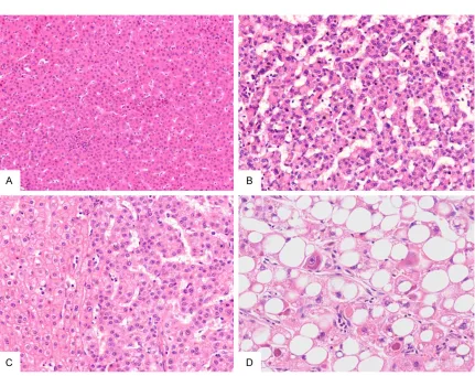
Grossly, vaguely nodular type was present in 16.4% (10/61) (Figure 1A and 1B) and distinct-ly nodular type in 86.6% (51/61), including sim-ple nodular type (35/61) (Figure 1C and 1D), extranodular extension type (14/61) (Figure 2A and 2B), and multinodular confluent type (2/51) (Table 2). Nodule in nodule appearance was present in 9 patients (14.8%) (Figure 2C and 2D). Fifty five patients (90.2%) had only one tumor mass. Six patients (9.8%) had more than one tumor mass and the largest mass was less than 2 cm. Histologically, 62.3% (38/61) were well differentiated type. 36.1% (22/61) were moderately differentiated type, and 1.6% (1/61) were poorly differentiated type. Vaguely nodular HCCs showed a thin trabecular pattern and no significant obvious cytological atypia. Comparison of vaguely nodular small HCC and distinctly nodular small HCC is shown in Table 3. The mean age of patients with vaguely nodu-lar type was 54.8 ± 8.79 (mean ± SD). The mean age of patients with distinctly nodular type was 57.6 ± 10.88. The vaguely nodular small HCCs had a mean diameter of 1.37 ± 0.41 cm, whereas distinctly nodular small HCCs had a mean diameter of 1.54 ± 0.40 cm. In the vaguely nodular small HCCs, the tumor cells were well differentiated in 70% (7/10) (Figure 3A-D) and moderately differentiated in 30% (3/10). In the distinctly nodular small HCCs, the tumor cells were well differentiated in 60.8% (31/51), moderately differentiated in 37.3% (19/51) (Figure 4A-C), and poorly differentiated in 1.9% (1/51) (Figure 4D). Fatty change within tumors was present in 24.6% (15/61). Vascular invasion was present in 8.2% (5/61). Vascular invasion was found only in distinctly nodular Table 1. Clinicopathologic features in small
hepatocellular carcinoma
Variables Small HCC (n=61)
Age (years) 37-75 (median, 57)
Sex
Male 48 (78.7%)
Female 13 (21.3%)
Underlying diseases
HBV 46 (75.4%)
HCV 7 (11.4%)
Alcoholic 4 (6.6%)
Unknown 4 (6.6%)
Cirrhosis
Present 47 (77.0%)
Absent 14 (23.0%)
Change
Large cell change 20 (32.8%) Small cell change 15 (24.6%) Dysplastic nodule
Low grade dysplastic nodule 8 (13.1%) High grade dysplastic nodule 10 (16.4%) Fatty change in tumor
Present 15 (24.6%)
Absent 46 (75.4%)
Vascular invasion
Present 5 (8.2%)
type. Cirrhosis was present in 77.0% (47/61). No statistically significant correlation was observed between gross type and age, tumor size, histologic grade, vascular invasion, fatty change, and cirrhosis, respectively (P > 0.05). In the non-neoplastic liver, large cell change was present in 32.8% (20/61) and small cell change was present in 24.6% (15/61). Low grade dysplastic nodule was present in 13.1% (8/61), and high grade dysplastic nodule was present in 16.4% (10/61).
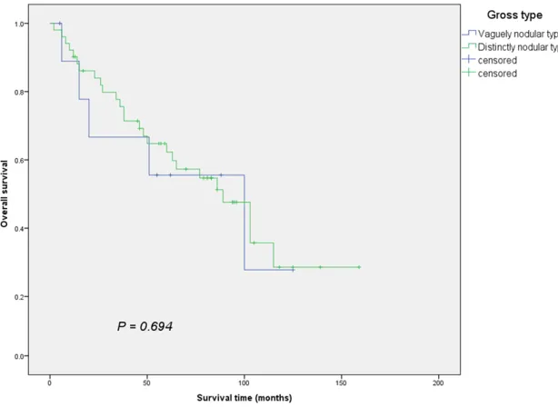
Survival of small hepatocellular carcinoma pa-tients based on gross type
There was no significant difference in overall survival between patients with vaguely nodular type and patients with distinctly nodular type (P=0.694) (Figure 5).
Discussion
Worldwide, hepatocellular carcinoma (HCC) is the fifth leading cause of death in males and
accounts for approximately 5.4% of all cancers, although its incidence varies widely in different parts of the world [11]. The highest incidences of HCC are found in Asian countries (southeast China, Korea, Taiwan) and sub-Saharan African countries. The main etiologic agents for HCC are chronic hepatitis B and C, alcoholic cirrho-sis, non-alcoholic fatty liver disease, and hemo-chromatosis. The chronic inflammation and cel-lular regeneration associated with viral hepati-tis or activation of the IL-6/JAK/STAT pathway may be predisposing factors for development of HCC [11]. In the current study, chronic hepa-titis B was the most common cause of small HCC in Korea, accounting for approximately 75% of cases. Liver cirrhosis was found in 77% of cases. Liver cirrhosis is known as a major clinical risk factor for HCC [4].
[image:3.612.91.524.72.399.2]female ratio was 3.69:1. The reason for the gender imbalance is not known. The median age was 57.
The evolution of dysplastic nodules to hepato-cellular carcinoma within several months to a few years of follow-up is well documented [8]. Vaguely nodular HCC has been identified as early well-differentiated neoplasm that usually measures less than 1.5 cm in greatest dimen-sion. As this lesion grows larger, it may
[image:4.612.89.524.72.395.2]trans-form into nodular HCC with a distinct margin [8]. In the current study, the distinctly nodular type tended to be larger than the vaguely nodu-lar type. In addition, the age of patients with distinctly nodular type tended to older com-pared with patients with vaguely nodular type. Small HCC is defined as HCC measuring less than 2 cm in diameter. More recent studies support the division of small HCC into two clini-copathological groups, termed early HCC and progressed HCC [4]. Well-differentiated small HCC of the vaguely nodular type appears to be early stage of HCC development and small HCCs of the distinctly nodular type represent progressed carcinoma [5]. In the current study, vaguely nodular HCCs were mostly well differ-entiated and had no vascular invasion. Distinctly nodular HCCs were moderately differ-entiated in 37.3% of cases and had vascular invasion. Early HCC has a longer time to recur-rence and a higher 5-year survival rate com-Figure 2. Macroscopic findings of small HCC. A, B. Distinctly nodular HCC, simple nodular type with extranodular growth. Extranodular growth is present. C, D. Distinctly nodular HCC, simple nodular type. Nodule in nodule appearance is present.
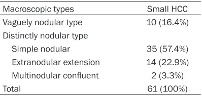
Table 2. Macroscopic type in small hepatocel-lular carcinoma
Macroscopic types Small HCC
Vaguely nodular type 10 (16.4%) Distinctly nodular type
Simple nodular 35 (57.4%)
Extranodular extension 14 (22.9%) Multinodular confluent 2 (3.3%)
[image:4.612.90.289.483.578.2]pared with progressed HCC [12]. In the present study, patients with vaguely nodular type had no significantly better overall survival than those with distinctly nodular type. Our study is limited by relatively small sample size. It is needed to investigate further.
[image:5.612.92.523.237.578.2]Small cell change, large cell change, low grade dysplastic nodule and high grade dysplastic nodule have been identified as precursor lesions to HCC. HBV-related large cell change is more consistent with dysplastic rather than merely reactive hepatocytes [13]. Small cell Table 3. Comparison of vaguely nodular type and distinctly nodular type in small hepatocellular carci-noma
Variables Vaguely nodular (n=10) Distinctly nodular (n=51) P
Age (years) (mean ± SD) 54.8 ± 8.79 57.6 ± 10.88 0.320
Tumor size (mean ± SD) 1.37 ± 0.41 cm 1.54 ± 0.40 cm 0.1987
Histologic grade 0.776
Well differentiated 7 (70%) 31 (60.8%)
Moderately differentiated 3 (30%) 19 (37.3%)
Poorly differentiated 0 (0%) 1 (1.9%)
Vascular invasion 0 (0%) 5 (9.8%) 0.580
Fatty change 3 (30%) 12 (23.5%) 0.696
Cirrhosis 7 (70%) 40 (78%) 0.683
change is considered to be a more advanced precursor lesion than large cell change [14]. Dysplastic nodules are classified as low and high grade according to the degree of atypia [3]. High grade dys-plastic nodule is the most advanced pre-cancerous lesion of the liver [15]. Dyspla- stic nodules may evol- ve over time to vaguely nodular HCC, which may then transform into distinctly nodular HCC. In the current study, 15% of cases showed small cell Figure 4. Microscopic findings of distinctly nodular HCC. A. Moderately differentiated HCC. The tumor cells are arranged in a trabecular pattern (hematoxylin-eosin stain, x100). B. Moderately differentiated HCC. The tumor cells are arranged in a pseudoglandular pattern (hematoxylin-eosin stain, x100). C. Moderately differentiated HCC. Sinusoidal blood vessels are replaced by fibrous connective tissue (hematoxylin-eosin stain, x100). D. Poorly dif -ferentiated HCC. The tumor cells show compact growth (hematoxylin-eosin stain, x100).
[image:6.612.92.399.476.700.2]change in the nontumorous liver and 16.4% of cases showed high grade dysplastic nodule in the nontumorous liver.
With advances in diagnostic imaging, detection of HCCs less than 2 cm in diameter has increased. Detection of premalignant lesions of HCC is important. Diagnosis of early HCC can improve patient survival. Histologically, it is sometimes difficult to distinguish early HCC from premalignant lesions. In recent studies, glypican-3, heat shock protein 70 and gluta-mine synthetase could be marker for diagnosis of early HCC [16, 17]. The diagnostic accuracy of this panel of markers has been tested in liver biopsy of hepatocellular nodules. Correlation of the histological features with clinical and radio-logical findings is important for accurate diagnosis.
Fatty change has been reported in premalig-nant nodules and early HCC [18]. Hypoxic con-dition or hypoperfusion in the tumor has been proposed as a possible mechanism of fatty change. In the current study, fatty change of tumor cells was found in 30% of vaguely nodu-lar type and 23.5% of distinctly nodunodu-lar type, respectively. The prevalence of fatty change decreases along with increasing tumor size [5]. When a well differentiated HCC reaches a size of approximately 1.0-1.5 cm, less differentiat-ed cancerous tissue evolve within it [19]. Such a phenomenon is often appreciated grossly and histologically as a nodule in nodule appear-ance. In the current study, 14.8% of HCCs were nodule in nodule appearance. Regarding mor-phologic evolution of HCC, HCCs arise either from premalignant lesions such as dysplastic nodule in liver cirrhosis or de novo lesions in noncirrhotic liver [20]. HCC is well differentiat-ed in the early stage, when it grows slowly. The growth rate accelerates when dedifferentiation occurs, with or without a nodular in nodule appearance, and the tumor develops to advanced HCC. In this study, multicentricity was present in 9.8% of cases. Multicentric development is relatively common in HCC [21]. In conclusion, a large percentage of small HCCs are well differentiated. The majority of small HCCs arise in cirrhotic liver. Hepatitis B virus infection is the most common cause of small HCCs in Korea. Further studies on molecular markers for precise diagnosis for early HCC and
mechanisms of hepatocarcinogensis are required.
Acknowledgements
This work was supported by the 2015 Yeungnam University Research Grant.
Disclosure of conflict of interest
None.
Address correspondence to: Dr. Joon Hyuk Choi, Department of Pathology, Yeungnam University College of Medicine, 170 Hyeonchung-ro, Namgu, Daegu City 705-703, Korea. Tel: 82-53-640-6754; Fax: 82-53-656-1429; E-mail: joonhyukchoi@ynu. ac.kr
References
[1] Matsui O. Detection and characterization of hepatocellular carcinoma by imaging. Clin Gas-troenterol Hepatol 2005; 3 Suppl 2: S136-140.
[2] Sakamoto M, Hirohashi S and Shimosato Y. Early stages of multistep hepatocarcinogene-sis: adenomatous hyperplasia and early hepa-tocellular carcinoma. Hum Pathol 1991; 22: 172-178.
[3] International Working Party. Terminology of nodular hepatocellular lesions. Hepatology 1995; 22: 983-993.
[4] Theise ND, Curado MP, Franceschi S, Hytiro-glou P, Judo M, Park YN, Sakamoto M, Torben-son M and Wee A. Hepatocellular carcinoma. In: Bosman FT, Carneiro F, Hruban RH, Theise ND, editors. WHO classification of tumours of the digestive system. 4th edition. Lyon: IARC Press; 2010. pp. 205-216.
[5] International Consensus Group for Hepatocel-lular NeoplasiaThe International Consensus Group for Hepatocellular Neoplasia. Patholog-ic diagnosis of early hepatocellular carcinoma: a report of the international consensus group for hepatocellular neoplasia. Hepatology 2009; 49: 658-664.
[6] Nakashima O, Sugihara S, Kage M and Kojiro M. Pathomorphologic characteristics of small hepatocellular carcinoma: a special reference to small hepatocellular carcinoma with indis-tinct margins. Hepatology 1995; 22: 101-105. [7] Sung CO, Choi SJ and Park CK. Clinicopatho-logic features of early hepatocellular carcino-ma. Korean J Pathol 2004; 38: 138-144. [8] Hytiroglou P. Morphological changes of early
[9] Kondo F. Histological features of early hepato-cellular carcinomas and their developmental process: for daily practical clinical application: Hepatocellular carcinoma. Hepatol Int 2009; 3: 283-293.
[10] Liver Cancer Study Group of Japan. General rules for the clinical and pathological study of primary liver cancer. 2nd edition. Tokyo: Kane-hara & Co., Ltd.; 2003. pp. 15-16.
[11] Theise ND. Liver and gallbladder. In: Kumar V, Abbas AK, Aster JC, editors. Robbins and Co-tran Pathologic Basis of Disease. 9th edition. Philadelphia: Elsevier Saunders; 2015. pp. 870-875.
[12] Takayama T, Makuuchi M, Hirohashi S, Saka-moto M, YamaSaka-moto J, Shimada K, Kosuge T, Okada S and Takayasu K, Yamasaki S. Early hepatocellular carcinoma as an entity with a high rate of surgical cure. Hepatology 1998; 28: 1241-1246.
[13] Kim H, Oh BK, Roncalli M, Park C, Yoon SM, Yoo JE and Park YN. Large liver cell change in hepatitis B virus-related liver cirrhosis. Hepa-tology 2009; 50: 752-762.
[14] Park YN. Update on precursor and early lesions of hepatocellular carcinomas. Arch Pathol Lab Med 2011; 135: 704-715.
[15] Di Tommaso L, Sangiovanni A, Borzio M, Park YN, Farinati F and Roncalli M. Advanced pre-cancerous lesions in the liver. Best Pract Res Clin Gastroenterol 2013; 27: 269-284.
[16] Libbrecht L, Severi T, Cassiman D, Vander Borght S, Pirenne J, Nevens F, Verslype C and Roskams T. Glypican-3 expression distinguish-es small hepatocellular carcinomas from cir-rhosis, dysplastic nodules, and focal nodular hyperplasia-like nodules. Am J Surg Pathol 2006; 30: 1405-1411.
[17] Di Tommaso L, Destro A, Seok JY, Balladore E, Terracciano L, Sangiovanni A, Iavarone M, Co-lombo M, Jang JJ, Yu E, Jin SY, Morenghi E, Park YN and Roncalli M. The application of markers (HSP70, GPC3 and GS) in liver biop-sies is useful for detection of hepatocellular carcinoma. J Hepatol 2009; 50: 746-754. [18] Kutami R, Nakashima Y, Nakashima O, Shiota
K and Kojiro M. Pathomorphologic study on the mechanism of fatty change in small hepa-tocellular carcinoma of humans. J Hepatol 2000; 33: 282-289.
[19] Kojiro M and Nakashima O. Histopathologic evaluation of hepatocellular carcinoma with special reference to small early stage tumors. Semin Liver Dis 1999; 19: 287-296.
[20] Kojiro M. Pathology of hepatocellular carcino-ma. Malden (Massachusetts): Blackwell Pub-lishing, Inc.; 2006. pp. 57-59.