ISOLATION, CHARACTERIZATION & BIOCHEMICAL ANALYSIS
OF MULTIDRUG-RESISTANT BACTERIA FROM SOIL SAMPLES
COLLECTED FROM HOSPITAL WASTE AND THEIR MOLECULAR
CHARACTERIZATION USING 16S RDNA
Iqbal Hussain Mir*1, Puja Baba1, Pratibha Singh1, Dr. Mukesh Sharma1 and Ajeet
Singh2
1
Department of Biotechnology, Maharaj Vinayak Global University, Jaipur, Rajasthan, India.
2
Helix Bio Genesis Pvt. Ltd., Noida, Utter Pradesh, India.
ABSTRACT
The antibiotic sensitivity test was performed for the isolated cultures
obtained from different hospital waste of Delhi & NCR. During the
study, out of 25 cultures, only 5 samples showed a remarkable
antibiotic resistance. The bacterial isolates were characterized
biochemically with the help of Bergey’s manual. They included;
Neisseria mucosa, Staphylococcus epidermidis and Morecoccus
cerebrosus. The bacterial isolates were further confirmed by molecular
characterization using 16s rDNA. The total of 11 antibiotics were used
included; Tetracycline, Ampicillin, Sparfloxacin, Co-timoxazole,
Gatifloxacin, Cefrizoxime, Amoxicillin, Ciprofloxacin, Nitrofurantoin,
Ofloxacin and Streptomycin. All strains were found resistant to Tetracycline, Ampicillin
Sparfloxacin, Co-timoxazole and Gatifloxacin. 8 antibiotics showed an intermediate
resistance. All isolates were found sensitive to Nitrofurantoin, Ofloxacin and Streptomycin.
KEYWORDS: MDRO’s, Hospital waste, Antibiotics, 16s rDNA.
INTRODUCTION
Microorganisms are everywhere a largely unseen world of activities that helped to create the
biosphere and that continue to support the life processes on earth. Microorganisms that are
resistant to one or more therapeutic classes of antimicrobial agents are called
Multidrug-resistant organisms (MDROs). Multiple drug resistance or Multidrug resistance is a
Volume 7, Issue 16, 1199-1208. Research Article ISSN 2277– 7105
Article Received on 09 July 2018,
Revised on 29 July 2018, Accepted on 18 August 2018
DOI: 10.20959/wjpr201816-13215
*Corresponding Author
Iqbal Hussain Mir
Department of
Biotechnology, Maharaj
Vinayak Global University,
condition enabling a disease-causing organism to resist distinct drugs or chemicals of a wide
variety of structure and function targeted at eradicating the organisms. Pathologic cells,
including bacterial and neoplastic cells can display multidrug resistance or multiple drug
resistance. Antibiotic resistance occurs when bacteria change in some way that reduces or
eliminates the effectiveness of drugs, chemicals, or other agents designed to cure or prevent
infectious. The bacteria survive and continue to multiply causing more harm. Bacteria can do
this through several mechanisms. Some bacteria develop the ability to neutralize the
antibiotic before it can do harm, others can rapidly pump the antibiotic out, and still others
can change the antibiotic attack site so it cannot affect the function of the bacteria.
The number of MDROs will increase if the selective pressure of antibiotic use continues
and the resistant organisms are able to spread from one person to another. Now,
multiple-drug resistant (MDR) bacteria are a big public health problem in our world. It is
really urgent to improve the current strategies to control this global public health threat.
Several studies have evaluated the microbiological content of hospital and household waste
quantitatively and qualitatively and found that general hospital waste contains
microorganisms with pathogenic potentials for humans comparable to household waste.[1]
Most of the microorganisms have also been reported to be resistant to the commonly used
antibiotics and as such have led to the outbreak of several diseases/infections.[2] Hospital
effluent could contain multidrug resistant (MDR) enterobacteria and enteric pathogens which
could pose a grave problem for communities. The antimicrobial selective pressure through
indiscriminate use of antibiotics has played a significant role in enriching the MDR strains in
the hospital practice. Present time the antibiotic resistance has become a major problem in the
clinical and public health prospects.[3] The main risk for public health is that resistant genes
are transferred from environmental bacteria to human pathogens. Waste effluent from
hospitals contains high numbers of resistance bacteria and antibiotic residues at concentration
able to inhibit the growth of susceptible bacteria.[4] Although sewage treatment processes
reduce the numbers of bacteria in wastewater, the effluent will still generally contain large
numbers of both resistant and susceptible bacteria.[5]
The present study is an attempt to isolate and characterize the multi-drug resistant (MDR)
bacteria from hospital waste which was obtained from various hospitals of Delhi & NCR.
The isolates were investigated for their sensitivity against eleven antibiotics and to check the
MATERIAL AND METHODS
Sample Collection
The soil samples were taken from hospital wastage dumping sites. Soil sample was chosen
because of higher probability of finding bacterial strains of localized zone mainly obtained
from dump hospital wastage which may include medicines, edibles, patient’s dressings etc, so
there might be probability of finding large amount of pathogenic bacteria. Nearly, twenty five
different soil samples were collected from various hospitals of Delhi & NCR. The
bacteriological analysis of these samples was done by serial dilution and agar plate culture
techniques. The obtained pure cultures were characterized based on their morphological and
biochemical characteristics as described in Bergey’s Manual for bacteriology.[6]
Serial Dilution and Agar Plating Technique
This method is based on the principle that when soil sample containing bacterial colonies are
cultured, bacterium develops into a visible colony on the nutrient agar plate. One gram of the
collected soil sample was suspended in 9ml of saline to obtain a 10-1 dilution (10 times
dilution). From the above dilution 1ml was transferred to a fresh 9ml saline solution to obtain
a 10-2 dilution. The process was repeated in order to produce 10-3, 10-4, 10-5 and 10-6 serial
dilutions. From the dilutions ranging from 10-3 to 10-6 0.1 ml of the suspensions were added
to nutrient agar plates (each dilution in 3 replicates) under sterile conditions and incubated at
37°C for 24 hours. The number of bacteria in the testes soil sample can be calculated by the
following formula:
Organisms per millilitre per gram soil = number of colonies (average of 3 replicates)/ volume
The isolated cultures were differentiated by their morphological characteristics and
transferred to fresh nutrient agar media to produce in pure form.
Antimicrobial resistance & susceptibility profiling of organisms using different
antibiotics
The standard method of Kirby-Bauer by disk diffusion was employed to investigate the
antibiotic susceptibility profiles of the bacterial isolates.[7] The antibiotic discs used in this
experiment are given in the following (Table 1). The Muller Hinton agar was allowed to
solidify in the Petri plates for the purpose of our experiment. Sample 1, 2 and 3 previously
inoculated into nutrient broths were spread evenly on the MH plates. Antibiotics discs of the
above mentioned concentration were placed carefully on the plates and left for diffusion for
some time. The plates were then incubated for 24 hours at 37°C.
The test antibiotic immediately begins to diffuse outward from the disks, creating a gradient
of antibiotic concentration in the agar such that the highest concentration is found close to the
disk with decreasing concentrations further away from the disk. After an overnight incubation
at 37°C, the bacterial growth around each disc is observed. If the test isolate is susceptible to
a particular antibiotic, a clear area of “no growth” will be observed around that particular
disk. The zone around an antibiotic disk that has no growth is referred to as the zone of
inhibition since this approximates the minimum antibiotic concentration sufficient to prevent
growth of the test isolate. Clear zones of growth inhibition were measured in millimetres by
Table 1: Commercial antibiotics (HiMedia Laboratories Pvt. Ltd.) used for
susceptibility of bacterial isolates.
S.
No. Antibiotics
Antibiotic Code
Antibiotic Disc Concentration
Antibiotic Class
Antibiotic Action
1 Ampicillin AM 10 mcg Penicillin Cidal*
2 Amoxicillin AX 25 mcg Penicillin Cidal
3 Ciprofloxacin CIP 5 mcg Fluorquinolones Cidal
4 Ofloxacin OF 5 mcg Fluorquinolones Cidal
5 Sparfloxacin SPX 5 mcg Fluorquinolones Cidal
6 Gatifloxacin GAT 5 mcg Fluorquinolones Cidal
7 Streptomycin STR 10 mcg Aminoglycosides Cidal
8 Cotrimoxazole CoT 25 mcg Sulfonamides Cidal
9 Ceftrizoxime CZX 30 mcg Cephalosporins Cidal
10 Nitroflurantoin NIT 300 mcg Nitrofurantoin Static
11 Tetracyclin TE 30 mcg Tetracycline Static
*Cidal= Bacteriocidal; Static = Bacteriostatic
Characterization of Bacterial Culture
Gram staining of bacteria
A smear of the microorganisms grown in NB was prepared on clean grease free glass slide by
a sterile/flamed inoculating loop after cooling it. Smear was allowed to air dry and then heat
fixed. Smear was flooded with crystal violet & allowed to stand for 1 minute and then gently
washed with tap water. Smear was flooded with Gram Iodine and allowed to stand for 1
minute. Smear was decolorised / washed with 95% ethyl alcohol until alcohol runs almost
clear. Smear was counterstained with Safranin for 1 minute. Gently washed with tap water,
air dried & observed under compound microscope at 40X magnifications.
Biochemical characterization of the isolated bacterial colonies and growth in
MacConkey agar
Total of twenty seven biochemical tests were performed according to Bergey’s Manual for
bacteriology to characterize the unknown bacterial cultures. Initially an unknown bacterial
culture was checked for catalase and oxidase activity.[8] The reagent used in oxidase test was
N, N-dimethyl-p-phenylenediamine (DMPD). The presence of catalase enzyme in the test
isolate was detected using hydrogen peroxide. The catalase test was done by placing a drop of
hydrogen peroxide on a microscope slide. Carbohydrate fermentation tests were used to
detect the ability of microorganisms to ferment a specific carbohydrate. In our experiment,
we used glucose, galactose, mannose, lactose, maltose glycerol, mannitol and sucrose with
drops of Kovac’s reagent in peptone water broth with bacterium incubated for 18-24 hours at
37°C. In Voges Proskauer (VP) test, 40% KOH and alpha-nephthol were added to test broth
after incubation and exposed to atmospheric oxygen. Nitrate reduction test was done by
adding 1ml of each sulphanilic acid and α-nepthylamine to the inoculated test organisms to
detect the production of enzyme nitrate reductase which reduces nitrate to nitrites. Starch
hydrolysis test was also carried out to check the presence of and exoenzyme amylase which
cleaves the starch into disaccharides and monosaccharides. This was done by flooding Iodine
reagent to the overnight culture medium containing starch. Similarly, gelatinase test was
carried out to check the presence of gelatinase.
With some knowledge about the nature of organisms we moved towards the task for growing
the selected MDR on the following media. For this, MacConkey agar culture medium was
used.[8] The bacterial colonies isolated were subjected to catalase and oxidase test and
observations were noted. The MDR’s obtained were made to grow in MacConkey colony and
characteristics were noted down.
Extraction of DNA and Molecular Characterization Using 16s rDNA
Bacterial genomic DNA was isolated as per the standard protocol.[9] The isolated cultures
were streaked on Luria Betani (LB) agar and grown overnight. Cells were harvested from 5
mL of the culture and to this 100μL of lysozyme was added and incubated at RT for 30 min,
followed by the addition of 700 μL of cell lysis buffer (Guanidium isothiocyanate, SDS,
TrisEDTA). The contents were mixed by inverting the vial for 5 min with gentle mixing till
the suspension looked transparent. 700 μL of isopropanol was added on top of the solution.
The two layers were mixed gently till white strands of DNA were seen. The DNA extracted
50μL of 1X TE buffer. The quality of the DNA was checked by running on 0.8% agarose gel stained with ethidium bromide (0.5 μg/μL). A single intense band with slight smearing was
noted. The extracted genomic DNA was used as template DNA for amplification of the 16S
rDNA gene. PCR amplification of 16S rRNA gene: PCR reaction was performed in a
gradient thermal cycler (Thermo Scientific). The reaction mixture of 50 μl consisted of 10 ng
of genomic DNA, 3 units of Taq DNA polymerase, 5 μl of 10X PCR amplification buffer
(100 mM TrisHCl, 500 mM KCl pH-8.3), 200μM dNTP, 10 p moles each of the two
universal primers and 1.5mM MgCl2. The 16S rDNA sequence was amplified using
universal primers (Forward primer 5'- AGAGTTTGATCCTGGCTCAG-3' and reverse
primer 5 (5’ GGTTACCTTGTTACGACTT 3’). The reaction condition include 1 min
denaturation (95°C) followed by 30 cycles of 96°C for 30 s, 48.5°C for 30 s and 72°C for
30 s and a final extension of 72°C for 10 min. PCR products were then separated and
visualized on 1% agarose gel electrophoresis to confirm amplification.
Samples thus were sent for DNA sequencing to determine the precise sequence of amplified
product. The amplified product was sequence using the same universal primers used for
amplification. The sequencing was done using ABI 3500 Genetic AnalyserTM with Big Dye
Terminator version 3.1. Applied Biosystems Micro Amp Optical 96-well reaction plate was
used for the sequencing. Bioinfomatic analysis of sequence data (FASTA format) was done
for genebank search and sequence alignment assay. Nucleotide BLAST was performed for
this purpose and E-Value, % similarity and local alignment were taken into consideration for
determining the homology and to identify the bacterial strains upto species level.
RESULTS AND DISCUSSION
Twenty five bacterial isolates were selected for antibiotic resistance, morphological,
biochemical and further investigations. These bacterial isolates were screened for
antimicrobial sensitivity profiles. Out of the twenty five different samples only five samples
showed a remarkable antibiotic resistance and these five samples were labelled as C2, C3,
C18, C22 and C24. Resistance range of the bacterial isolates for 11 antibiotics examined in
downward order was separately tetracyclin, Ampicillin, Sparfloxacin, Co-timoxazole,
Gatifloxacin, Cefrizoxime, Amoxicillin, Ciprofloxacin, Nitrofurantoin, Ofloxacin and
Streptomycin. The resistance of the 5 bacterial isolates to the 11 commonly used antibiotics
revealed that for all antibiotics, all the cultures were showing resistance against Tetracycline,
intermediate resistant to 8 antibiotics out of total 11 investigated. All isolates were found
sensitive to Nitrofurantoin, Ofloxacin and Streptomycin. Antibiotic susceptibility pattern of
the bacterial isolates is summarized below in Table 2.
Five MDR bacterial cultures were isolated from soil samples collected from hospital waste of
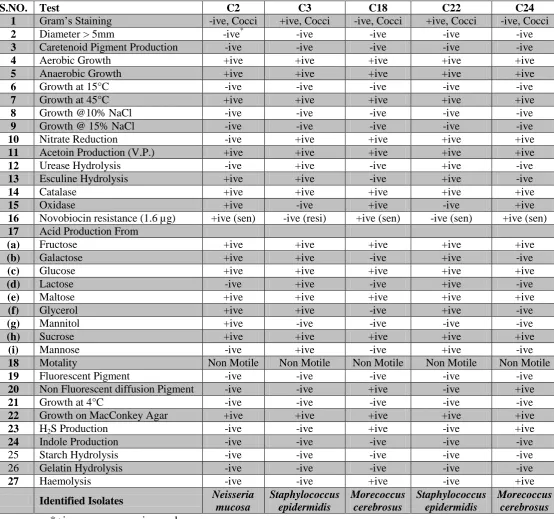
Delhi & NCR which were selected for further characterization. Based upon Gram staining,
three isolates (C2, C18 and C24) were identified as Gram negative which are cocci and two
isolates (C3 and C22) were Gram positive which are cocci also. All isolated bacterial cultures
were aerobic. Based upon the biochemical analysis (Table, 3), bacterial isolate labbled as C2
was identified as Neisseria mucosa, bacterial isolates labbled as C3 and C22 was identified as
Staphylococcus epidermidis and bacterial isolates labbled as C18 and C24 was identified as
Morecoccus cerebrosus. The use of 16S rDNA gene sequences to identify new strains of
bacteria is gaining momentum in recent years. Based upon the molecular characterization
while using 16S rDNA gene sequence to characterize the bacterial isolate, the homology
search made using BLAST showed 97% identity with that of Neisseria mucosa strain labelled
as C2, NCBI Gene Bank Accession No: CP028150.1 and E-value equal to 0 for all closely
related taxa, 98% maximum similarity with Staphylococcus epidermidis strains labelled asC3
and C22, NCBI Gene Bank Accession No: CP014119.1 and E-value equal to 0 for all closely
related texa and 98% similarity with Morecoccus cerebrosus strains labelled asC18 and C24,
NCBI Gene Bank Accession No: LN899799.1 and E-value equal to 0 for all closely related
texa.
Table 2: Antibiotic susceptibility test of 5 bacterial cultures.
Antibiotic discs C2 C3 C18 C22 C24
Tetracyclin R* R R R R
Ampicillin R R R R R
Sparfloxacin R R R R R
Cotrimoxazole R R R R R
Gatifloxacin R R R R R
Cefrizoxime R R R S R
Amoxicillin R R R S S
Ciprofloxacin R S S R S
Nitroflurantoin S S S S S
Ofloxacin S S S S S
Streptomycin S S S S S
Table 3: Characteristic tests of multidrug-resistant bacterial isolates.
S.NO. Test C2 C3 C18 C22 C24
1 Gram’s Staining -ive, Cocci +ive, Cocci -ive, Cocci +ive, Cocci -ive, Cocci
2 Diameter > 5mm -ive* -ive -ive -ive -ive
3 Caretenoid Pigment Production -ive -ive -ive -ive -ive
4 Aerobic Growth +ive +ive +ive +ive +ive
5 Anaerobic Growth +ive +ive +ive +ive +ive
6 Growth at 15°C -ive -ive -ive -ive -ive
7 Growth at 45°C +ive +ive +ive +ive +ive
8 Growth @10% NaCl -ive -ive -ive -ive -ive
9 Growth @ 15% NaCl -ive -ive -ive -ive -ive
10 Nitrate Reduction -ive +ive +ive +ive +ive
11 Acetoin Production (V.P.) +ive +ive +ive +ive +ive
12 Urease Hydrolysis -ive +ive -ive +ive -ive
13 Esculine Hydrolysis +ive +ive -ive +ive -ive
14 Catalase +ive +ive +ive +ive +ive
15 Oxidase +ive -ive +ive -ive +ive
16 Novobiocin resistance (1.6 µg) +ive (sen) -ive (resi) +ive (sen) -ive (sen) +ive (sen)
17 Acid Production From
(a) Fructose +ive +ive +ive +ive +ive
(b) Galactose +ive +ive -ive +ive -ive
(c) Glucose +ive +ive +ive +ive +ive
(d) Lactose -ive +ive -ive +ive -ive
(e) Maltose +ive +ive +ive +ive +ive
(f) Glycerol +ive +ive -ive +ive -ive
(g) Mannitol +ive -ive -ive -ive -ive
(h) Sucrose +ive +ive +ive +ive +ive
(i) Mannose -ive +ive -ive +ive -ive
18 Motality Non Motile Non Motile Non Motile Non Motile Non Motile
19 Fluorescent Pigment -ive -ive -ive -ive -ive
20 Non Fluorescent diffusion Pigment -ive -ive +ive -ive +ive
21 Growth at 4°C -ive -ive -ive -ive -ive
22 Growth on MacConkey Agar +ive +ive +ive +ive +ive
23 H2S Production -ive -ive +ive -ive +ive
24 Indole Production -ive -ive -ive -ive -ive
25 Starch Hydrolysis -ive -ive -ive -ive -ive
26 Gelatin Hydrolysis -ive -ive -ive -ive -ive
27 Haemolysis -ive -ive +ive -ive +ive
Identified Isolates Neisseria mucosa Staphylococcus epidermidis Morecoccus cerebrosus Staphylococcus epidermidis Morecoccus cerebrosus *+ive = presence; -ive = absence
CONCLUSION
A major factor in the emergence of Multi Drug Resistant Organisms (MDRO’s) is
overuse of antibiotics in any setting, the hospital or the community. Many human
pathogenic bacteria are examples of this emerging crisis, and they are extremely difficult to
treat with present existing extended-spectrum antibiotics, leading to increased morbidity and
day. The main source of the Multi Drug Resistant Organisms is the hospital wastes. In the
metro cities like New Delhi, the hospitals and the clinics are found in every knock and corner
and are increasing. Thus Multi Drug Resistant Organisms are increasing in such cities and is
becoming a serious threat in the whole world. It is really urgent to improve the current
strategies to control this global public health threat.
In our present study we have tried to isolate and identify using both biochemical and
molecular methods to ascertain the MDR microbes upto species level. We have also done the
detailed analysis of MDR to understand and decide the possible control methods for the
above mentioned strains/microbes.
REFERENCES
1. Saini S, Bimal K.D, Kapil A, Shyama S.N. and Sarma R.K. The study of bacterial flora of
different types in hospital waste: Evaluation of waste treatment at AIIMS hospital, New
Delhi. Southeast Asian Journal of Tropical Medicine and Public Health, 2004; 35(4):
986-88.
2. Chitnis V, Chitnis D, Patil S and Kant R. Hospital effluent: A source of multiple
drug-resistant bacteria. Current Science, 2000; 79: 989-91.
3. Stuart. B. Factors impacting on the problem of antibiotic resistance. Journal of
Antimicrobial Chemotherapy, 2002; 1: 25-30.
4. Grabow, W and Prozesky, O. Drug resistance of coliform bacteria in hospital and city
sewage. Antimicrob. Agents Chemother, 1973; 3: 175-80.
5. Schwartz T, Kohnen W, Jansen B and Obst U. Detection of antibiotic-resistant bacteria
and their resistance genes in wastewater, surface water and drinking water biofilms.
FEMS Microbiol Ecol., 2003.
6. G.M. Garrity, D.R. Boone and R.W. Castenholz, “Bergey’s Manual of Systematic
Bacteriology”, 2nd
ed., vol. 1, Springer-Verlag, New York, NY, 2001.
7. NCCLS, National Committee for Clinical Laboratory Standards 2012. NCCLS document
M100-S9. Performance standards for antimicrobial susceptibility testing, 9th ed.
Informational supplement. NCCLS, Wayne, Pa.
8. Cappuccino JG, Sherman N. Microbiology: A laboratory manual. 7th ed., New Delhi;
Pearson education Inc. and Dorling Kindersley, 2005: 71-73.