ORIGINAL RESEARCH
Evaluation of Carotid Artery Stenosis with
Multisection CT and MR Imaging: Influence of
Imaging Modality and Postprocessing
M. Lell C. Fellner U. Baum T. Hothorn R. Steiner W. Lang W. Bautz F.A. Fellner
BACKGROUND AND PURPOSE: We prospectively evaluated the influence of different imaging tech-niques (time-of-flight MR angiography [TOF-MRA], contrast-enhanced MR angiography [CE-MRA], multisection CT angiography [CTA]) and postprocessing methods (maximum intensity projection [MIP], multiplanar reformation [MPR]) on carotid artery stenosis grading.
MATERIALS AND METHODS: Fifty patients (34 men, 16 women) with symptomatic stenosis of the internal carotid artery were examined with a 16-section spiral CT and a 1.5T MR unit. Two MRA techniques were applied: 3D-TOF and CE-MRA. MPR was used for postprocessing with all modalities; MIP was used only with MRA. Four readers measured and calculated the percentage diameter stenosis independently according to NASCET criteria. The Wilcoxon test was used to measure interobserver variability, and the Friedman test was used to test the null-hypothesis of equality of the modalities.
RESULTS: The hypothesis for global equality was rejected (P⬍.001). TOF-MRA and CTA assessed with MPR showed the highest concordance (difference, 0.6%; confidence interval [CI],⫺3.0, 4.3%), and CE-MRA with MIP and CTA showed the lowest concordance in stenosis grading (difference, 7.0%; CI, 3.4, 10.6%). MPR resulted in lower degrees of stenosis than MIP for both MRA sequences, although not statistically significant (CE,⫺3.0%; CI,⫺6.6, 0.6%; TOF,⫺2.2%; CI,⫺5.8, 1.4%). When only studies with good or excellent image quality were considered, the differences decreased, but the trends remained.
CONCLUSION:Stenosis grading is dependent on the examination method and postprocessing tech-nique. CTA and TOF-MRA evaluated with MPR revealed highest concordance.
T
herapeutic decisions in the large clinical trials (North American Symptomatic Carotid Endarterectomy Trial Collaborators, European Carotid Surgery Trial, and Asymp-tomatic Carotid Atherosclerosis Study)1-4 were based on maximal internal carotid artery (ICA) stenosis depicted with conventional angiography. Selective intra-arterial digital sub-traction angiography (DSA) still provides the highest spatial resolution and dynamic information, but it is associated with the risk of neurologic complications.5-8Furthermore, diag-nostic strategies with DSA were found to be inferior consider-ing cost-effectiveness calculations in patients with symptom-atic ICA stenosis.9The limited number of projections may be a limitation of DSA in the evaluation of high-grade stenosis leading to underestimation of severity.10,11The standard de-viation associated with percent diameter stenosis measure-ment for 60% stenosis at carotid x-ray angiography across several published studies was reported to be 8%.12 Noninva-sive tests like sonography, MR angiography (MRA), and spiral CT angiography (CTA) are increasingly used in clinical rou-tine. They offer multiple projections and the possibility to use cross-sectional images to measure the stenotic lumen. As many as 80% of patients with carotid artery stenosis in the United States were estimated to have undergone carotidend-arterectomy without x-ray angiography13; in addition, this ap-proach is considered controversial.14,15
Despite numerous studies comparing MRA or CTA to DSA, only few data have been published concerning the influ-ence of imaging technique and postprocessing technique on stenosis grading in vivo.16-21The aim of this study was to in-traindividually compare the results of high-resolution MRA and multisection CTA and the effect of different postprocess-ing techniques on stenosis measurement.
Methods
Patients
Sixty-nine consecutive patients who had symptoms related to atheroscle-rotic carotid artery disease within a 6-month period and ICA stenosis at sonography were screened for inclusion in this prospective study. Pa-tients with contraindications to MR imaging or iodinated contrast mate-rial were excluded from the study. Written informed consent was ob-tained from all patients and the ethics committee approved the study. CTA and MRA were performed within 24 hours of each other.
Nineteen patients were excluded from the study because of the following reasons: technical failure of the CT scanner (2 patients), incomplete MR examinations (2 patients due to claustrophobia, 2 patients rejecting IV contrast material [CM]), only 1 study performed within 24-hour interval (13 patients) as a result of scanner availability or withdrawal from study. Therefore, the study population consisted of 50 patients (34 men, aged 50 – 84 years; median, 66.5 years; 16 women: 53– 82 years; median, 74.5 years).
CT Angiography
CTA was performed with a 16-section-spiral CT scanner (Somatom Sensation 16; Siemens, Erlangen, Germany). An 18-gauge
intrave-Received December 24, 2005; accepted after revision March 13, 2006.
From the Institute of Diagnostic Radiology (M.L., U.B., R.S., W.B.) and Departments of Medical Informatics, Biometry, and Epidemiology (T.H.) and Vascular Surgery (W.L.), University Erlangen-Nuremberg, Erlangen, Germany; Institute of Radiology (C.F.), Landes-Nervenklinik Wagner Jauregg, Linz, Austria; and Institute of Radiology (F.A.F.), AKh Linz, Linz, Austria.
nous catheter was placed in an antecubital vein and 60 mL nonionic CM (Ultravist 370; Schering, Berlin, Germany) were injected with a power injector at a rate of 4 mL/s followed by a saline flush of 80 mL. The start delay (TDelay) was individually adjusted with the test bolus method (10 mL of CM, 50 mL of saline, 4 mL/s). The scan volume included the circle of Willis to the aortic arch; scanning was per-formed in craniocaudal direction. Scanner settings were: 120 kV, 110 eff䡠mAs, detector collimation, 16⫻0.75 mm; table speed, 18 mm/ rotation; rotation time, 0.5 seconds. The patients were instructed to hold their breath during the scan; total scan time was 9 seconds. Sec-tions (1.0 mm) were reconstructed with 0.5-mm increment. The field of view (FOV) was 120 mm, and the resulting voxel size was 0.2⫻ 0.2⫻0.5 mm3. Average effective dose was 2.4 mSv for female patients and 2.2 mSv for male patients (International Commission on Radio-logical Protection publication 60, 1990).22
For each carotid artery a thin slab maximum intensity projection (MIP) (10 –15-mm slab thickness) in a sagittal oblique projection was
created to display vascular anatomy. Based on this MIP, cross-sec-tional images perpendicular to the longitudinal axis of the ICA at the site of maximum stenosis and a distal reference site were generated with the multiplanar reformation (MPR) technique. On these cross-sectional images, the lumen diameters were measured. Images were displayed with a window/level setting of 700/200 HU; in cases of mas-sive calcification, a wider setting (1100/200 HU) was applied.23
MR Angiography
Two different MRA sequences were performed with a 1.5T MR unit (Symphony; Siemens) with the use of head and neck array coils.
3D time-of-flight (TOF) MRA of the carotid bifurcation settings were: TR, 35 ms; TE, 6.95 ms; flip angle, 25°; rFOV, 150⫻200 mm; matrix, 192⫻512; slab thickness, 57.6 mm; 72 partitions; voxel size after zero-filling, 0.8⫻0.4⫻0.8 mm3; first-order flow compensa-tion; total scan time, 4:02 minutes.
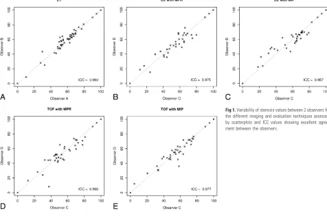
Contrast-enhanced (CE) MRA, coronal 3D fast low-angle shot Fig 1.Variability of stenosis values between 2 observers for the different imaging and evaluation techniques assessed by scatterplots and ICC values showing excellent agree-ment between the observers.
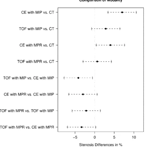
Mean difference in stenosis value and 95% confidence intervals of all symptomatic stenosis for each imaging modality (cases of local signal loss assigned as 90% stenosis)
Modality
All Examinations (Signal Loss⫽90%) Examinations Without Local Signal Loss Only
Difference (%) 95% Simultaneous CI Difference (%) 95% Simultaneous CI
CE-MIP vs CT 7.0 3.4 10.6 2.8 ⫺0.8 6.4
TOF-MIP vs CT 2.8 ⫺0.8 6.5 ⫺0.8 ⫺4.4 2.8
CE-MPR vs CT 4.0 0.4 7.6 ⫺0.8 ⫺4.4 2.7
TOF-MPR vs CT 0.6 ⫺3.0 4.3 ⫺3.9 ⫺7.5 ⫺0.4
TOF-MIP vs CE-MIP ⫺4.2 ⫺7.8 ⫺0.6 ⫺3.6 ⫺7.2 0
CE-MPR vs CE-MIP ⫺3.0 ⫺6.6 0.6 ⫺3.6 ⫺7.2 ⫺0.1
TOF-MPR vs TOF-MIP ⫺2.2 ⫺5.8 1.4 ⫺3.1 ⫺6.7 0.4
TOF-MPR vs CE-MPR ⫺3.4 ⫺7.0 0.3 ⫺3.1 ⫺6.7 0.5
Note:—CI indicates confidence interval; CE, contrast-enhanced; MIP, maximum intensity projection; TOF, time-of-flight; MPR, multiplanar reformation. When only studies without local signal loss are included, the differences between the modalities decrease.
BRAIN
ORIGINAL
[image:2.585.53.529.46.361.2](FLASH) sequence settings were: TR, 3.78 ms; TE, 1.54 ms; flip angle, 35°; rFOV, 188⫻300 mm; matrix, 160⫻512; slab thickness, 64 mm; 80 partitions; voxel size after zero-filling, 1.2⫻0.6⫻0.8 mm3; scan time, 21 seconds per scan. The patients were instructed to hold their breath during the scan. TDelay was measured with the test bolus method. The 3D-FLASH sequence was performed before and after the injection of the full CM bolus (20 mL of gadolinium-diethylene-tri-aminepentaacetic acid; flow, 2 mL/s) for subtraction.
The minimal residual lumen diameter and the nonstenotic diam-eter of the ICA were measured on the MIP image, selecting the out-ermost margin of the vessel lumen perpendicular to the longitudinal axis of the vessel. In a second session, MRA was analyzed with the MPR method as described above for CTA.
Image Analysis
To reduce errors by measuring the reference lumen at different sites, a senior radiologist reviewed the TOF-MRA studies and specified for each patient the point of reference measurement (normal ICA lumen) by determining the distance from the carotid flow divider to the point of measurement. Four investigators (4 – 6 years experience in neuro-vascular imaging) were blinded to the clinical data and each other’s results; the examinations were anonymized and randomly ordered. Each technique was evaluated by 2 investigators independently. CE-MRA and TOF-MRA were evaluated at different sessions with a delay of at least 2 months. CTA and MRA were evaluated interactively on separate workstations with the Syngo-3D platform (Leonardo VD10B; Siemens); the ICA lumen was measured with a digital ruler (resolution, 0.01 cm). The degree of stenosis was calculated with the following equation: stenosis⫽(1⫺minimal residual lumen/distal ICA diameter)⫻100%. Because we did not categorize stenosis, we had to assign cases of local signal intensity loss, where no measure-ments could be taken, an arbitrary value; we chose 90% as the degree of stenosis in these cases.24,25Cases of near occlusion (defined as a
continuously enhanced lumen or a short segment of nonenhanced lumen due to a very tight stenosis and significant narrowing of the poststenotic ICA) were assigned 95% as the degree of the stenosis according to the literature.26Each investigator subjectively classified the image quality as excellent (no artifacts, no venous enhancement), good (moderate venous enhancement or minimal artifacts but suffi-cient for diagnostic purposes), poor (reduced contrast-to-noise ratio [CNR] or major artifacts) and insufficient for diagnosis (low CNR or severe artifacts). Examinations with poor image quality were not ex-cluded from the study. Three imaging acquisition methods (CTA, TOF-MRA, CE-MRA) and 2 postprocessing techniques for MRA re-sulted in 5 different modalities for each carotid artery.
Statistical Analysis
For each of the 5 modalities (CT, CE-MIP, CE-MPR, TOF-MIP, TOF-MPR), the discrepancy between the 2 independent observers was analyzed by scatterplots and intraclass correlation coefficients (ICC). Stenosis values by 2 observers were averaged within the mo-dalities for comparison. Only the symptomatic side was used for sta-tistical analysis. The global null-hypothesis of the equality of the dis-tributions of the stenosis values derived by the 5 modalities was tested using the Friedman test. The assessment of the deflections from the null-hypothesis was performed by simultaneous 95% Tukey confi-dence intervals (CI) for the mean difference in stenosis in a 1-way layout for the aligned stenosis values. The alignment of the stenosis values by centering the stenosis values for each patient by their mean allows for a comparison of the stenosis differences between patients at a common measurement scale.27Hypotheses were rejected when the Pvalue of the corresponding test statistic was less than␣⫽0.05. The relationship of stenosis values (of the symptomatic side only) for 2 modalities or observers are depicted by scatterplots and Bland-Alt-man plots with slightly jittered values as recommended for the anal-ysis of carotid artery stenosis measurements by Rothwell.28,29Mutual dependence of image quality and imaging technique was tested with the Fisher exact test. To examine the influence of image quality on differences on stenosis measurement the estimates and 95% CIs were computed for studies with excellent and good image quality only. All computations were performed in the R system for statistical comput-ing;30simultaneous confidence intervals were computed using the multcomp add-on package.31
Results
All 3 acquisition methods provided arterial phase images with-out significant venous enhancement. Overall image quality of the CTA and MRA examinations differed significantly (P⬍ .001, Fisher exact test); in 17 patients, the image quality for CTA was rated 1 or more grades higher than the best MRA sequence, and in 8 patients, the best MRA sequence was rated higher than CTA. Mean grade of stenosis was 60% measured with CT, 67% with CE-MIP, 63% with TOF-MIP, 64% with CE-MPR, and 61% with TOF-MPR, respectively. For the grading of stenosis, no systematic deviations between the 2 observers could be found for any technique. The ICC values and the scatterplots are shown in Fig 1. To test bias from the examiners, CTA and TOF-MIP were evaluated by 1 group; the estimated difference was 0.3% for CTA and ⫺0.5% for TOF-MIP on average.
The 5 modalities differ with respect to the stenosis values; the Friedman test was able to reject the hypothesis of global equality (P⬍.001). The degree and direction of the deviations Fig 2.Comparison of modalities by differences between stenosis values. Pairs of
[image:3.585.55.286.46.280.2]were assessed by simultaneous confidence intervals for the dif-ference of stenosis values (Table).
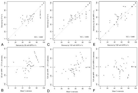
The highest agreement was found for TOF-MPR and CTA with an estimated difference of 0.6%. CE-MRA differed signif-icantly from CTA, the estimated difference of CE-MIP and CTA was 7.0%, and that of CE-MPR and CTA was 4.0%. TOF-MIP differed significantly from CE-TOF-MIP; the estimated differ-ence was⫺4.2%, indicating that the stenosis values were 4.2% lower with TOF-MIP compared with CE-MIP. The data indi-cate a trend toward higher degrees of stenosis for MIP com-pared with MPR for both TOF-MRA and CE-MRA and higher degrees of stenosis with CE-MRA compared with TOF-MRA and CTA. A graphical representation of the confidence inter-vals is shown in Fig 2, and the stenosis values are depicted in Fig 3.
When only patients with excellent and good image qual-ity in all of the 3 different image acquisition methods (CTA, CE-MRA, TOF-MRA) were included, the differences be-tween the methods decreased, but the direction of the de-viations remained, except for the comparison of TOF-MPR and CTA, where an estimated difference of ⫺0.5% was found.
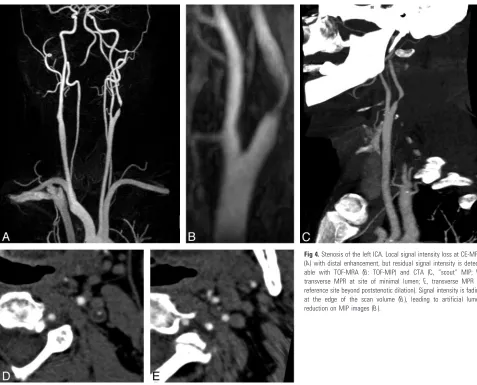
We recorded local signal intensity losses in 12 cases with CE-MRA (Fig 4) and 8 with TOF, whereas on the cross-sec-tional images of CTA, a residual lumen was depicted in all but 1 case without total occlusion.
Excluding cases with local signal intensity loss to avoid bias by arbitrarily assigning a stenosis value resulted in equalizing
the estimated differences in stenosis values across the meth-ods, and only borderline significances resulted for TOF-MRA versus CTA and CE-MPR versus CE-MIP (Table).
All 3 image acquisition methods depicted total occlusion in 5 carotid arteries. Another artery was suggested to be occluded on TOF-MRA, but both CE-MRA and CTA could demon-strate distal vessel enhancement; high-grade stenosis was con-firmed at surgery.
Discussion
[image:4.585.56.531.42.360.2]sity loss is caused by different factors: spin dephasing effects, imaging parameters, and partial volume effects.25,33-36 How-ever, assigning these cases as 90% stenosis may exaggerate the differences between the modalities (see Table, right part). Be-cause we did not categorize the stenoses, we had to determine a certain value as the degree of stenosis.
Besides the acquisition technique, postprocessing can in-fluence the assessment. Lumen measurement can be inaccu-rate on source images. If the minimal diameter is not perpen-dicular to the image plane,37subtle vascular signals may not be distinguished from background signals with the MIP algo-rithm.38Calcified plaques preclude MIP evaluation of CTA; therefore, we did not introduce this method in our study. Heavy calcifications or calcified plaque on both sides of the lumen can lead to overestimation of the stenosis in CT39 be-cause of the so called “blooming” artifact. This is why we changed the level setting in these cases. The window-level setting is critical; we chose settings proposed in the liter-ature23,40and validated them with phantom measurements simulating calcified and noncalcified stenosis.
Image quality was found to influence the concordance but to a lesser extent than the type of technique applied. CTA provided higher overall image quality compared with CE-MRA and TOF-CE-MRA. 3D-TOF-CE-MRA was more susceptible to motion artifacts because of the prolonged acquisition time and the limited volume coverage. Tapering vessel walls at the
edge of the scan volume may be found on MIP images because of reduction of signal intensity due to increased spin satura-tion. Image quality in CE-MRA is highly dependent on correct contrast bolus timing and imaging parameters.
Contrary to our study, the only multicenter study includ-ing data from multisection CT,41comparing the results of ul-trasound, CTA, and CE-MRA, revealed no significantly differ-ent overall concordance rates, but in the subgroup of surgical asymptomatic patients, CTA led to underestimation in 11 of 64 patients. However, the study had some limitations: the study was performed on different scanner types; CTA data acquisition and postprocessing were not standardized, unlike Doppler ultrasound and CE-MRA; and 81.8% of the misclas-sification came from one center. According to the proposal of Rothwell,28we did not categorize stenosis but compared the percentage values. These values differed significantly for CTA and CE-MRA, especially if assessed with MIP, whereas the difference between CTA and TOF-MRA was in a range of 0.6 – 2.8%. The trend toward higher stenosis values in CE-MRA compared with CTA is in agreement with the CARMEDAS study.41
[image:5.585.53.530.39.424.2]85%–95% and specificities of 93%–98% for detecting severe stenosis (⬎70%) with CTA.42,43
The largest study comparing DSA and TOF-MRA was pub-lished by Nederkoorn et al44; the sensitivity for detecting se-vere stenosis was 92.2% and specificity was 75.7% for TOF-MRA. CE-MRA tended to overestimate stenosis compared with DSA by a mean bias of 2.4 –3.8% according to U-King-Im et al.35A multivariable receiver operating characteristic curve analysis45demonstrated that the presence of verification bias predicted the performance of ultrasound, whereas the type of scanner predicted the performance of MRA. Performance of MRA is dependent on multiple factors including spatial reso-lution, type of sequence, interpolation algorithms, and imple-mentation details,25making the comparison of results with different MRA techniques difficult.
A limitation of our study is the relatively small patient pop-ulation; to reduce bias, we analyzed only the symptomatic side of each patient. We did not analyze the performance of duplex sonography, because the examinations were performed in dif-ferent laboratories with nonuniform equipment and exper-tise. DSA or—preferably—3D-rotational DSA as a reference standard would have been desirable, but our primary goal was to intraindividually compare the results of high-resolution MRA and multisection CTA and the effect of different post-processing techniques on stenosis measurement. Defining the site of the distant reference lumen measurement (denomina-tor in the formula) conflicts with clinical practice but was nec-essary for this study to reduce potential method-independent errors.
In conclusion, we found the grade of stenosis in carotid angiography by CT and MR to be dependent on the examina-tion method (CT, CE-MRA, TOF-MRA) and the postprocess-ing method (MIP, MPR). The highest agreement was found for CTA and TOF-MRA evaluated with the MPR method, ir-respective of assigning cases of local signal intensity loss as 90% stenosis. A separate analysis of all studies without local signal intensity loss indicates that those differences vanish. Further trials will be needed to determine whether the differ-ences are clinically significant in patient outcome.
References
1. Barnett HJ, Taylor DW, Eliasziw M, et al.Benefit of carotid endarterectomy in patients with symptomatic moderate or severe stenosis. North American Symptomatic Carotid Endarterectomy Trial Collaborators.N Engl J Med
1998;339:1415–25
2. Randomised trial of endarterectomy for recently symptomatic carotid stenosis: final results of the MRC European Carotid Surgery Trial (ECST).Lancet1998; 351:1379 – 87
3. Anonymous.Endarterectomy for asymptomatic carotid artery stenosis. Exec-utive Committee for the Asymptomatic Carotid Atherosclerosis Study.JAMA
1995;273:1421–28
4. Rothwell PM, Eliasziw M, Gutnikov SA, et al.Analysis of pooled data from the randomised controlled trials of endarterectomy for symptomatic carotid ste-nosis.Lancet2003;361:107–16
5. Willinsky RA, Taylor SM, TerBrugge K, et al.Neurologic complications of cerebral angiography: prospective analysis of 2,899 procedures and review of the literature.Radiology2003;227:522–28
6. Dion JE, Gates PC, Fox AJ, et al.Clinical events following neuroangiography: a prospective study.Stroke1987;18:997–1004
7. Citron SJ, Wallace RC, Lewis CA, et al.Quality improvement guidelines for adult diagnostic neuroangiography: cooperative study between ASITN, ASNR, and SIR.J Vasc Interv Radiol2003;14:S257– 62
8. Connors JJ 3rd, Sacks D, Furlan AJ, et al.Training, competency, and creden-tialing standards for diagnostic cervicocerebral angiography, carotid stent-ing, and cerebrovascular intervention: a joint statement from the American Academy of Neurology, the American Association of Neurological Surgeons,
the American Society of Interventional and Therapeutic Neuroradiology, the American Society of Neuroradiology, the Congress of Neurological Surgeons, the AANS/CNS Cerebrovascular Section, and the Society of Interventional Radiology.Radiology2005;234:26 –34
9. Buskens E, Nederkoorn PJ, Buijs-Van Der Woude T, et al.Imaging of carotid arteries in symptomatic patients: cost-effectiveness of diagnostic strategies.
Radiology2004;233:101–12
10. Elgersma OE, Buijs PC, Wust AF, et al.Maximum internal carotid arterial stenosis: assessment with rotational angiography versus conventional in-traarterial digital subtraction angiography.Radiology1999;213:777– 83 11. Anzalone N, Scomazzoni F, Castellano R, et al.Carotid artery stenosis:
intra-individual correlations of 3D time-of-flight MR angiography, contrast-en-hanced MR angiography, conventional DSA, and rotational angiography for detection and grading.Radiology2005;236:204 –13
12. Heiserman JE.Measurement error of percent diameter carotid stenosis deter-mined by conventional angiography: implications for noninvasive evalua-tion.AJNR Am J Neuroradiol2005;26:2102– 07
13. Grant EG, Benson CB, Moneta GL, et al.Carotid artery stenosis: gray-scale and Doppler US diagnosis—Society of Radiologists in Ultrasound Consensus Conference.Radiology2003;229:340 – 46
14. Collins P, McKay I, Rajagoplan S, et al.Is carotid duplex scanning sufficient as the sole investigation prior to carotid endarterectomy?Br J Radiol2005;78: 1034 –37
15. Rothwell PM.For severe carotid stenosis found on ultrasound, further arterial evaluation prior to carotid endarterectomy is unnecessary: the argument against.Stroke2003;34:1817–19; discussion 1819
16. Skutta B, Furst G, Eilers J, et al.Intracranial stenoocclusive disease: double-detector helical CT angiography versus digital subtraction angiography.
AJNR Am J Neuroradiol1999;20:791–99
17. Leclerc X, Godefroy O, Lucas C, et al.Internal carotid arterial stenosis: CT angiography with volume rendering.Radiology1999;210:673– 82
18. Vanninen RL, Manninen HI, Partanen PK, et al.How should we estimate ca-rotid stenosis using magnetic resonance angiography?Neuroradiology1996; 38:299 –305
19. De Marco JK, Nesbit GM, Wesbey GE, et al.Prospective evaluation of extracra-nial carotid stenosis: MR angiography with maximum-intensity projections and multiplanar reformation compared with conventional angiography.AJR Am J Roentgenol1994;163:1205–12
20. Alvarez-Linera J, Benito-Leon J, Escribano J, et al.Prospective evaluation of carotid artery stenosis: elliptic centric contrast-enhanced MR angiography and spiral CT angiography compared with digital subtraction angiography.
AJNR Am J Neuroradiol2003;24:1012–19
21. Bash S, Villablanca JP, Jahan R, et al.Intracranial vascular stenosis and occlu-sive disease: evaluation with CT angiography, MR angiography, and digital subtraction angiography.AJNR Am J Neuroradiol2005;26:1012–21 22. Kalender WA, Schmidt B, Zankl M, et al.A PC program for estimating organ
dose and effective dose values in computed tomography.Eur Radiol1999;9: 555– 62
23. Bartlett ES, Walters TD, Symons SP, et al.Quantification of carotid stenosis on CT angiography.AJNR Am J Neuroradiol2006;27:13–19
24. Patel SG, Collie DA, Wardlaw JM, et al.Outcome, observer reliability, and patient preferences if CTA, MRA, or Doppler ultrasound were used, individ-ually or together, instead of digital subtraction angiography before carotid endarterectomy.J Neurol Neurosurg Psychiatry2002;73:21–28
25. Fellner C, Lang W, Janka R, et al.Magnetic resonance angiography of the carotid arteries using three different techniques: accuracy compared with in-traarterial X-ray angiography and endarterectomy specimens.J Magn Reson Imaging2005;21:424 –31
26. Anonymous.North American Symptomatic Carotid Endarterectomy Trial. Methods, patient characteristics, and progress.Stroke1991;22:711–20 27. Ha´jek J, Sida´k Z, Sen PK.Theory of Rank Tests.London: Academic Press; 1999 28. Rothwell PM.Analysis of agreement between measurements of continuous
variables: general principles and lessons from studies of imaging of carotid stenosis.J Neurol2000;425:825–34
29. Rothwell PM, Pendlebury ST, Wardlaw J, et al.Critical appraisal of the design and reporting of studies of imaging and measurement of carotid stenosis.
Stroke2000;31:1444 –50
30. Ihaka R, Gentleman R.R: A language for data analysis and graphics.J Comput Graphical Stat1996;5:299 –314
31. Bretz F, Hothorn T, Westfall P.On multiple comparisons in R.R News2002;2: 14 –17
32. Eliasziw M, Fox AJ, Sharpe BL, et al.Carotid artery stenosis: external validity of the North American Symptomatic Carotid Endarterectomy Trial measure-ment method.Radiology1997;204:229 –33
33. Lev MH, Romero JM, Gonzalez RG.Flow voids in time-of-flight MR angiog-raphy of carotid artery stenosis? It depends on the TE!AJNR Am J Neuroradiol
2003;24:2120
34. Heiserman JE.Flow voids and carotid MR angiography.AJNR Am J Neurora-diol2003;24:1727
angiog-raphy for carotid disease: diagnostic and potential clinical impact.Neurology
2004;62:1282–90
36. Nederkoorn PJ, van der Graaf Y, Eikelboom BC, et al.Time-of-flight MR an-giography of carotid artery stenosis: does a flow void represent severe steno-sis?AJNR Am J Neuroradiol2002;23:1779 – 84
37. Josephson SA, Bryant SO, Mak HK, et al.Evaluation of carotid stenosis using CT angiography in the initial evaluation of stroke and TIA.Neurology2004;63: 457– 60
38. Anderson CM, Saloner D, Tsuruda JS, et al.Artifacts in maximum-intensity-projection display of MR angiograms.AJR Am J Roentgenol1990;154:623–29 39. Woodcock RJ Jr, Goldstein JH, Kallmes DF, et al.Angiographic correlation of
CT calcification in the carotid siphon.AJNR Am J Neuroradiol1999;20:495–99 40. Mahnken AH, Buecker A, Wildberger JE, et al.Coronary artery stents in mul-tislice computed tomography: in vitro artifact evaluation.Invest Radiol2004; 39:27–33
41. Nonent M, Serfaty JM, Nighoghossian N, et al.Concordance rate differences of
3 noninvasive imaging techniques to measure carotid stenosis in clinical rou-tine practice: results of the CARMEDAS multicenter study.Stroke2004;35: 682– 86
42. Koelemay MJ, Nederkoorn PJ, Reitsma JB, et al.Systematic review of computed tomographic angiography for assessment of carotid artery disease.Stroke
2004;35:2306 –12
43. Hollingworth W, Nathens AB, Kanne JP, et al.The diagnostic accuracy of com-puted tomography angiography for traumatic or atherosclerotic lesions of the carotid and vertebral arteries: a systematic review.Eur J Radiol2003;48:88 – 102
44. Nederkoorn PJ, Mali WP, Eikelboom BC, et al.Preoperative diagnosis of carotid artery stenosis: accuracy of noninvasive testing.Stroke2002;33: 2003– 08