Original Article
Decrease in circulating myeloid dendritic cell precursors
in patients with intracranial large artery atherosclerosis
Jin-Xia Zhang1*, Bing-Ling Li2*, Zhong-Qiu Lin3, Ni Zhang1, Xiong Peng1, Zhi-Hua Gong1, Liu-Cheng Long1, Xuan Zhou1, Ding-Cheng Xiang1
Departments of 1Cardiovascular, 2Pharmacy, 3Elderly Cardiovascular, Guangzhou General Hospital of Guangzhou Military Command, Guangzhou 510010, China. *Equal contributors.
Received June 13, 2015; Accepted July 23, 2015; Epub September 1, 2015; Published September 15, 2015 Abstract: Intracranial large artery atherosclerosis (ILAA) is a major cause of ischemic cerebrovascular disease. The aim of this study was to investigate whether the levels of circulating dendritic cell precursors (DCP) could reflect the severity of intracranial large artery atherosclerosis (ILAA). For this purpose, a series of angiography were taken to determine the severity and extent of coronary artery and intracranial large artery stenosis, and flow cytometry were taken to determine the levels of circulating mDC precursors and pDC precursors in patients with severe intracranial large artery atherosclerosis (ILAA) (n = 101) and mild intracranial large artery atherosclerosis (ILAA) (n = 123) ac-cording to the angiography. Circulating mDC precursors were lower in patients with severe intracranial large artery atherosclerosis (ILAA) than in mild intracranial large artery atherosclerosis (ILAA) (P < 0.05), but circulating pDC precursors were not significant differences (P > 0.05). According to these data, circulating mDC precursors could predict the severity of ILAA, which also could be able to reflect the severity of ILAA.
Keywords: Intracranial large-artery atherosclerosis (ILAA), dendritic cell (DC), coronary atherosclerosis, angiogra-phy
Introduction
Atherosclerosis (AS) is the most common path-ological basis of coronary artery and cerebro-vascular disease, which are one of the leading cause of disability and death in developed country and china [1, 2]. Recent epidemiologi-cal studies shown that coronary atherosclero-sis is strongly associated with cerebral athero-sclerosis, and have strongly prognostic signi- ficance for each other [3, 4]. Intracranial large artery atherosclerosis (ILAA), the main cause of ischemic cerebrovascular disease, is an inde-pendent predictor of coronary heart disease [5]. However, Dionesia et al. [6] showed that significant atherosclerotic disease in the carot-id arteries could not predict significant athero-sclerotic disease in the coronary arteries, ver-tebral arteries, or aorta in patients with acute ischemic stroke.
Inflammation plays a pivotal role in the develop-ment and progression of atherosclerosis. Re- cently emerging evidences have suggested that the immune system integrated with
(DCs) are present and accumulate preferential-ly within the vulnerable plaque shoulder by colocalizing with T cells [13-15]. Additionally, DCs accumulate in parallel to plaque complexi-ty and inflammation in human atherosclerotic lesions [15], and with statin treatment, the number of DCs in atherosclerotic plaques was lowered. Correspondingly, several previous study have demonstrated that in patients with acute coronary syndrome, the number of myeloid DCs was increased in atherosclerotic plaques, but the number of circulating myeloid DC precursors was significantly decreased [15, 16]. The decreased circulating myeloid DC pre-cursors may be recruited from blood into ath-erosclerotic lesions and subsequently develop into myeloid DCs which play a role in plaque progression and destabilization [17].
Since atherosclerosis is a systemic disease, the degree of atherosclerosis in different vas-cular systems may be consistent. However, recent study showed that the presence and degree of atherosclerosis in different types of arteries are not completely simultaneous and consistent [6]. It has been demonstrated that the levels of circulating DC precursors may reflect the coronary atherosclerotic burden and plaque destabilization [17].The purpose of the present study was to assess whether the levels of circulating DC precursors could reflect the severity of intracranial large artery atheroscle-rosis (ILAA). For this purpose, digital subtrac-tion angiography protocol was used to evaluate the stenosis of intracranial large artery athero-sclerosis (ILAA), and coronary artery by angiog-raphy simultaneously, 4-color flow cytometry assay was used to determine the levels of the circulating DC precursors (mDC precursors and pDC precursors) in peripheral blood mononu-clear cells.
Materials and methods
Patients and controls
The study protocol conforms to the principles of the Declaration of Helsinki and was performed with approval of the Ethics Committee of South Medical University. Subjects were selected from individuals who simultaneous underwent angiography for coronary artery, cerebral artery and another artery to investigate ischemic heart disease and ischemic cerebrovascular disease based on clinical indications (typical and atypical chest or head discomfort) and
exclude another artery stenosis from December 2006 to October 2010. All subjects are Han Chinese, which were gave informed consent both verbally and in writing for participation in the study, and underwent angiography at Zhujiang Hospital of South Medical University before entering the study. Based on coronary artery angiography, the severity of coronary ste-nosis was evaluated by Genisi score. Based on cerebral angiography, the severity of intracra-nial large-artery atherosclerotic stenosis was characterized by measuring the degree of maxi-mal diameter stenosis. According to the criteria of Warfarin-Asprin Symptomatic Intracranial Disease Study for Stroke, the severe stenosis were defined as at least one stenosis > 50% in a major intracranial artery (carotid artery, mid-dle cerebral artery, vertebral artery, and basilar artery). Patients who had angiographic check were further divided into the severe intracranial large artery atherosclerosis (ILAA) group (with different degrees of coronary artery stenosis) and the mild intracranial large artery athero-sclerosis (ILAA) group (with different degrees of coronary artery stenosis). Finally, 224 subjects (162 men and 62 women, age range from 32 to 84 years with mean age of 63.5 ± 8.84) were included in the present study.
In addition, patients with extracranial artery stenosis, other artery stenosis (such as kidney artery, aorta) and cerebral hemorrhage were exclude in the present study. Patients with autoimmune, neoplastic, liver, hematological or renal diseases, diabetes mellitus, surgery or trauma within the present, valvular heart dis-ease, nonischemic cardiomyopathy, and chron-ic inflammatory conditions were also exclud- ed from the study. In addition, patients who took medications, such as immunosuppressive agent, statins, angiotensin converting enzyme inhibitors, and angiotensin receptor blockers before enrollment were also excluded. The study was approved by the local ethics commit-tee. Each participant gave informed written consent.
Analysis of the DC precursors’ percentage in peripheral blood mononuclear cells by fluorescence-activated cell sorting (FACS)
flow cytometry (FACS-CALIBUR, CellQuest soft-ware, BD, USA). The four-color Dendritic Value Bundle Kit (BD Biosciences San Jose, California, USA) was used for dendritic cells (DCs) analysis according to the manufacturer’s instructions. The four-color Dendritic Value Bundle Kit includes FITC-conjugated anti-lineage 1 (lin1) cocktail antibodies, human leukocyte gen (HLA)-DR-PerCP, CD11c-APC, anti-CD123-PE, and isotype control mouse IgG2a-APC and mouse IgG1-PE antibodies. DCs were defined as cells positive for PerCP-conjugated HLA-DR, negative for FITC-conjugated lin1 and positive for either PE-conjugated anti-CD11c (myeloid DCs precursor or mDC precur-sors) or APC-conjugated anti-CD123 (plasma-cytoid DCs precursor or pDC precursors) mAb (Figure 1). According to the clinic standards, routine blood analyses were performed in our hospital clinical laboratory.
Determination the severity of coronary artery lesions by Gensini score
Selective coronary and cerebrovascular angiog-raphy was conducted by two experienced
inter-Statistical analysis
[image:3.612.92.345.95.359.2]Statistical analysis was performed by SPSS software. Continuous variables were expressed as mean ± SD and categorical were express- ed as counts and percentages. Differences between the severe intracranial artery stenosis and the mild intracranial artery stenosis groups were evaluated with the independent t-test for continuous variables and the nonparametric Mann-Whitney U method for categorical vari-ables except for the levels of mDC precursors and pDC precursors, which were analyzed with analysis of covariance. Binary logistic regression analysis was conducted to identify variables independently associated with the severity of intracranial artery stenosis in all patients. A P-value < 0.05 was considered statistically significant. According to the criteria of Warfarin-Asprin Symptomatic Intracranial Disease Study for Stroke, the severe stenosis were defined as at least one stenosis > 50% in a major intracranial artery (carotid artery, mid-dle cerebral artery, vertebral artery, and basilar artery).
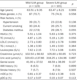
Table 1. The clinical characteristics and laboratory data of severe ILAA group and the mild ILAA group
Mild ILAA group
(n = 123) Severe ILAA group (n = 101) P Age (years) 63.51 ± 9.35 63.47 ± 8.21 0.969 Male gender, n (%) 87 (70.7) 75 (74.3) 0.558 Risk factors, n (%)
Hypertension 39 (31.7) 23 (22.8) 0.138 Current smoking 30 (24.4) 26 (25.7) 0.816 Diabetes mellitus 22 (17.9) 29 (28.7) 0.055 HbA1c (%) 5.51 ± 1.04 5.63 ± 0.96 0.375 TC (mmol/L) 4.87 ± 1.25 5.19 ± 1.20 0.056 LDL-C (mmol/L) 2.73 ± 0.80 2.92 ± 0.78 0.069 TG ( mmol/L ) 1.38 ± 0.90 1.49 ± 0.93 0.384 Leukocytes (G/L) 7.42 ± 2.15 7.72 ± 3.08 0.401 Creatinine (μmol/L) 102.54 ± 66.92 105.60 ± 60.65 0.723 Uric acid (μmol/L) 320.63 ±137.86 318.25 ± 134.96 0.897 Genisi score 41.00 ± 37.02 48.59 ± 38.99 0.137 AMI history (%) 8 (6.5) 7 (6.9) 0.899 Stroke history (%) 7 (5.7) 8 (7.9) 0.507 mDC-p (%) 0.81 ± 0.37 0.62 ± 0.38 0.01 pDC-p (%) 0.17 ± 0.07 0.15 ± 0.07 0.113
Values are expressed as percentages or mean ± SD. AMI, acute myocar-dial infarction; HbA1c, haemoglobin A1C; TC, total cholesterol; LDL-C, low-density lipoprotein cholesterol; TG, triglycerides; mDC-p, myeloid dendritic cell precursors; pDC-p, plasmacytoid dendritic cell precursors.
Results
Baseline characteristics
The clinical characteristics and laboratory data of subjects are summarized in Table 1. In our study, 123 patients with severe ILAA were com-pared with 101 mild ILAA group, we found no significant differences patients with major clini-cal (age, hypertension, current smoking, diabe-tes mellitus, AMI history, stroke history, HbA1c, TC, LDL-c, TG, leukocytes, creatinine, uric acid) and coronary artery angiographic data (Genisi score).
Immunohistochemical analysis
The clinical characteristics and laboratory data of both groups of ILAA patients had no signifi-cant differences (Table 2). In the present study, the occurrence of dendritic cells was analyzed by four-color flow cytometry. For analysis of mDCs, the number of HLA-DR+CD123+ cell expressed by pDC precursors and HLA-DR+CD- 11c+ cell expressed by mDC precursors was
evaluated. Immunostaining with these markers showed a significantly higher cells number of immature as well as mature mDCs in both groups of ILAA patients (Figure 1).
Additionally, immunostaining with HLA-DR, a functional marker expressed by activated anti-gen-presenting cells, revealed a significantly higher cells number in femoral plaques of ILAA patients (P = 0.01) (Table 1).
Decrease in circulating mDCPs and pDCPs in ILAA
The levels of mDC precursors were significantly lower in severe ILAA group than in mild ILAA group [0.375% (0.60-0.695) vs. 0.75% (0.61-0.695)] (Table 1), but the levels of pDC precur-sors were similar between in severe intracranial large artery atherosclerosis (ILAA) group and in mild intracranial large artery atherosclerosis (ILAA) group [0.16% (0.13-0.21) vs. 0.14% ( 0.12-0.20)] (Table 1).
Comparison of circulating DCPs in ILAA
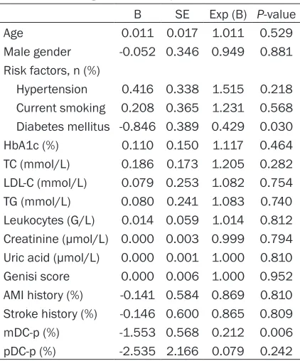
In our data, we found no significant differences patients with major clinical (age, hypertension, current smoking, diabetes mellitus, AMI history, stroke history, HbA1c, TC, LDL-c, TG, leuko-cytes, creatinine, uric acid) and coronary artery angiographic data (Genisi score). At binary logistic regression analysis, we also found that the levels of mDC precursors and diabetes mel-litus were the independent predictor of the severity of intracranial artery stenosis (Table 2).
Discussion
In this study, we demonstrate that the levels of circulating mDC precursors are lower in severe intracranial large artery atherosclerosis group than in mild intracranial large artery atheroscle-rosis group. Decreased circulating mDC precur-sors could predict the severity of ILAA, suggest-ing a new contributory mechanism to ILAA. Previous clinical studies have demonstrated mDC precursors decrease in peripheral circula-tion in ACS [17, 18] and increase in vulnerable carotid plaques [15]. The decreased circulating mDCs precursor may be recruited into the ath-erosclerotic lesion. Several chemokines, such as monocyte chemoattractant protein-1 (MCP-Table 2. Predictors of the severity of ILAA in
peripheral blood mononuclear cells at linear multivariate regression analysis: all patients
B SE Exp (B) P-value Age 0.011 0.017 1.011 0.529 Male gender -0.052 0.346 0.949 0.881 Risk factors, n (%)
Hypertension 0.416 0.338 1.515 0.218 Current smoking 0.208 0.365 1.231 0.568 Diabetes mellitus -0.846 0.389 0.429 0.030 HbA1c (%) 0.110 0.150 1.117 0.464 TC (mmol/L) 0.186 0.173 1.205 0.282 LDL-C (mmol/L) 0.079 0.253 1.082 0.754 TG (mmol/L) 0.080 0.241 1.083 0.740 Leukocytes (G/L) 0.014 0.059 1.014 0.812 Creatinine (μmol/L) 0.000 0.003 0.999 0.794 Uric acid (μmol/L) 0.000 0.001 1.000 0.810 Genisi score 0.000 0.006 1.000 0.952 AMI history (%) -0.141 0.584 0.869 0.810 Stroke history (%) -0.146 0.600 0.865 0.809 mDC-p (%) -1.553 0.568 0.212 0.006 pDC-p (%) -2.535 2.166 0.079 0.242
[image:4.612.90.301.106.360.2]1) and fractalkine, which could be induced by several atherogenic factors, typically oxidized LDL-cholesterol [19], may contribute to recruit-ment of circulating mDC precursors into the atherosclerotic lesion. Monocyte chemoattrac-tant protein-1 (MCP-1), a member of the che-mokine family, was found to be highly expressed
[image:5.612.93.386.72.534.2]bers of circulating mDCPs, pDCPs and DCPs were significantly reduced, and a dense infiltra-tion of mDCs co-localized with T-cells, single pDCs and high HLA-DR expression were obs- erved in brain infract area. However, the distri-butions of circulating DC subsets in ILAA are still unknown and require further investigation. Figure 1. Detection of dendritic cell precursors (mDC precursors and pDC
pre-cursors) in peripheral blood by four-color flow cytometry. R1: region based on forward and side light scatter properties to exclude debris. R2: region contain-ing DC, defined as HLA-DR+ and lineage cells. R4 and R5: regions containing
cells gated on R1 and R2. R4 identifies HLA-DR+CD123+ cell (pDC precursors),
R5 identifies HLA-DR+CD11c+ cell (mDC precursors).
in human atherosclerotic lesions [20]. Deletion of MCP-1 or its corresponding receptor CCR2 could atten-uate atherosclerosis in experimental mouse mod-els [21-23]. Clinical evi-dence also has shown that the plasma levels of MCP-1 have independent prognos-tic value in the acute and chronic phases after ACS [24, 25]. Additionally, the role of MCP-1/CCR2 in DCs biology is classically seen as being critical for cell migration and maturation [26]. Deletion of the fract- alkine receptor CX3CR1 resulted in decreased ath-erosclerosis and a decre- ased number of DC in ath-eromas in ApoE-/- mouse [27].
num-Intracranial large-artery atherosclerosis is a major cause of ischemic stroke worldwide [32, 33], especially in Asians [33]. It is noteworthy that ILAA may be associated with coronary ath-erosclerosis and another athath-erosclerosis [34-36]. To exclude the effect of the coronary ath-erosclerosis and another athath-erosclerosis on the distribution of circulating DC precursor sub-sets, angiography, the gold standard for artery stenosis detection, was used to determine the extent and severity of the artery stenosis. We found that the levels of circulating mDC precur-sors were decreased in severe ILAA, but the levels of circulating pDC precursors were not significant changed. This result along with pre-vious studies may indicate that the percentage of mDCs precursors reflects the total athero-sclerotic burden, the decreased circulating mDCs precursors are recruited from blood into the atherosclerotic lesions.
We also found that diabetes mellitus was the predictor of the severity of ILAA at binary logis-tic regression analysis. It has been shown that diabetes mellitus is a more important determi-nant for intracranial atherosclerosis related stroke than extracranial atherosclerosis or non-atherosclerosis in a multi-ethnic community-based cohort [37] Diabetes mellitus was also considered as a significant risk factors for intra-cranial artery stenosis in asymptomatic popula-tions [38]. Seifarth et al. [39] found that the levels of circulating mDCs and pDCs are decreased in patients with type 2 diabetes mel-litus, especially for the levels of circulating mDC in these patients with poor blood glucose con-trol. However, whether circulating mDC impli-cated in the pathogenesis of intracranial ath-erosclerosis in diabetes mellitus are still unclear.
There are some limitations in our study. First, because of abiding by the necessarily stringent inclusion and exclusion criteria, the relatively small sample size is the main limitation of our study. Second, we did not quantitative evaluate the severity and extent of intracranial athero-slerotic lesions based on present study, it could help to better understand the relation of circu-lating DC subset and the severity and extent of ILAA.
In conclusion, we found that a significant decrease in circulating mDCs in patients with ILAA. In addition, we also demonstrated
mark-ers indicative for mDCs are negatively correlat-ed with the severity and extent of ILAA. Therefore, further studies are required to dem-onstrate whether regulation of the percentage of circulating mDC precursors in ILAA might yield new therapies, and the association of inflammation and DCs in ILAA.
Acknowledgements
This work was funded by the science and tech-nology plan of Guangzhou City (No. 2014Y2-00068), the major project of Guangdong Province science and technology plan (No. 2012A080104020), the Guangzhou Key Labo- ratory of medical Internet Network (2013-163-15) and the Guangdong Provincial Information Industry Development Special Fund (2014- 975).
Disclosure of conflict of interest
None.
Address correspondence to: Dr. Ding-Cheng Xiang, Department of Cardiovascular, Guangzhou General Hospital of Guangzhou Military Command, 111 Liuhua Road, Guangzhou 510010, China. E-mail: [email protected]
References
[1] Liu L. Cardiovascular diseases in China. Bio-chem Cell Biol 2007; 85: 157-163.
[2] Marzegalli M, Lunati M, Landolina M, Perego GB, Ricci RP, Guenzati G, Schirru M, Belvito C, Brambilla R, Masella C, Di Stasi F, Valsecchi S, Santini M. Remote monitoring of CRT-ICD: the multicenter Italian CareLink evaluation--ease of use, acceptance, and organizational impli-cations. Pacing Clin Electrophysiol 2008; 31: 1259-1264.
[3] Li AH, Chu YT, Yang LH, Chen KC, Chu SH. More coronary artery stenosis, more cerebral artery stenosis? A simultaneous angiographic study discloses their strong correlation. Heart Vessels 2007; 22: 297-302.
[4] Bae HJ, Yoon BW, Kang DW, Koo JS, Lee SH, Kim KB, Lee J, Roh JK. Correlation of coronary and cerebral atherosclerosis: difference be-tween extracranial and intracranial arteries. Cerebrovasc Dis 2006; 21: 112-119.
[6] Adraktas DD, Brasic N, Furtado AD, Cheng SC, Ordovas K, Chun K, Chien JD, Schaeffer S, Wintermark M. Carotid atherosclerosis does not predict coronary, vertebral, or aortic ath-erosclerosis in patients with acute stroke symptoms. Stroke 2010; 41: 1604-1609. [7] Hansson GK. Inflammation, atherosclerosis,
and coronary artery disease. N Engl J Med 2005; 352: 1685-1695.
[8] Elkind MS. Inflammatory mechanisms of stroke. Stroke 2010; 41: S3-8.
[9] Bobryshev YV. Dendritic cells and their role in atherogenesis. Lab Invest 2010; 90: 970-984. [10] Bobryshev YV, Lord RS. Ultrastructural recogni-tion of cells with dendritic cell morphology in human aortic intima. Contacting interactions of Vascular Dendritic Cells in athero-resistant and athero-prone areas of the normal aorta. Arch Histol Cytol 1995; 58: 307-322.
[11] Jongstra-Bilen J, Haidari M, Zhu SN, Chen M, Guha D, Cybulsky MI. Low-grade chronic in-flammation in regions of the normal mouse arterial intima predisposed to atherosclerosis. J Exp Med 2006; 203: 2073-2083.
[12] Paulson KE, Zhu SN, Chen M, Nurmohamed S, Jongstra-Bilen J, Cybulsky MI. Resident intimal dendritic cells accumulate lipid and contribute to the initiation of atherosclerosis. Circ Res 2010; 106: 383-390.
[13] Yilmaz A, Lochno M, Traeg F, Cicha I, Reiss C, Stumpf C, Raaz D, Anger T, Amann K, Probst T, Ludwig J, Daniel WG, Garlichs CD. Emergence of dendritic cells in rupture-prone regions of vulnerable carotid plaques. Atherosclerosis 2004; 176: 101-110.
[14] Bobryshev YV, Lord RS. Co-accumulation of dendritic cells and natural killer T cells within rupture-prone regions in human atherosclerot-ic plaques. J Histochem Cytochem 2005; 53: 781-785.
[15] Kawahara I, Kitagawa N, Tsutsumi K, Nagata I, Hayashi T, Koji T. The expression of vascular dendritic cells in human atherosclerotic carot-id plaques. Hum Pathol 2007; 38: 1378-1385. [16] Paul K, Kretzschmar D, Yilmaz A, Barthlein B,
Titze S, Wolf G, Busch M, Investigators GC-S. Circulating dendritic cell precursors in chronic kidney disease: a cross-sectional study. BMC Nephrol 2013; 14: 274.
[17] Fukunaga T, Soejima H, Irie A, Fukushima R, Oe Y, Kawano H, Sumida H, Kaikita K, Sugiyama S, Nishimura Y, Ogawa H. High ratio of myeloid dendritic cells to plasmacytoid dendritic cells in blood of patients with acute coronary syn-drome. Circ J 2009; 73: 1914-1919.
[18] Yilmaz A, Weber J, Cicha I, Stumpf C, Klein M, Raithel D, Daniel WG, Garlichs CD. Decrease in circulating myeloid dendritic cell precursors in coronary artery disease. J Am Coll Cardiol 2006; 48: 70-80.
[19] Lowery DE, Pasternack JM, Gonzalez-DeWhitt PA, Zurcher-Neely H, Tomich CC, Altman RA, Fairbanks MB, Heinrikson RL, Younkin SG, Greenberg BD. Alzheimer’s amyloid precursor protein produced by recombinant baculovirus expression. Proteolytic processing and prote-ase inhibitory properties. J Biol Chem 1991; 266: 19842-19850.
[20] Yilmaz A, Lipfert B, Cicha I, Schubert K, Klein M, Raithel D, Daniel WG, Garlichs CD. Accumulation of immune cells and high ex-pression of chemokines/chemokine receptors in the upstream shoulder of atherosclerotic carotid plaques. Exp Mol Pathol 2007; 82: 245-255.
[21] Gu L, Okada Y, Clinton SK, Gerard C, Sukhova GK, Libby P, Rollins BJ. Absence of monocyte chemoattractant protein-1 reduces athero-sclerosis in low density lipoprotein receptor-deficient mice. Mol Cell 1998; 2: 275-281. [22] Inoue S, Egashira K, Ni W, Kitamoto S, Usui M,
Otani K, Ishibashi M, Hiasa K, Nishida K, Takeshita A. Anti-monocyte chemoattractant protein-1 gene therapy limits progression and destabilization of established atherosclerosis in apolipoprotein E-knockout mice. Circulation 2002; 106: 2700-2706.
[23] Boring L, Gosling J, Cleary M, Charo IF. Decreased lesion formation in CCR2-/- mice reveals a role for chemokines in the initiation of atherosclerosis. Nature 1998; 394: 894-897.
[24] de Lemos JA, Morrow DA, Sabatine MS, Murphy SA, Gibson CM, Antman EM, McCabe CH, Cannon CP, Braunwald E. Association between plasma levels of monocyte chemoattractant protein-1 and long-term clinical outcomes in patients with acute coronary syndromes. Circulation 2003; 107: 690-695.
[25] de Lemos JA, Morrow DA, Blazing MA, Jarolim P, Wiviott SD, Sabatine MS, Califf RM, Braunwald E. Serial measurement of mono-cyte chemoattractant protein-1 after acute coronary syndromes: results from the A to Z trial. J Am Coll Cardiol 2007; 50: 2117-2124. [26] Jimenez F, Quinones MP, Martinez HG, Estrada
CA, Clark K, Garavito E, Ibarra J, Melby PC, Ahuja SS. CCR2 plays a critical role in dendritic cell maturation: possible role of CCL2 and NF-kappa B. J Immunol 2010; 184: 5571-5581. [27] Liu P, Yu YR, Spencer JA, Johnson AE, Vallanat
CT, Fong AM, Patterson C, Patel DD. CX3CR1 deficiency impairs dendritic cell accumulation in arterial intima and reduces atherosclerotic burden. Arterioscler Thromb Vasc Biol 2008; 28: 243-250.
stroke: time course, activation state, and ori-gin. Brain Behav Immun 2010; 24: 724-737. [29] Gelderblom M, Leypoldt F, Steinbach K,
Behrens D, Choe CU, Siler DA, Arumugam TV, Orthey E, Gerloff C, Tolosa E, Magnus T. Temporal and spatial dynamics of cerebral immune cell accumulation in stroke. Stroke 2009; 40: 1849-1857.
[30] Reichmann G, Schroeter M, Jander S, Fischer HG. Dendritic cells and dendritic-like microglia in focal cortical ischemia of the mouse brain. J Neuroimmunol 2002; 129: 125-132.
[31] Yilmaz A, Fuchs T, Dietel B, Altendorf R, Cicha I, Stumpf C, Schellinger PD, Blumcke I, Schwab S, Daniel WG, Garlichs CD, Kollmar R. Transient decrease in circulating dendritic cell precur-sors after acute stroke: potential recruitment into the brain. Clin Sci (Lond) 2010; 118: 147-157.
[32] Sacco RL, Kargman DE, Gu Q, Zamanillo MC. Race-ethnicity and determinants of intracra- nial atherosclerotic cerebral infarction. The Northern Manhattan Stroke Study. Stroke 1995; 26: 14-20.
[33] Wong KS, Huang YN, Gao S, Lam WW, Chan YL, Kay R. Intracranial stenosis in Chinese pa-tients with acute stroke. Neurology 1998; 50: 812-813.
[34] Sen S, Lynch DR Jr, Kaltsas E, Simmons J, Tan WA, Kim J, Beck J, Rosamond W. Association of asymptomatic peripheral arterial disease with vascular events in patients with stroke or tran-sient ischemic attack. Stroke 2009; 40: 3472-3477.
[35] Hoshino A, Nakamura T, Enomoto S, Kawahito H, Kurata H, Nakahara Y, Ijichi T. Prevalence of coronary artery disease in Japanese patients with cerebral infarction: impact of metabolic syndrome and intracranial large artery athero-sclerosis. Circ J 2008; 72: 404-408.
[36] Fu JH, Chen YK, Chen XY, Mok V, Wong KS. Coexisting small vessel disease predicts poor long-term outcome in stroke patients with in-tracranial large artery atherosclerosis. Cere- brovasc Dis 2010; 30: 433-439.
[37] Rincon F, Sacco RL, Kranwinkel G, Xu Q, Paik MC, Boden-Albala B, Elkind MS. Incidence and risk factors of intracranial atherosclerotic stroke: the Northern Manhattan Stroke Study. Cerebrovasc Dis 2009; 28: 65-71.
[38] Bae HJ, Lee J, Park JM, Kwon O, Koo JS, Kim BK, Pandey DK. Risk factors of intracranial ce-rebral atherosclerosis among asymptomatics. Cerebrovasc Dis 2007; 24: 355-360.