RESEARCH ARTICLE
AN EFFICIENT WAY TO ENHANCE MAMMOGRAM IMAGE USING NONSUBSAMPLED CONTOURLET
TRANSFORM
1
Sangeetha, T. A. and
2Dr. Saradha, A.
1
CS
Kongu Arts and Science college, Erode. (Research Scholar-Mother Teresa Women’s University),Kodaikkanal, TN, India
2CSE, Institute of Road and Techology, Erode, TN, India
ARTICLE INFO ABSTRACT
Breast cancer is one of the most important causes of increased women death rate in the world. Mammography is the most efficient approach for the early identification of breast diseases. The major objective of mammography is to identify small, non-palpable cancers during its premature stage. On the other hand, mammograms are extremely complicated to interpret being the fact that the pathological transformations of the breast are slight and their visibility is very poor with low contrast and noise. Mammograms has the valuable information such as microcalcifications and masses, which are extremely complicated to identify because mammograms are of low-contrast. Since the mammogram images are very noisy, low-contrast, blur and fuzzy, it is necessary to enhance the mammogram images for accurate identification and early diagnosis of breast cancer. In this paper, proposed an efficient technique to enhance mammogram image using nonsubsampled contourlet transform. The nonsubsampled contourlet transform is built upon nonsubsampled pyramids and nonsubsampled directional filter banks and provides a shiftinvariant directional multiresolution image representation. Existing methods for image enhancement cannot capture the geometric information of images and tend to amplify noises when they are applied to noisy images since they cannot distinguish noises from weak edges. In contrast, the nonsubsampled contourlet transform extracts the geometric information of images, which can be used to distinguish noises from weak edges. Experimental results show the proposed method achieves better enhancement results than other enhancement method.
Copy Right, IJCR, 2012, Academic Journals. All rights reserved.
INTRODUCTION
REAST cancer is a “malignant neoplasm of the breast” [1]. The characteristic of cancer affected cells completely differs from the normal tissue cells in terms of the cell outline, shape, structure of nucleus and most significantly, its ability to metastasize and infiltrate. This disease is quite common. Because of its well exposed nature and possible for lethality, breast cancer is perhaps the most severe kind of cancer. Also, it is to be noted that, if identified and correctly treated during its early stages, breast cancer can be cured. Breast cancer continues to be a significant public health problem in the world, which not only endangers the life of the patient, but also causes damage to the female sexual characteristic organ [1-4]. It’s one of the most rapidly increasing malignant tumors. Early detection of breast cancer is of utmost importance: localized cancer leads to a 5-year survival rate of 97.5%, whereas cancer that has spread to distant organs has a 5-year survival rate of only 20.4% [5]. Breast cancer can be either invasive or noninvasive [2].
Noninvasive Breast Cancer
Non-invasive cancers stay within the milk ducts or lobules in
*Corresponding author: [email protected], [email protected]
the breast. They do not grow into or overrun normal tissues within or beyond the breast. Non-invasive cancers are sometimes called carcinoma in situ (“in the same place”) or pre-cancers.
Fig 1. Non-Invasive Cell
Invasive Breast cancer
Invasive cancers grow into normal healthy tissues. Most breast cancers are invasive. Whether the cancer is non-invasive or B
ISSN: 0975-833X
International Journal of Current Research
Vol. 4, Issue, 12, pp. 385-390, December,2012
INTERNATIONAL JOURNAL OF CURRENT RESEARCH
Article History:
Received 09th September, 2012
Received in revised form
15th October, 2012
Accepted 23rd November, 2012
Published online 28th December, 2012
Key words: Breast Cancer, Mammography, Image Enhancement,
invasive will determine your treatment choices and how you might respond to the treatments that you receive.
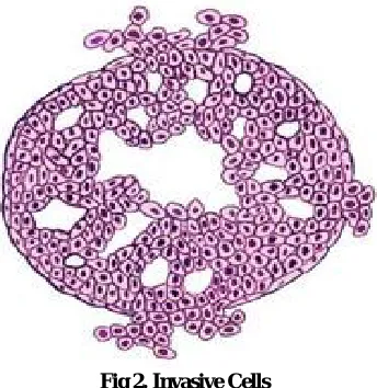
Fig 2. Invasive Cells
In some cases, a breast cancer may be both invasive and non-invasive. This means that part of the cancer has grown into normal tissue and then a part of the cancer has stayed inside the milk ducts or milk lobules. It would be treated as an invasive cancer. A breast cancer also may be a “mixed tumor,” that means it contains a fusion of cancerous ductal cells and lobular cells. This type of cancer is also called “invasive mammary breast cancer” or “infiltrating mammary carcinoma.” It would be treated as a ductal carcinoma. If there is more than one tumor in the breast, the breast cancer is described as either multifocal or multicentric. In multifocal breast cancer, all of the tumors arise from the original tumor, and they are usually in the same section of the breast. If the cancer is multicentric, it means that all of the tumors formed separately, and they are often in different areas of the breast.
Mammogram
Mammograms are used as a screening tool to detect early stages of breast cancer in women experiencing no symptoms. It can also be used to detect and diagnose breast disease in women experiencing symptoms such as a lump, pain or nipple discharge. Mammography is the process of using low-energy-X-rays (usually around 30 kVp) to examine the human breast and is used as a diagnostic and a screening tool. The goal of mammography is the early detection of breast cancer, through detection of characteristic masses and/or microcalcifications. Like all x-rays, mammograms use doses of ionizing radiation to create images. But the digital mammography is very noisy, low-contrast, blur and fuzzy and hence there is a requirement for enhancing images [3]. This is essential for enhancing the Peak Signal-to-Noise Ratio (PSNR) and reducing the Mean Squared Error (MSE) for accurate identification. One of the most important objectives of mammogram image enhancement is to enhance the contrast between regions of interest and the background. Also the medical images fluctuate extensively in terms of acquisition, noise characteristics and quality [4]. Thus, there is a requirement to process an image on the image basis. This motivates the design and construction of effective mammogram image enhancement method using nonsubsampled contourlet transform. The remainder of this paper is organized as follows. The next section presents some basic concepts of mammography Section 3 provides a brief revision of nonsubsampled contourlet transform. Section 4
describes the proposed model, and some experimental results are illustrated in Sect. 5. Finally, the conclusions are drawn in Sect. 6.
Related Work
Mammography is one of the most reliable methods for early detection of breast carcinomas [5]. And it is currently the best way that can detect the clinical asymptomatic concealed breast cancer. It can reveal pronounced substantiation of abnormality, such as masses and calcifications, as well as slight signs such as bilateral asymmetry and architectural distortion [6]. Reports from the American Cancer Society (ACS) shows that early diagnosed breast cancer patient can achieve a survival rate as high as 97% before the spreading of the carcinoma cells [7]. Therefore, early detection and diagnosis of the breast cancer is very important in saving the life of the patient and it’s the most effective way to increase the cure rate and lower the mortality rate. However, because of the particularity of the breast tissue, mammograms are always lack of hierarchy, low-contrast, leaving the misdiagnosis rate and neglected diagnosis rate too high. Since diagnostic features in mammograms, such as masses and calcifications, may be small and have low contrast with respect to the surrounding breast tissues.
These attributes could render the diagnostic features hard to detect. Contrast enhancement techniques can improve the ability of a radiologist to perceive subtle diagnostic features, leading to earlier, more accurate diagnosis of breast cancer. Contrast enhancement can improve the quality of an otherwise unsatisfactory mammogram, as stated by Ram [8], who further indicated that the application of contrast enhancement techniques in a clinical situation may reduce the radiation dose by about 50%. The enhancement of mammographic images could improve the accuracy of detection of early signs of breast features. To highlight the features of the lesion region and improve the visual effect of the mammogram, the most commonly used method is imposing enhancement preprocessing on the image [9, 10]. For reviews on image enhancement in mammography, see Rangayyan [11], Morrow
et al.,[12], and Rangayyan et al. [13]. Currently, contrast
stretching, histogram equalization spatial domain filtering, frequency domain filtering, Wavelet Transform (WT), mathematical morphology etc. are the major commonly used image preprocessing techniques [14-23].
representation. Based on the NSCT, we propose a new method for image enhancement.
A Revision of Nonsubsampled Contourlet Transform
Contourlet Transform
Do and Vetterli proposed a “true” two-dimensional image representation scheme -- Contourlet Transform [29] in 2002. It is a multidirectional and multiscale transform that can capture the intrinsic geometrical structure that is key in visual information. Unlike other approaches, such as Ridgelet and curvelet, that first develop a transform in the continuous domain and then discrete for sampled data, contourlet transform starts with a discrete-domain construction and then studies its convergence to an expansion in the continuous domain. Do and Vetterli [29] constructed a multidirectional and multiscale transform in the discrete domain by combining the Laplacian pyramid (LP) and the directional filter bank (DFB) using nonseparable filter banks, as shown in Figure 3, the same like wavelet. The Laplacian pyramid is first used to capture the point discontinuities, and then followed by a directional filter bank (DFB) to link point discontinuities into linear structures.
Nonsubsampled Contourlet Transform
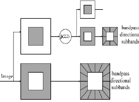
[image:3.595.317.555.53.217.2]Due to downsamplers and upsamplers present in both the LP and the DFB, the contourlet transform is not shiftinvariant, which will cause pseudo-Gibbs phenomena [30] around singularities. In 2006, L. da Cunha and J. Zhou [31] improved the Contourlet Transform and developed the nonsubsampled contourlet transform. Figure 4 displays an overview of the proposed NSCT . The NSCT is a fully shift-invariant, multiscale, and multidirection expansion that has a fast implementation. It is composed of two shift-invariant parts: 1) a nonsubsampled pyramid structure that ensures the multiscale property; and 2) a nonsubsampled DFB structure that gives directionality, without sampling as that in the Contourlet Transform. Because of its shiftinvariant property, we can achieve better results in the image processing tasks where redundancy is not a major issue, such as image denoising and enhancement. Besides, it is more flexible for the design of the filter.
[image:3.595.39.280.553.730.2]Fig. 3. The flowchart of Contourlet transform
Fig. 4. The flowchart of NSCT
Proposed Model
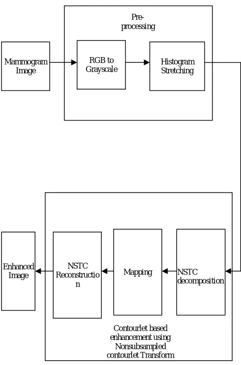
In this paper, proposed an efficient technique to enhance mammogram image using non subsampled contourlet transform. The following section discusses about the architecture and the use of non subsampled contourlet transform in mammogram image enhancement. Fig:5 shows the system architecture of the proposed scheme. Initially, the mammogram images are transformed from RGB to grayscale. Then the histogram stretching is carried out to the grey image for preliminary enhancement. Then the output of histogram stretching is given as input to the non subsampled contourlet transform . In this transform, the histogram equalization is not used as the preliminary step, even though it is more efficient. Because histogram equalization enhances the image contrast by transforming the pixel distribution as a result they can conform to uniform distribution. The NSCT is shift-invariant so that each pixel of the transform subbands corresponds to that of the original image in the same spatial location.
Fig. 5. Diagram for the Proposed Mammogram Image Enhancement using non subsampled Contourlet Transform
Mammography Images
Mammography is a specific kind of imaging that utilizes a low-dose x-ray system to check breasts. A mammography exam is called as mammogram, used to assist in the premature detection and early diagnosis of breast cancer and related diseases in women. An x-ray is noninvasive medical tests that assist physicians in diagnosing the disease. Imaging with x-rays involves exposing a part of the body to a tiny amount of ionizing radiation to generate pictures of the inside of the body. X-rays are the traditional and most commonly used form of medical imaging. Two recent advances in mammography include [32]
Digital mammography and
Computer-aided detection
Digital mammography is also called as Full-Field Digital Mammography (FFDM), a mammography system in which the x-ray film is substituted with solid-state detectors that transform x-rays into electrical signals. These detectors are like the detectors found in digital cameras. The electrical signals are utilized to generate images of the breast that is visible on a computer screen or printed on special film as like traditional mammograms. Computer-Aided Detection (CAD) systems utilize a digitized mammographic image that can be acquired from either a traditional film mammogram or a digitally acquired mammogram. The computer software then
examines for irregular areas of density, mass or calcification that may specify the occurrence of cancer. The CAD system points out the places of interest on the images, alerting the radiologist to the need for additional analysis. But these mammogram images are very noisy, low-contrast, blur and fuzzy, hence there is a requirement for image enhancement. In this paper, mammogram images are enhanced using non subsampled contourlet transform
Image enhancement algorithm
Existing image enhancement methods amplify noises when they amplify weak edges since they cannot distinguish noises from weak edges. In the frequency domain, both weak edges and noises lead to low-value coefficients. The nonsubsampled contourlet transform provides not only multiresolution analysis, but also geometric and directional representation. Since weak edges are geometric structures, while noises are not, we can use this geometric representation to distinguish them. The NSCT is shift-invariant such that each pixel of the transform subbands corresponds to that of the original image in the same location. Therefore, we gather the geometric information pixel by pixel from the NSCT coefficients. We observe that there are three classes of pixels: strong edges, weak edges, and noises. First, the strong edges correspond to those pixels with big-value coefficients in all subbands. Second, the weak edges correspond to those pixels with big-value coefficients in some directional subbands but small-value coefficients in other directional subbands within the same scale. Finally, the noises correspond to those pixels with small-value coefficients in all subbands. Based on this observation, we can classify pixels into three categories by analyzing the distribution of their coefficients in different subbands. One simple way is to compute the mean (denoted by mean) and the maximum (denoted by max) magnitude of the coefficients for each pixel, and then classify it by
where c is a parameter ranging from 1 to 5, and _ is the noise standard deviation of the subbands at a specific level. We first estimate the noise variance of the input image with the robust median operator [7] and then compute the noise variance of each subband [8]. The goal of image enhancement is to amplify weak edges and to suppress noises. To this end, we modify the NSCT coefficients according to the category of each pixel by a nonlinear mapping function (similar to [9])
where the input x is the original coefficient, and 0 < p < 1 is the amplifying ratio. This function keeps the coefficients of strong edges, amplifies the coefficients of weak edges, and zeros the coefficients of noises. We summarize our enhancement method using the NSCT in the following algorithm:
1) Compute the NSCT of the input image for N levels. 2) Estimate the noise standard deviation of the input image.
Mammogram Image
RGB to
Grayscale Histogram Stretching
Pre-processing
NSTC Reconstructio
n
Mapping NSTC
decomposition Enhanced
Image
Contourlet based enhancement using
3) For each level DFB,
Estimate the noise variance.
Compute the threshold and the amplifying ratio.
At each pixel, compute the mean and the maximum magnitude of all directional subbands at this level, and classify it by (1) into strong edges, weak edges, or noises.
For each directional subband, use the nonlinear mapping function given in (2) to modify the NSCT coefficients according to the classification.
4) Reconstruct the enhanced image from the modified NSCT coefficients.
Enhancement Evaluation Measures
Several measures that are capable of quantifying the relative utility of enhancement techniques in digital mammography are studied in [34]. Experiments show that the measure, Target to Background Contrast Ratio using Variance, keeps good accordance with the visual effect of the algorithm. Therefore, we take this measure as an evaluation of our algorithm. Denote and as the mean grey of target T and background B in the original mammographic image, and
as the mean grey of target T and background B in the enhanced mammographic image, respectively.
The difference between the ratios of the mean grey in the target and border areas in the original and enhanced images can be denoted as
A good enhancement method should aim to enhance the contrast between target T and background B by increasing the mean grey level of the target area T and reducing that of the background area B, thereby increasing the value of . In addition, the enhancement technique should at the same time aim to reduce the spread of grey scales in the enhanced target area compared with the target area in the original image. This reduction can be measured by the ratio of the grey level variances as
where and are the standard deviations of the target in the in the original and enhanced image. The resulting target to background contrast ratio using variance now be computed as
An effective enhancement technique will lead to a large value of TBc.
Experimental Result
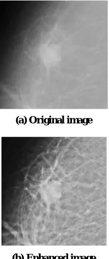
From the diagram, observe that the proposed algorithm suppresses noises while enhancing weak edges in the textures
and boosting the contrast between the lesion area and the background. Existing image-enhancement methods amplify noise when they amplify weak edges since they cannot distinguish noise from weak edges. But in nonsubsampled contourlet transform extracts the geometric information of images, which can be used to distinguish noises from weak edges.
(a) Original image
[image:5.595.382.494.145.412.2](b) Enhanced image
Fig. 6. Resultant Enhanced mammogram image Obtained by nonsubsampled contourlet transform
Conclusion
We present the nonsubsampled contourlet transform constructed by iterated nonsubsampled filter banks. This transform provides shift-invariant directional multiresolution image representation. We propose a new algorithm for image enhancement using the nonsubsampled contourlet transform. Experimental results show that the proposed algorithm achieves better enhancement results than the undecimated wavelet transform and other transform. The enhanced image is viewed clearly and free from noise.
REFERENCE
[1] Temidayo O Ogundiran, Samuel A Ademola, Odunayo M Oluwatosin, Effiong E Akang and Clement A Adebamowo, “Primary osteogenic sarcoma of the breast”, World Journal of Surgical Oncology, Published online:
http://www.biomedcentral.com/content/pdf/1477-7819-4-90.pdf, 2006.
[2] Breast Cancer In-Depth Report, The Newyork Times, http://health.nytimes.com/health/guides/disease/breast-cancer/print.html.
Digital Mammography, Toronto, Canada, M.J. Yaffe (ed.), Medical Physics Publishing, Pp. 547-553, 2000. [4] R. Krishnamoorthy, N. Amudhavalli and M.K.
Sivakkolunthu, “An Adaptive Mammographic Image Enhancement in Orthogonal Polynomials Domain”, International Journal of Computer and Information Engineering, Vol. 4, No. 2, Pp. 120-128, 2010.
[5] C. D. Maggio, "State of the art of current modalities for thediagnosis of breast lesions," European Journal of Nuclear Medicine and Molecular Imaging, vol. 31, pp. 56-69, 2004
[6] M. J. Homer, Mammographic Interpretation: A Practical Approach. New York: McGraw-Hill Companies, 1991. [7] ACS, "Cancer Prevention & Early Detection Facts &
Figures 2008," 2008
[8] G. Ram, "Optimization of ionizing radiation usage in medical imaging by means of image enhancement
techniques," Medical Physics, vol. 9, pp. 733-737, 1982. [9] H. D. Cheng, X. Cai, X. Chen, L. Hu, and X. Lou,
"Computer-aided detection and classification of microcalcifications in mammograms: a survey," Pattern Recognition, vol. 36, pp. 2967-2991, 2003
[10]H. D. Cheng, X. J. Shi, R. Min, L. M. Hu, X. P. Cai, and H.N. Du, "Approaches for automated detection and classification of masses in mammograms," Pattern Recognition, vol. 39, pp. 646-668, 2006.
[11]R. M. Rangayyan, Biomedical Image Analysis. Boca Raton,FL: CRC Press, 2005.
[12]W. M. Morrow, R. B. Paranjape, R. M. Rangayyan, and J.E. L. Desautels, "Region-based contrast enhancement of mammograms," IEEE Trans. Med. Imag., vol. 11, pp. 392-406, 1992.
[13]R. M. Rangayyan, L. Shen, Y. Shen, J. E. L. Desautels, H.Bryant, T. J. Terry, N. Horeczko, and M. S. Rose, "Improvement of sensitivity of breast cancer diagnosis with adaptive neighborhood contrast enhancement of mammograms," Information Technology in Biomedicine,IEEE Transactions on, vol. 1, pp. 161-170, 1997.
[14]D. C. Chang and W. R. Wu, "Image contrast enhancement based on a histogram transformation of local standard deviation," IEEE Trans. Med. Imag., vol. 17, pp. 518-531,1998.
[15]P. Sakellaropoulos, L. Costaridou, and G. Panayiotakis, "A wavelet-based spatially adaptive method for mammographic contrast enhancement," Physics in Medicine and Biology, vol. 48, pp. 787-803, 2003. [16]V. E. Pera, E. L. Heffer, H. Siebold, O. Schütz, S.
Heywang-K?brunner, L. G?tz, A. Heinig, and S. Fantini, "Spatial second-derivative image processing: an application to optical mammography to enhance the detection of breast tumors," Journal of Biomedical Optics, vol. 8, p. 517,2003.
[17]A. Laine, J. Fan, and W. Yang, "Wavelets for contrast enhancement of digital mammography," Engineering in Medicine and Biology Magazine, IEEE, vol. 14, pp. 536-550, 1995.
[18]H. Li, K. J. Liu, S. C. B. Lo, O. T. Inc, and M. D. Jessup, "Fractal modeling and segmentation for the enhancement of microcalcifications in digital mammograms," IEEE Trans. Med. Imag., vol. 16, pp. 785-798, 1997.
[19]A. Laine, M. Lewis, and F. Taylor, "A wavelet based mammographic system," in IEEE International Conference on Acoustics, Speech, and Signal Processing(ICASSP1994), 1994.
[20]H. K. Kang, Y. M. Ro, and S. M. Kim, "A Microcalcification Detection Using Adaptive
[21]Contrast Enhancement on Wavelet Transform and Neural Network," IEICE Transactions on Information and Systems, pp. 1280-1287, 2006.
[22]H.-K. Kang, N. N. Thanh, S.-M. Kim, and Y. M. Ro, "Robust Contrast Enhancement for Microcalcification in Mammography," in Computational Science and Its Applications – ICCSA 2004, 2004, pp. 602-610.
[23]H.-K. Kang, S.-M. Kim, N. N. Thanh, Y. M. Ro, and W.- H. Kim, "Adaptive Microcalcification Detection in Computer Aided Diagnosis," in Computational Science -ICCS 2004, 2004, pp. 1110-1117.
[24]Ö. Özsen, "Early Detection of Breast Cancer Using Mathematical Morphology," in Knowledge-Based Intelligent Information and Engineering Systems, 2004, pp.583-590.
[25]J. K. Romberg, M. B. Wakin, and R. G. Baraniuk, "Multiscale geometric image processing," in Proceedings of the SPIE: Visual Communications and Image Processing 2003, 2003, pp. 1265-1272.
[26]J. Li-Cheng and T. Shan, "Development and prospect of image multiscale geometric analysis," Acta Electronica Sinica, vol. 31, pp. 1975-1981, 2003.
[27]M. N. Do and M. Vetterli, "The finite ridgelet transform for image representation," IEEE Trans. Image Process, vol.12, pp. 16-28, 2003.
[28]J. L. Starck, E. J. Candes, and D. L. Donoho, "The curvelet transform for image denoising," IEEE Trans. Image Process, vol. 11, pp. 670-684, 2002
[29]E. Le Pennec and S. Mallat, "Sparse geometric image representations with bandelets," IEEE Trans. Image Process, vol. 14, pp. 423-438, 2005.
[30]M. N. Do and M. Vetterli, "The contourlet transform: an efficient directional multiresolution image representation," IEEE Trans. Image Process, vol. 14, pp. 2091-2106, 2005.
[31]R. R. Coifman and D. L. Donoho, Translation invariant de-noising: Wavelets and statistics. NewYork:
Springer-Verlag, 1995
[32]A. L. Da Cunha, J. Zhou, and M. N. Do, "The Nonsubsampled Contourlet Transform: Theory, Design, and Applications," IEEE Trans. Image Process, vol. 15, pp. 3089-3101, 2006.
[33]Giovanni Luca Masala, “Computer Aided Detection on Mammography”, World Academy of Science, Engineering and Technology, Vol. 15, Pp. 1-6, 2006.