CHARACTERIZATION, GROWTH AND CONTROL OF
*1
Arun prasath
1
Department of Botany and Microbiology, A.V.V.M Sri Pushpam College
Poondi
2
Department of Botany, A.V.C College (Autonomous), Mannampandal, Mayiladuthurai, Nagapattinam,
ARTICLE INFO ABSTRACT
Cryptococcosis is an acute, subacute or chronic fungal disease caused by an encapsulated yeast
Cryptococcus neoformans.
infected individuals a
antibiotics especially chloramphenicol was more effective in controlling
variety of plants can be screened which are potentially antimicrobial. Morphological and characteristics of
direct observation under the microscope by using wet mount and negative staining techniques. The cultural characteristics of the organism were studied
influence of different carbon sources. The influence of some commercial antibiotics and plant extracts were assessed for their antimycotic activities. The identity of the fungus was confirmed by nigrosin staining and biochemical tests. Effect of some physico
salinity, sugar (dextrose) and peptone on the growth of
requirements for its growth have been determined. Antibiotic act available 11 antibiotics were assayed against
growth of the test organism. The presences of antimicrobial properties in 12 tested against
(leaf) was more effective than other plant extracts tested.
Copyright © Arun prasath, V. et al. This is an open access article distributed under the Creative Commons Attribution License, which permits unrestricted use, distribution, and reproduction in any medium, provided the original work is properly cited.
INTRODUCTION
Cryptococcosis is an acute, subacute or chronic fungal disease caused by encapsulated yeast belonging to the genus
Cryptococcus. C. neoformans is significantly pathogenic to
human beings and animals. A rapid increase of Cryptococcosis
worldwide has followed the pandemic of AIDS.
Cryptococcosis is the second most common fungal infection after cadidiasis in HIV-infected individuals and potentially most serious infections. In India, Cryptococcus neoformans var. gatti has been reported from Northern India for the first time in 1993 followed by reports from other states also. It has been reported from Ferozepur district of Punjab, Chandigarh, Karnataka and Tamilnadu. Cryptococcosis is more prevalent among the male patients as compared to females. The disseminated form of disease is frequently found in the immune-compromised patients. The clinical features of cryptococcosis may be described depending
anatomical sites involved such as pulmonary cryptococcosis,
*Corresponding author:Arun prasath, V.,
Department of Botany and Microbiology, A.V.V.M Sri Pushpam College (Autonomous), Poondi -613503, Thanjavur, Tamilnadu, India
ISSN: 0975-833X
Vol.
Article History:
Received 16th September, 2013 Received in revised form 04th October, 2013
Accepted 27th November, 2013 Published online 02nd December, 2013
Key words: Cryptococcosis, Cryptococcus neoformans, Pedalium murex.
RESEARCH ARTICLE
CHARACTERIZATION, GROWTH AND CONTROL OF Cryptococcus neoformans
CAUSING CRYPTOCOCCOSIS
Arun prasath, V.,
1Kumar, T., and
2Saravanamuthu,
Department of Botany and Microbiology, A.V.V.M Sri Pushpam College (Autonomous),
Poondi -613503, Thanjavur, Tamilnadu, India
Department of Botany, A.V.C College (Autonomous), Mannampandal, Mayiladuthurai, Nagapattinam,
Tamilnadu, India
ABSTRACT
Cryptococcosis is an acute, subacute or chronic fungal disease caused by an encapsulated yeast
Cryptococcus neoformans. It is the second most common fungal infection after candidiasis in HIV
infected individuals and it is consider as the potentially most serious infections. Commercial antibiotics especially chloramphenicol was more effective in controlling
variety of plants can be screened which are potentially antimicrobial. Morphological and
characteristics of C. neoformans Sanf. were studied. Morphology of the organism was studied by direct observation under the microscope by using wet mount and negative staining techniques. The cultural characteristics of the organism were studied in Sabouraud’s dextrose agar and broth under the influence of different carbon sources. The influence of some commercial antibiotics and plant extracts were assessed for their antimycotic activities. The identity of the fungus was confirmed by nigrosin ining and biochemical tests. Effect of some physico-chemical factors such as temperature, pH, salinity, sugar (dextrose) and peptone on the growth of C. neoformans
requirements for its growth have been determined. Antibiotic act
available 11 antibiotics were assayed against C. neoformans and chloramphenical alone inhibited the growth of the test organism. The presences of antimicrobial properties in 12
tested against C. neoformans. Among the plant extracts tested Coleus forskohlii (leaf) was more effective than other plant extracts tested.
is an open access article distributed under the Creative Commons Attribution License, which permits unrestricted use, distribution, and reproduction in any medium, provided the original work is properly cited.
Cryptococcosis is an acute, subacute or chronic fungal disease caused by encapsulated yeast belonging to the genus is significantly pathogenic to human beings and animals. A rapid increase of Cryptococcosis
worldwide has followed the pandemic of AIDS.
Cryptococcosis is the second most common fungal infection infected individuals and potentially the
Cryptococcus neoformans
has been reported from Northern India for the first time in 1993 followed by reports from other states also. It has been reported from Ferozepur district of Punjab, Chandigarh, taka and Tamilnadu. Cryptococcosis is more prevalent among the male patients as compared to females. The disseminated form of disease is frequently found in the compromised patients. The clinical features of cryptococcosis may be described depending upon the anatomical sites involved such as pulmonary cryptococcosis,
Department of Botany and Microbiology, A.V.V.M Sri Pushpam College 613503, Thanjavur, Tamilnadu, India.
CNS cryptococcosis or cryptococcal meningitis, visceral or systematic cryptococcosis, osseous cryptococcosis, cutaneous
cryptococcosis. Candida albicans
neoformans infections have been increased dramatically over
the last few years, due to the
epidemics as well as solid organ transplant recipients around the world (Basha et al., 2010; Jafari
and Sittambalam et al. (2012)
induced cryptococcal infection in a non
person.The medicinal properties of plants have been investigated in the light of recent scientific developments throughout the world due to their potent pharmacological activities, low toxicity and economic viability. India is one of the richest countries in biological diversity, with a variety of climatic conditions and different ecological habits. The world health organization (WHO) has approved the use of herbal medicines (Kamboj, 2000). Ayurvedic and Siddha (Hindu) physicians practicing thousand
Lewis, 1978). The objective of present study is microscopic and macroscopic identification of
studies and antifungal activity of plant extracts towards the understanding the control of pathogenic fungus and
Available online at http://www.journalcra.com
International Journal of Current Research Vol. 5, Issue, 12, pp. 3594-3600, December,2013
INTERNATIONAL
Cryptococcus neoformans Sanf.
Saravanamuthu, R.
(Autonomous),
Department of Botany, A.V.C College (Autonomous), Mannampandal, Mayiladuthurai, Nagapattinam,
Cryptococcosis is an acute, subacute or chronic fungal disease caused by an encapsulated yeast It is the second most common fungal infection after candidiasis in HIV nd it is consider as the potentially most serious infections. Commercial antibiotics especially chloramphenicol was more effective in controlling C. neoformans. A wide variety of plants can be screened which are potentially antimicrobial. Morphological and cultural Sanf. were studied. Morphology of the organism was studied by direct observation under the microscope by using wet mount and negative staining techniques. The in Sabouraud’s dextrose agar and broth under the influence of different carbon sources. The influence of some commercial antibiotics and plant extracts were assessed for their antimycotic activities. The identity of the fungus was confirmed by nigrosin chemical factors such as temperature, pH,
C. neoformans was studied and the optimum
requirements for its growth have been determined. Antibiotic activities of some commercially and chloramphenical alone inhibited the growth of the test organism. The presences of antimicrobial properties in 129 plant extracts were also
Coleus forskohlii and Pedalium murex
is an open access article distributed under the Creative Commons Attribution License, which permits unrestricted use,
cryptococcosis or cryptococcal meningitis, visceral or systematic cryptococcosis, osseous cryptococcosis, cutaneous
Candida albicans and Cryptococcus
infections have been increased dramatically over explosion of AIDS and cancer epidemics as well as solid organ transplant recipients around ., 2010; Jafari et al., 2011). Chariman (2012) reported a case of steroid-induced cryptococcal infection in a non-HIV-infected person.The medicinal properties of plants have been investigated in the light of recent scientific developments throughout the world due to their potent pharmacological activities, low toxicity and economic viability. India is one of untries in biological diversity, with a variety of climatic conditions and different ecological habits. The world health organization (WHO) has approved the use of herbal medicines (Kamboj, 2000). Ayurvedic and Siddha (Hindu) physicians practicing thousand years ago (Lewis and Elwin Lewis, 1978). The objective of present study is microscopic and macroscopic identification of C. neoformans, growth studies and antifungal activity of plant extracts towards the understanding the control of pathogenic fungus and diseases.
MATERIALS AND METHODS
Collection of culture
Pure culture of Cryptococcus neoformans sanf. was obtained from Institute of Microbial Technology (MTCC), Chandigarh, India.
Macroscopic examination
Culture medium
The culture was streaked on Sabouraud’s dextrose agar medium for the cultural identification of organism.
Microscopic examination
The culture was examined microscopically by Wet mount preparation, Simple staining (Kannan, 1996), Nigrosin staining, LCB mounting (Sivamani, 1999) and Saline wet mount preparation.
Biochemical tests (Conventional identification method), (Aneja, 2001)
(Aneja, 2001) Carbohydrates utilization test, TSI test, Urease test, Phenol oxidase test, Nitrate reduction test were tested in
C. neoformans for identification of biochemical characters.
Screening of suitable medium
Agar media
Six different types of media viz., Sabouraud’s dextrose agar, Potato dextrose agar, Nutrient agar, Cornmeal agar, Malt extract agar, Rose Bengal agar, were prepared for the selection of suitable medium for the growth of Cryptococcus
neoformans.
Sabouraud’s dextrose agar (SDA) medium
The media were distributed separately in 90 mm diameter Petriplates and allowed to solidify. The Cryptococcus
neoformans was inoculated and incubated at room temperature
for 4 days. After incubation, the cultural characters and colony morphology were studied.
Different broth media
Six different types of media viz., Sabouraud’s dextrose broth, Potato dextrose broth, Nutrient broth, Cornmeal broth, Malt extract broth, Rose Bengal broth were also prepared for the selection of suitable culture broth. The organism was inoculated in 100 ml conical flask containing the broth. The
flasks were incubated at 30± 2°C. The growth rate was
determined by UV-spectrophotometer 119 (Systronics, India) at 560 nm for 12 hrs intervals.
Influence of pH, temperature, salinity, sugar (dextrose) and peptone concentration of the growth of C. neoformans
pH
Sabouraud’s dextrose broth was prepared and the pH was adjusted to 4, 5, 6, 7, 8 and 9 using HCl and 0.1 N NaOH
respectively. The different pH adjusted broth was taken in test
tubes and the organism was inoculated and incubated at 30±
2°C for 2-4 days. The growth rate (OD value) of the organism was measured.
Temperature
C. neoformans was inoculated in test tubes containing
Saboraud’s dextrose broth and were incubated at different
temperature viz., 10, 22 and 30± 2°C upto 54 hours. The
growth rate (OD value) was calculated for 24 hrs intervals.
Salinity
SD broth was prepared with different concentrations of salinity (40, 60, 80 and 100 ppt) by adding NaCl and measuring with
Refractometer, and (Atago, Japan). The pathogen was
inoculated an incubated at room temperature for 12 hrs intervals and the growth rate (OD value) of the organism was determined.
Sugar-g/l
Different concentration of sugar (dextrose) viz., 0, 2, 4, 6 and 8 g/ml of Sabouraud’s dextrose broth were prepared. C.
neoformans was inoculated and incubated at 30± 2°C and the growth rate (OD value) of the organisms was determined.
Peptone
Different concentration of peptone viz., 0, 0.5, 1.0, 1.5 and 2.0/100 ml of Sabouraud’s dextrose broth were prepared.
Culture of C. neoformans inoculated and incubated at 30± 2°C
upto 54 hrs. Every 12 hrs intervals the growth rate (OD value) of the organism was determined.
Antimycotic activities
Commercial antibiotics
Sabouraud’s dextrose agar medium was prepared and poured into sterile Petriplates. After solidification the organism was swabbed over the medium with the help of sterile cotton swabs.
The commercial antibiotic discs of Amphotericin-B,
Chloramphenicol, Clotrinazole, Cotrimoxazole, Flucanazole,
Gentamycin, Griscofeclein, Itraconazole, Ketocotazole,
Micanazole, and Roxithromycin were placed over the medium in the petriplates and incubated at room temperature for 2-4 days. The plates were then examined for the development of inhibition zone.
Plant extracts
Totally 129 plants were collected from in and around Karur and Thanjavur districts. Their plant extracts were taken and assayed for antimycotic activity.
Preparation of plant extracts (Damodaran and Venkatraman, 1993)
The collected plant parts were washed thoroughly in tap water and 0.1 percent mercuric chloride solution followed by sterile distilled water. 50 g of plant parts (Leaves, Bulbs, Rhizomes,
Seeds, and Roots) were weighed and ground in individually with 100 ml of sterile distilled water, by using mortar and pestle. The extract was filtered through a nylon cloth and centrifuged at 5,000 rpm for 10 minutes. The supernatant was taken and 25, 50 and 100 per cent concentrations of the extract were prepared. Extracts were assayed by disc diffusion method (Kirby-Bauer method, 1996). 5 mm diameter sterile discs were dipped in extracts and placed over the medium swabbed with the organism in the Petriplates. The plates were incubated at room temperature and observed and the measured the zone of inhibition (diameter).
RESULTS
Culture characters of Cryptococcus neoformans
In the present investigation lyophilized Cryptococcus
neoformans Sanf. was obtained from the institute of Microbial
technology, Chandigarh and regenerated on sabouraud’s dextrose agar. After 3 days of incubation, white with tan cream colour colonies were developed with smooth pasty and yeast like appearance. Microscopic examination of the nigrosin staining revealed encapsulated, round to oval shaped yeast cells (upto 6 µ in size), so as to confirmed that the culture was yeast-like fungi.
Biochemical test
[image:3.595.326.539.466.586.2]Twelve biochemical characters were tested for C. neoformans and recorded. The organism grew in seven carbon sources such as dextrose, glucose, lactose, maltose, melibiose, rhamnose, sucrose, xylose and TSI and grew at 37°C The organism produced urease and phenoloxidase and could not reduce nitrate to nitrite.
Table 1. Biochemical tests for C. neoformans
S.No. Biochemical tests Results
1. Dextrose Positive
2. Glucose Positive
3. Lactose Negative
4. Maltose Positive
5. Melibiose Negative
6. Rhamnose Positive
7. Sucrose Positive
8. Xylose Positive
9. Triple sugar iron test Positive
10. Urea test Positive
11. Phenol oxidase test Positive 12. Nitrate reduction test Negative
Screening of suitable medium (solid and liquid medium)
Among the different medium tested the pathogen grew very well in Sabouraud’s dextrose agar (SDA) and broth. Cornmeal agar (CMA) and broth and PDA and broth showed good growth, but nutrient agar (NA) and and Rose Bengal agar (RBA) and broth recorded only moderate growth.
Influence of pH, temperature, salinity, sugar (dextrose), and peptone concentration on the growth of C. neoformans Effect of pH
C. neoformans grew in all the pH tested. However, better
growth was observed at pH 7.0 to 9.0.
Effect of temperature
Temperature influenced the growth of C. neoformans Among
the three temperatures viz., 10, 22 and 30 ± 2°C Maximum
growth rate was observed only at 30 ± 2°C.
Effect of salinity
Among the various salinity (40, 60, 80 and 100 ppt) tested the growth rate was high at 60 ppt of salinity concentration.
Effect of sugar (dextrose)
C. neoformans inoculated in Sabouraud’s dextrose broth
amended with different concentration of sugar (Dextrose – 0, 2, 4, 6 and 8g/100 ml) the maximum growth was observed in the broth containing 2g/100 ml concentration of sugar.
Effect of peptone
C. neoformans inoculated in Sabouraud’s dextrose broth
amended with different concentration of peptone (0, 0.5, 1.0, 1.5, 2.0 g/100 ml). The maximum growth was observed in 1.5g/100 ml after the incubation period.
Antimycotic activity
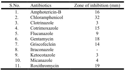
Effect of commercial antibiotics
The commercial antibiotics showed zone of inhibition on the growth when placed on SDA medium swabbed with C.
neoformans. But observed in there was no zone of inhibition.
In vitro inhibitory effect of antimicrobial agents against C. neoformans
S.No. Antibiotics Zone of inhibition (mm)
1. Amphotericin-B 16
2. Chloramphenicol 32
3. Clotrinazole 3
4. Cotrimoxazole 15
5. Flucanazole 9
6. Gentamycin 18
7. Griscofeclein 14
8. Itraconazole -
9. Ketocotazole 3
10. Micanazole 4
11. Roxithromycin 19
Effect of plant extracts
Aqueous extract of 129 medicinally important plant parts (leaves, bulbs, rhizomes, seeds and roots) were individually tested. Among the 129 plants, 37 plants were showed inhibitory effect on C. neoformans. The most effective plants were Pedalium murex (19 mm), Coleus fovskohlii (19 mm),
Hibiscus esculentus (17 mm), Vetiveria zizanoides (16 mm), Solanum nigrum (15 mm), and Andrographis paniculata (16
mm).
DISCUSSION
Zone of inhibition of Cryptococcus neoformans to aqueous medicinal plant extracts
S.No. Botanical Name Family Using Part Zone of inhibition (mm) 100%
1. Abrus precatorius Linn. Fabaceae Seed - 2. Acalypha indica Linn. Euphorbiaceae Leaf 9 3. Achyranthes aspera Linn. Amarantaceae Leaf 10 4. Acorus calamus Linn. Araceae Leaf - 5. Adhotoda vasica Nees. Acanthaceae Leaf 10 6. Aegle marmelos Correa. Rutaceae Leaf 12 7. Aleurites molucanna Wild. Euphorbiaceae Seed 8 8. Allium cepa Linn. Liliaceae Bulb 14 9. Allium sativum Linn. Liliaceae Bulb 15 10. Aloe vera Linn. Liliaceae Leaf - 11. Alpinia galanga Sw. Zingiberaceae Leaf - 12. Alternanthera sessilis Br. Amarantaceae Leaf 5 13. Andrographis paniculata Nees. Acanthaceae Leaf 17 14. Anisochilus carnosus Wall. Lamiaceae Leaf - 15. Annona squamosa Linn. Annonaceae Leaf 8 16. Arachis hypogea Linn. Fabaceae Leaf - 17. Aristolochia indica Linn. Aristolochiaceae Leaf - 18. Artemisia vulgaris Linn. Asteraceae Leaf - 19. Artocarpus integrifolia Linn. Moraceae Leaf - 20. Azadirachta indica Linn. Meliaceae Leaf 6 21. Barleria cuspidata Heyne. Acanthaceae Leaf - 22. Bassia longifolia Linn. Sapotaceae Seed - 23. Cajanus indicus Spreng. Fabaceae Leaf - 24. Calophyllum inophyllum Linn. Clusiaceae Leaf - 25. Capsicum frutescens Linn. Solanaceae Leaf - 26. Cardiospermum halicacabum Linn. Sapindaceae Leaf - 27. Carica papaya Linn. Caricaceae Leaf - 28. Carum bulboehastranum Koch. Apiaceae Seed - 29. Carum copticum Benth. Apiaceae Leaf - 30. Carum copticum Benth. Apiaceae Leaf and
Seed
12 - 31. Cassia auriculata Linn. Fabaceae Leaf - 32. Cassia obtusifolia Linn. Caesalpiniaceae Leaf 8 33. Celosia cristata Linn. Amarantaceae Leaf - 34. Cinnamomum zeylonium Fr. Lauraceae Bark - 35. Cissus quadrangularis Wall. Vitaceae Leaf 10 36. Citrus acidia Roxb. Rutaceae Leaf 10 37. Coriandrum sativum Linn. Apiaceae Leaf - 38. Coriandrum sativum Linn. Apiaceae Leaf and Seed - 39. Crataeva religiosa Forsk. Capparidaceae Rhizome - 40. Cucumis sativus Linn. Cucurbitaceae Fruit - 41. Cuminum cyminum Linn. Apiaceae Seed - 42. Cymopsis psoralioides Dc. Fabaceae Leaf - 43. Cynodan dactylon Pers. Poaceae Leaf - 44. Cyperus rotundus Linn. Cyperaceae Leaf - 45. Delonix regia Raf. Caecalpinaceae Leaf - 46. Dolichos biflorus Linn. Caesalpinaceae Seed - 47. Eclipta alba Linn. Asteraceae Leaf - 48. Eugenia caryophyllata Thumb. Myrtaceae Seed - 49. Eugenia jambolana Lam. Myrtaceae Flower - 50. Ferula asafetida Linn. Apiaceae Root 15 51. Ferula asafoetida Linn Moraceae Leaf - 52. Ficus religiosa Linn. Moraceae Leaf - 53. Gossypium arboretum Linn. Malvaceae Leaf - 54. Gossypium arboreum Linn Asteraceae Leaf - 55. Helianthus annus Linn Asclepiadaceae Root - 56. Hibiscus canabinus Linn. Malvaceae Leaf - 57. Hibiscus esculentus Linn. Malvaceae Leaf 17 58. Hibiscus rosasinensis Linn. Malvaceae Leaf - 59. Hydrocotyle asiatica Linn. Apiaceae Leaf - 60. Ilicium verum Linn. Magnoliaceae Flower - 61. Indigofera tinctoria Linn. Fabaceae Leaf 12 62. Jasminum sambac Ait. Oleaceae Leaf - 63. Jatropha curcas Linn. Euphorbiaceae Leaf - 64. Lablap purpureus (L.) Sweet. Fabaceae Leaf - 65. Lagenaria vulgaris Serrnge. Cucurbitceae Leaf - 66. Lavendula vera Dc.. Cucurbitaceae Leaf 7 67. Leucas aspera Spreng. Lamiaceae Leaf - 68. Luffa acutangula Roxb. Cucurbitaceae Leaf - 69. Magnifera indica Linn. Anacardiaceae Leaf - 70. Mentha piperita Linn. Lamiaceae Leaf -
71. Mimosa pudica Linn. Mimosaceae Leaf - 72. Moringa pterigosperma Goerth. Moringaceae Leaf 8 73. Mukia scarbrella Ann. Cucurbitaceae Leaf 10 74. Murraya koenigii Spr. Rutacaeae Leaf 6 75. Musa paradisiace Linn. Musaceae Flower, Stem - 76. Musa paradisiaca Houtt. Myristioaceae Seed - 77. Nyctanthes arbor-tristis Linn. Oleaceae Leaf - 78. Ocimum basilicum Linn. Lamiaceae Leaf - 79. Ocimum gratissium Linn. Lamiaceae Leaf - 80. Ocimum sanctum Linn. Lamiaceae Leaf - 81. Ocimum var. thyrsiflorum, Benth. Lamiaceae Leaf 11 82. Papaver somniferum Linn. Papaveraceae Seed - 83. Pedalium murex Linn. Pedaliaceae Leaf 19 84. Phascelus radiatus Linn. Fabaceae Leaf - 85. Phaseolus radiatus Linn Fabaceae Seed - 86. Phyllathus distichus Muell. Euphorbiaceae Leaf - 87. Phyllathus niruri Linn. Euphorbiaceae Leaf 15 88. Physalis minima Linn. Solanaceae Leaf - 89. Pimpinella hyneana Wall. Apiaceae Seed 7 90. Piper betle Linn. Piperaceae Leaf - 91. Piper nigrum Linn. Piperaceae Seed - 92. Pisidium gaujava Linn. Myrtaceae Leaf - 93. Pisidium gujava Linn. Fabaceae Leaf - 94. Polyalthia longifolia Benth. Annonaceae Leaf - 95. Polygala telephoides Wild. Polygalaceae Leaf - 96. Portulaca quadrifida Linn. Portulacaceae Leaf 10 97. Prunus amygdalus Bail. Rosaceae Leaf and Seed - 98. Prunus amygdalus Basl. Rosaceae Seed - 99. Psoralea corylifolia Linn. Fabaceae Leaf - 100. Punica granatum Linn. Punicaceae Leaf - 101. Querus incana Roxb. Fagaceae Seed - 102. Raphanus sativus Linn. Brassicaceae Leaf - 103. Ricinus communis Linn. Euphorbiaceae Seed - 104. Rubia cordifolia Linn. Rubiaceae Seed - 105. Sesamum indicum Dc. Pedaliaceae Leaf 11 106. Sesbania grandiflora Pers. Fabaceae Leaf 7 107. Sida caprinifolia Linn. Malvaceae Leaf - 108. Sida spinosa Linn. Malvaceae Leaf - 109. Solanum mélangena Linn. Solanaceae Leaf - 110. Solanum nigrum Linn. Solanaceae Leaf 16 111. Solanum tarvum Swantz. Solanaceae Leaf 11 112. Solanum torvum Swantz. Solanaceae Leaf - 113. Solanum tuberosum Linn. Solanaceae Leaf - 114. Solanum viarum Dunal. Solanaceae Leaf - 115. Sphaeranthus amaronthoides burm. f. Amarantaceae Leaf - 116. Tabernaemontana coronacia Br. Apocynaceae Leaf 14 117. Tamarindus indicus Linn. Casalpiniaceae Leaf - 118. Tectona grandis Linn. Caesalpinaceae Leaf - 119. Terminalia chebula Retz. Combretaceae Seed - 120. Thespesia populnea Corr. Malvaceae Leaf - 121. Trigonella foenum-graceum Linn. Fabaceae Leaf 11 122. Vetiveria zizanoides Nash. Poaceae Root 16 123. Vetiveria zizanoides Nash. Poaceae Leaf - 124. Vitex negundo Linn. Euphorbiaceae Leaf - 125. Wedelia pauciflora W.A. Asteraceae Leaf - 126. Wrightia tinctoria Br. Apocynaceae Leaf 14 127. Zinzifer officinalis Zosa. Zingiberaceae Rhizome - 128. Zingifer officinalis Zosa. Rhamnaceae Leaf -
(-) Indicates no zone formation
Zone of inhibition of C. neoformans to selected aqueous plant extracts in various concentrations
S.No. Botanical name Family Using part Zone of inhibition (mm) 25% 50% 100% 1. Pedalium murex Pedaliaceae Leaf 9 11 19 2. Coleus fovskohlii Laminaceae Leaf 9 15 19 3. Vetiveria zizanoides Poaceae Leaf 8 12 16 4. Andrographis paniculata Acanthaceae Leaf 6 8 17
5. ` Hibiscus esculentus Malvaceae Leaf 8 10 `17
Cryptococcosis is caused by an encapsulated yeast belonging to the genus Cryptococcus and it can cause disease in apparently immuno competent, as well as immuno compromised hosts. Most susceptible to infection are patients with T-cell deficiencies (Kwon-Chung, 1992; Mitchel and Perfect, 1995). Immuno-compromised patients are more susceptible to mycotic diseases than the healthy people. However, it was emphasized that the type of fungus and the nature of the diseases are to be considered in the study of mycoses (Gopalakrishnan, 2003).
Methamphetamine enhances Cryptococcus neoformans
pulmonary infection and dissemination to the brain. Notably, C. neoformans modifies its capsular polysaccharide after METH exposure, highlighting the fungus’s ability to adapt to environmental stimuli, a possible explanation for its pathogenesis. The findings may translate into new knowledge and development of therapeutic and public health strategies to deal with the devastating complications of METH abuse. (Patel, 2013). The identification of the isolate was confirmed by microscopic examination for the presence of yeast like cells. Direct observation of yeast in KOH preparation Winn and Westernfeld (1997) and Kozel and Cazin (1971) and McGinnis (1980) reported an Indian-ink determined the capsule negative stain of C. neoformans. In the present investigation also pathogen (C. neoformans) was confirmed by negative staining and methylene blue staining method. The biochemical characters C. neoformans was described by Phaff and Fell (1970). In the present study, C. neoformans grew in seven carbon sources did not reduced nitrate to nitrite.
Growth and morphology of fungi is controlled by various physico-chemical characteristics and composition of the culture media (Rai, 1989). Hence, the standardization of the media is essential to grow the organism for any kind of investigation. In the present investigation, C. neoformans was cultured in six different nutrient media and their broth. Of them, in the yeast like fungi grew in all the six media and their broth were
slightly decreased compared to the others.
Temperature, pH and salinity are the major factors found to affect the growth of any fungi. Maximum growth rate of M.
furfur was at range of PH 7-9. However the fungus was able to
grow at all PH levels (Moore, 1938; and Vijayakumar 2003).
But, in the present investigation C. neoformans grew well at pH ranges from 6.0 to 8.0 indicated that the pH requirements may vary from medium to medium. In vitro study of growth rate of C. neoformans does not grow at 40°C or higher, but they grew at 37°C is normal (Fromtling et al. 1988). Roberts (1969) and Vijayakumar (2003) revealed that the growth rate of Malassezia furfur was well at 37°C on Malt extract agar or Sabouraud’s dextrose agar medium. In the present study also
revealed the pathogen (C. neoformans) grew well at 30± 2°C.
Likewise the salinity also had its own varying impact on the growth of different species. Most of the pathogenic fungal species are well adapted to higher salinity concentration (Vasuki et al., 2002; Gopalakrishnan, 2003; Vijayakumar, 2003). In the present investigation, the concentration of salinity was increased and the growth rate of pathogen was slightly
increased. Vijayakumar (2003) used five different
concentration of glucose and peptone (0, 1, 2, 3 and 4%) in sabourad’s dextrose agar and noticed that the growth of M.
furfur was observed at 12 hrs intervals. In glucose the
concentration was increased at same time, growth rate was increase at 4 percent. In the present work it was found that C.
neoformans grew well at 2.0g/10 ml in dextrose and 1g/100 ml
in peptone. Weindling (1934), Florey et al. (1946), Dobos and Hirsch (1966) Bava et al. (1989) reported that the antibiotics, viz., amphotericin B, nystatin, ketoconazole and fluconozole were effective to control the growth of many fungal pathogen who also stated that many yeast like fungi were resistant many commercial antibiotics. In the present study the maximum and minimum effect to control the growth of C. neoformans was
observed in chloramphenicol (32 mm), ketaconazole and
clotrimazole (3 mm) are respectively. Out of 11 commercial antibiotics intraconazole had no effect on the growth C.
neoformans. The antifungal effect of an aqueous extract of
garlic was tested against 10 strains of C. neoformans. In the present study, 129 locally available medicinal plants extract examined for antimycotic activity against C. neoformans. Of which 37 plants were showed the inhibitory effect. Pedalium
murex (19 mm) and Coleus fovskohliii (19 mm) were showed
the maximum inhibition and minimum was in Alternanthera
sessilis (5 mm).
Acknowledgement
We are grateful to Mr.K.Thulasiah vandayar, Secretary and correspondent, A.V.V.M Sri Pushpam College (Autonomous), Poondi-613 503, Thanjavur Dt, Tamilnadu, India for providing
necessary facilities to carryout this research work.
REFERENCES
Aneja, K. R., 2001. Experiments in Microbiology, Plant pathology, Tissue Culture and Mushroom Production
Technology, 3rd edition, New Age International (P)
Limited, New Delhi, pp.303-309.
Basha, S. G., Shareif, S.D. and Shankar, S.G. 2010. Comparison of In vitro susceptibility of non-melanised
Cryptococcus neoformans to Cinnamonum zeylanicum and Murraya paniculata. Int.J. of Biol.Tech., 1(3): 5-9.
Bauer A W, Kirby W M M, Sherris J C & Turck M. Antibiotic susceptibility testing by a standardized single disk method. Amer. I. C/in. Pathol. 45:493-6, 1966.
Bava, A. J., C. Rovanitii and R. Negroni, 1989. Comparative study of four antifungal drugs in an experimental model of marine cryptococcosis,, Mycopathologica, 108: 81-82. Charmian D. Sittambalam, Heidi Hanna, Justin Martello,
Dimitr mitsani, 2012. Cryptococcus infection in a non-HIV patient: a case report. Journal of Community Hospital
Internal Medicine Perspectives, 2012, 2: 19254.
Damodaran, S.and Venkataraman, S., 1993. A study of the therapeutic efficacy of Cassia alata Linn. leaf extract against Pityriasis versicolor. J. Etanopharmacology, 42(1): 19-23.
Dobos, R. J. and Hirsch, J.G., 1966. Bacterial and mycotic
infection in man, Asian edition, 4th edn., pp. 835-875.
Florey, H.W., M.A. Jenings, K. Gilliver and A.G. Sanders, 1946. Mycophenolic acid: An antibiotic from Penicillium
brevi, Compactum Dierckx. Lancet. 1: 46-49.
Fromtling, R. A., George K. Abruzzo and Alejandro Ruiz,
1988. Virulence and antifungal susceptibility of
environmental and clinical isolates of Cryptococcus
neoformans fro puecto rico, Mycopathlogica, 106:163-166.
Gopalakrishnan, K., 2003. Studies on Candida albicans, M.Sc.,
Dissertation, Bharathidasan University, Tiruchirapalli, pp. 1-32.
Jafari A.H.A., Vila, R., Freixa, B., Tomi, F., Casanova, J., Costa, J. and Canigueral, S. 2011. Composition and antifungal activity of the essential oil from the rhizome and roots of Ferula hermonis. Phytochemistry 72: 1406-1413. Kamboj, V.P., 2000. Herbal medicine, Curr. Sci., 78(1): 35-39.
Kannan, N.,1996.Laboratory Manual in General Microbiology,
1st edition, Palani Paramount Publications, Palani.
Kozel, T. R. and Cazin, Jr. J., 1971. Nonencapsulated variant of Cryptococcus neoformans. Virulence studies and characterization of soluble polysaccharide, infect. Immun., 3: 287-294.
Kwan-Chung, K. J., 1992. A new genus Filobasidiella the perfect state at Cryptococcus neoformans, Mycologica, 67: 1197-1200.
Lewis, W.H. and Elwin-Lewis, M.P.H., 1978. Medical Botany: Plants Affecting Man’s Health, John Wiley and Sons, New York.
McGinnis, M. R., 1980. Laboratory Handbook of Medical Mycology, Academic Press, New York, pp. 203-204. Mitchell, T.G. and J.R. Perfect, 1995. Cryptococcosis in the
era of AIDS-100 years after the discovery of Cryptococcus
neoformans, Clin. Microbiol. Rev., 8: 515-48.
Moore, M. 1938. Cultivation of Malassezia furfur, etiological
agent of Pityriasis (tinea) versicolor. Mycopathology, 1: 53-61.
Patel D, Desai GM, Frases S, Cordero RJB, DeLeon-Rodriguez CM, Eugenin EA, Nosanchuk JD, Martinez LR.
2013. Methamphetamine enhances Cryptococcus
neoformans pulmonary infection and dissemination to the
brain. mBio 4(4):e00400-13. doi:10.1128/mBio.00400-13.
Phaff, R.H. and J.W. Fell, 1970. Cryptococcus kutzing emenol. Phaff et spencer. In: The Yeasts, a Taxonomic study [J. Lodder (ed.)], North-Holland Publishing Company, Amsterdom, pp. 1088-1145.
Rai, M.K., 1989. The Effect of different media o the morphology of cultural characteristics of Candida
albicans. Curr. Sci., 58(15): 861-863.
Roberts, S.O., 1969. Pityrosporum orbiculare; Incidence and distribution on clinically normal skin. British J.
Dermatology, 81: 264-269.
Sivamani, P., 1999. Medical Mycology, 1st edition, Siva
Publications, Arcot.
Vasuki, S., Nadimuthu, N. and Kannan, L., 2002. Distribution of a fungal pathogen, Fusarium solani (MART) SACC, in the marine environs of Tamilnadu and its growth response to different salinity and pH. International Symposium on
Mycotic Infections in the 21st century, Rajah Muthaiah
Medical College, Annamalai Nagar, Abstract, 99 p. Vijayakumar, R., 2003. Studies on the characterization, growth
and control of Malassezia furfur Baillon, A Dandruff fungus, M.Phil., Dissertation, Bharathidasan University, Tiruchirapalli, pp. 1-51.
Weindling, R., 1934. Studies on the lethal principle effective in the parasitic action of Trichoderma lignorum on
Rhizoctonia solani and other soil fungi, Phytopath., 24:
1153-1179.
Winn, W.S. and Westernfeld, F.W., 1997. Mycotic diseases, Medical Microbiology, pp. 1211-1251.