A STUDY OF MANAGEMENT OF FRACTURES OF BOTH BONES FOREARM USING LOCKING
*Dr. Chirra Chakradhar Reddy, Dr. B. Shaik nazeer
Department of Orthopaedics, Sri Devaraj Urs Medical College, Tamaka, Kolar
ARTICLE INFO ABSTRACT
Locking compression plating is a popular method of fixation for the bones of the forearm and is associated with superior outcomes and less complication compared to external fixation and non operative methods. Hence an attempt is made to evaluate its manage
up. This study is a hospital based prospective study conducted in the Department of Orthopaedics at R.L. J. Hospital and Research Centre, Kolar, from November 2014 to February 2016. 30 patients with both bone fractures of
locking compression plating and were followed up for 6 months at regular intervals. The average age of the patients was 33.8 years and majority were male, right side was common (60%),
fractures of both bones forearm are located in the middle third and the fracture pattern transverse/short oblique was commonest. The results were based on Anderson
study, there were 25 (87 %) patients wi
superficial infection which resolved with antibiotics and two cases of posterior interosseous nerve injury immediately following surgery, which recovered spontaneously are observed. Open reduct and internal fixation with locking compression plating is one of the ideal methods of fixation of both bones forearm shaft fractures as there is stable fixation and earlyunion. Most of the fractures in this study united within 4 months of surgery. It i
adherence to AO principles and adequate post provides excellent results.
Copyright©2017, Chakradhar Reddy et al. This is an open access article
use, distribution, and reproduction in any medium, provided the original work is properly cited.
INTRODUCTION
Forearm bone fractures are commonly encountered in today’s industrialera. (Morgan et al., 1994; Anderson
Crenshaw, Andrew, 2013)Various treatment modalities were
introduced from time to time and each of them had some edge over the previous one. (Crenshaw, Andrew, 2013; Hertle 2006) Continuing this process of revolution and based on many years of experience with compression plating, promising results obtained (Sevitt Simon, 1981; Thakur, 2015; Colton, 1994; Chandler, 2015; Crenshaw, Andrew, 2013; Stern and Drury,
1983; Chapman et al., 1989). The forearm, being a component
of upper limb serves important movements that are important in activities of daily living. The forearm, in combination with the proximal and distal radioulnar joints, allows pronation and supination which in turn helps hand, to perform multi axial movements. (Snell and Richard, 2012; Benjamin, 2000; McRae
et al., 2002; Hoppenfeld, 2011)Fracture of the forearm both
bones may result in severe loss of function unless adequately
*Corresponding author: Dr. Chirra Chakradhar Reddy,
Department of Orthopaedics, Sri Devaraj Urs Medical College, Tamaka, Kolar
ISSN: 0975-833X
Article History:
Received 18th December, 2016
Received in revised form 05th January, 2017
Accepted 05th February, 2017
Published online 31st March,2017
Key words:
Bone forearm shaft fractures, Open reduction
and Internal fixation, Locking compression plate.
Citation: Dr. C. H. Chakradhar Reddy, Dr. B. Shaiknazeer
locking compression plates”, International Journal of Current Research
RESEARCH ARTICLE
A STUDY OF MANAGEMENT OF FRACTURES OF BOTH BONES FOREARM USING LOCKING
COMPRESSION PLATES
*Dr. Chirra Chakradhar Reddy, Dr. B. Shaik nazeer
and Dr. Arun, H. S.
Department of Orthopaedics, Sri Devaraj Urs Medical College, Tamaka, Kolar
ABSTRACT
Locking compression plating is a popular method of fixation for the bones of the forearm and is associated with superior outcomes and less complication compared to external fixation and non operative methods. Hence an attempt is made to evaluate its manage
up. This study is a hospital based prospective study conducted in the Department of Orthopaedics at R.L. J. Hospital and Research Centre, Kolar, from November 2014 to February 2016. 30 patients with both bone fractures of the forearm were managed with open reduction and internal fixation with locking compression plating and were followed up for 6 months at regular intervals. The average age of the patients was 33.8 years and majority were male, right side was common (60%),
fractures of both bones forearm are located in the middle third and the fracture pattern transverse/short
oblique was commonest. The results were based on Anderson et al
study, there were 25 (87 %) patients with excellent results and 5 (13 %) with satisfactory. A case of superficial infection which resolved with antibiotics and two cases of posterior interosseous nerve injury immediately following surgery, which recovered spontaneously are observed. Open reduct and internal fixation with locking compression plating is one of the ideal methods of fixation of both bones forearm shaft fractures as there is stable fixation and earlyunion. Most of the fractures in this study united within 4 months of surgery. It is concluded that with proper preoperative planning, adherence to AO principles and adequate post-operative rehabilitation locking compression plate provides excellent results.
is an open access article distributed under the Creative Commons Attribution License, which use, distribution, and reproduction in any medium, provided the original work is properly cited.
Forearm bone fractures are commonly encountered in today’s ., 1994; Anderson et al., 1975; Various treatment modalities were of them had some edge (Crenshaw, Andrew, 2013; Hertle et al., Continuing this process of revolution and based on many years of experience with compression plating, promising results Sevitt Simon, 1981; Thakur, 2015; Colton, 1994; drew, 2013; Stern and Drury, The forearm, being a component of upper limb serves important movements that are important in activities of daily living. The forearm, in combination with allows pronation and supination which in turn helps hand, to perform multi axial Benjamin, 2000; McRae Fracture of the forearm both bones may result in severe loss of function unless adequately
Dr. Chirra Chakradhar Reddy,
Department of Orthopaedics, Sri Devaraj Urs Medical College,
treated. Hence good anatomical re
of these fractures is necessary to restore functions.
reduction which was employed in earlier days yielded unsatisfactory results from either non
(Stern and Drury, 1983; Schemitsch
complex forces acting on the forearm bone that makes reduction and its maintenance of displaced fracture fragments difficult (Crenshaw and Andrew, 2013; Hertle
Union may be achieved with any of the methods available, however severe loss of function may be the end result unless adequately treated with proper technique and implants.
and Chow, 2006)The functional outcome was assessed using Anderson et al, (1975) scoring system. The variables taken into consideration are a) Union of the fractures and b)
elbow and wrist movements.
Study objective
To evaluate the functional outcome following the use of locking compression plate in the fractures of both bone forearm.
International Journal of Current Research Vol. 9, Issue, 03, pp.47593-47598, March, 2017
INTERNATIONAL
OF CURRENT RESEARCH
Dr. C. H. Chakradhar Reddy, Dr. B. Shaiknazeerand Dr. Arun, H. S. 2017. “A study of management of fractures of both bones forearm using
International Journal of Current Research, 9, (03), 47593-47598.
A STUDY OF MANAGEMENT OF FRACTURES OF BOTH BONES FOREARM USING LOCKING
and Dr. Arun, H. S.
Department of Orthopaedics, Sri Devaraj Urs Medical College, Tamaka, Kolar
Locking compression plating is a popular method of fixation for the bones of the forearm and is associated with superior outcomes and less complication compared to external fixation and non-operative methods. Hence an attempt is made to evaluate its management in our patients in rural set up. This study is a hospital based prospective study conducted in the Department of Orthopaedics at R.L. J. Hospital and Research Centre, Kolar, from November 2014 to February 2016. 30 patients with the forearm were managed with open reduction and internal fixation with locking compression plating and were followed up for 6 months at regular intervals. The average age of the patients was 33.8 years and majority were male, right side was common (60%), most of the fractures of both bones forearm are located in the middle third and the fracture pattern transverse/short
et al. (1975) scoring system and in this
th excellent results and 5 (13 %) with satisfactory. A case of superficial infection which resolved with antibiotics and two cases of posterior interosseous nerve injury immediately following surgery, which recovered spontaneously are observed. Open reduction and internal fixation with locking compression plating is one of the ideal methods of fixation of both bones forearm shaft fractures as there is stable fixation and earlyunion. Most of the fractures in this s concluded that with proper preoperative planning, operative rehabilitation locking compression plate
ribution License, which permits unrestricted
treated. Hence good anatomical reduction and internal fixation of these fractures is necessary to restore functions. Closed reduction which was employed in earlier days yielded unsatisfactory results from either non-union or loss of motion. (Stern and Drury, 1983; Schemitsch et al., 1992)Also there are complex forces acting on the forearm bone that makes reduction and its maintenance of displaced fracture fragments Crenshaw and Andrew, 2013; Hertle et al., 2006). Union may be achieved with any of the methods available, however severe loss of function may be the end result unless adequately treated with proper technique and implants. (Leung
The functional outcome was assessed using , (1975) scoring system. The variables taken into Union of the fractures and b) Range of
To evaluate the functional outcome following the use of locking compression plate in the fractures of both bone
INTERNATIONAL JOURNAL OF CURRENT RESEARCH
MATERIALS AND METHODS
The present study includes treatment of 30 cases of fracture both bones of forearm by open reduction and internal fixation with 3.5 mm Locking Compress Locking Compression Plate (LCP) between November 2014 to February 2016 at R. L. Jalappa Hospital attached to Sri Devaraj URS Medical College, Tamaka, Kolar - 563-101, India. The Criteria included in this case study are Patient above the age of 18 years, Patients with diaphyseal fractures of both bones forearm and both open and closed type fractures. The exclusion criteria adopted in the present study are Patients with diaphyseal fractures of type-III B and III C and Patients medically unfit for surgery.
Patient Clinical History
On admission of the patient, a careful history was elicited from the patient and/or attendants to reveal the mechanism of injury and the severity of trauma. The patients were then assessed clinically to evaluate their general condition and the local injury. The patient vital signs were recorded. Methodical examination was done to rule out fractures at other sites. Local examination of injured forearm revealed swelling, deformity and loss of function. Any nerve injury was looked for and noted. Palpation revealed abnormal mobility, crepitus and shortening of the forearm. Distal vascularity was assessed by radial artery pulsations, capillary filling, pallor and paraesthesia at finger tips. Radiographs of the radius and ulna i.e., anterio-posterior and lateral views, were obtained. The elbow and wrist joints were included in each view. The limb was then immobilized in above elbow Plaster of Paris slab with sling. The patient was taken for surgery after routine investigations and after obtaining fitness towards surgery. The investigations are as follows: Hb%, Urine for sugar, FBS, Blood urea, Serum creatinine, HIV, HBSAg and ECG. Proximal radius was approached by volar Henry approach was
used for middle and distal radius. 17 A narrow 3.5mm LCP was
used and a minimum of 6 cortices were engaged with screw fixation in each fragment. 21, 22
Statistical analysis
Data was entered into Microsoft excel data sheet and was analyzed using SPSS 22 version software. Categorical data was represented in the form of frequencies and proportions. Continuous data was represented as mean and standard deviation. Statistical software: MS Excel, SPSS version 22 (IBM SPSS Statistics, Somers NY, USA) was used to analyze data. EPI Info (CDC Atlanta), Open Epi, Med calc and Medley’s desktop were used to estimate sample size, odds ratio and reference management in the study.
Operative procedure
Pneumatic tourniquet was applied: time noted. Painting and draping of the part done. The radius was approached using volar henrys and Ulna was approached directly over the subcutaneous border. (Crenshaw and Andrew, 2013; Hertle and Dominique, 2006; Texhammer, 1994) The bone which was less comminuted and more stable was fixed first and later the other bone was fixed. After identifying the fracture ends, periosteal was not elevated and fracture ends were cleaned. With the help of reduction clamps fracture was reduced and held inposition. The plate was then applied after contouring if required. A plate of at least 6 holes was chosen and longer plates were used
inspiral, segmental and comminuted fractures. For upper third radial fractures, the plate was fixed dorsally. Formiddle third, the plate was fixed dorsolateral and for distal radialfractures the plate was fixed on the volar aspect. In ulnar fractures, plate was applied over the posterior surface of ulna (Crenshaw and Andrew, 2013; Hertle and Dominique, 2006). A drill sleeve for locking screw is fixed in the hole, near the fracturesite, and 2.7 mm drill bit is use to drill both the cortex of the bone, the sleeve is removed and the screw length is measured with depth gauge. A 3.5 mm locking screws are then inserted, as the locking screws are of self tapping, tapping of the screw hole is not done (Thakur, 2015; Moed et al., 1986; Gouda et al., 2016). After adaptation of the fragments, a screw hole for axial compression is drilled in the fragment which forms an acute angle near the plate. Here the load guide is used with the arrow pointing towards the fracture line to be compressed. At this position, a lag screw will be inserted for axial compression. The lag screw is applied by subsequently over drilling (3.5mm) the near cortex to create a gliding hole (Moed et al., 1986). The lag screw and remaining screws are inserted. Once stable fixation is achieved haemostasis secured after release of tourniquet, the wound is closed with suction drain and sterile dressing is applied. Postoperatively a crepe bandage was applied over the affected forearm and arm pouch was given advised to perform shoulder, elbow, wrist and finger movements. The patient was instructed to keep the limb elevated and move their fingers and elbow join. Wound was inspected after 3 days postoperatively. Antibiotics and analgesics were given to the patient till the time of suture removal. Suture/staples removed on 10th postoperative day. patients were followed up at monthly intervals for first 6 months and evaluation was done based on "Anderson et al” scoring system.
RESULTS AND ANALYSIS
Age- 36.7% were below 25 years, 26.7% were between 26 to 35 years, 16.7% were between 36 to 45 years and 20% were >45 years. The youngest patient was 19 years old and oldest was 65 years.
Sex- Majority of subjects were males (93.3%) and 6.7% were
females.
Nature of trauma- 76.7% had injury due to road traffic accident and 23.3% had injury due to self-fall.
Side involved-40% of subjects had fracture on left side and 60% of them had fracture on right side.
Site of fracture-Distal 3rd of Fracture was seen in 36.6% of radius and ulna, Middle 3rd of fracture was seen in 46.6% of radius # and 50% of ulna, Proximal 3rd fractures were seen in 16.8% of radius and 13.4% of ulna fractures.
Type of fracture- 63.3% of fractures radius and 40% of ulna fractures were transverse, 20% of radius and 43.3% of ulna fractures were comminuted, 6.7% of radius and 10% of ulna fractures were oblique, 10% of radius and 6.7% of ulna fractures were spiral. All the fractures were closed type.
Associated injuries-73.3% of subjects had no associated injuries and 3.3% of subjects had other associated injuries.
Range of movement- After surgery was full among 86.7% of subjects, good among 10% and loss of 10 degree pronation was seen in 3.3% of subjects.
Complication- PIN palsy was seen in 6.7% of subjects and infection was seen in 3.3% of subjects. Outcome of surgery excellent results (83.3%) and satisfactory results (16.7%) results.
DISCUSSION
In this study 30 both bone fractures of the forearm were treated surgically with open reduction and internal fixation with locking compression plates. The patients were followed up for 6 months. The cases in this study belonged to different age groups, both the sexes and included both open and closed fractures.
Age distribution
[image:3.595.335.532.210.254.2]In our study 36.7% were below 25 years, 26.7% were between 26 to 35 years, 16.7% were between 36 to 45 years and 20% were >45 years. The youngest patient was 19 years old and oldest was 65 years. In our study, fracture was commoner in the second and third decade, with average age of 33.8 years (18-55 years).
Table 1. Age distribution
Number %
Age < 25 years 11 36.7%
26 to 35 years 8 26.7%
36 to 45 years 5 16.7%
> 45 years 6 20.0%
Total 30 100.0%
Series Year Average age (years)
Michael W.Chapman et al 1989 33
Frankle Leung 2003 36
Goudetal 2016 33.5
Boussakri et al 2016 34.52
Present study 2016 33.8
Table 1 shows the age distribution and its comparison with other referred works in this area of research.
Table 2 to 9 shows the results of comparative study based on various parameters.
Sex distribution of patients
Majority of the patients in our study were male 93.3%. It correlated with other studies quoted below.
Table 2. Sex distribution
Series Years Males (%) Females (%)
Michael Chapman 1989 78 22
Frankie Leung 2003 82.6 17.4
Gouda et al 2016 70 30
Present study 2016 93.3 6.7
Mode of injury
In our series 76.7% of cases were due to road traffic accidents and 23.3% due to fall as compared to studies quoted below.
Table 3. Mode of injury
Series Year Accident
(%)
Fall (%)
Direct blow/ Miscellaneous (%)
Moed 1986 70 14 16
Gouda et al 2016 50 40 10
Present study 2016 76.7 23.3 0
Extremity affected
We had 60% incidence of fracture both bone on right extremity. It correlated with other studies as shown below.
Table 4. Extremity affected
Series Years Right (%) Left (%)
Chapman 1989 55 45
Babu et al 2015 50 50
Present study 2016 60 40
Fracture anatomy
a) Type of fracture
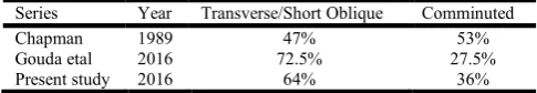
[image:3.595.311.554.380.422.2]In our series and 64% of fractures were transverse/short oblique and 36 % were comminuted .The other series are as shown below.
Table 5. Fracture pattern
Series Year Transverse/Short Oblique Comminuted
Chapman 1989 47% 53%
Gouda etal 2016 72.5% 27.5%
Present study 2016 64% 36%
b)Level of fracture
In our series 48.4% of fractures were in middle third, 36.6 % lower third and 15% in proximal third. The incidence fracture of middle third are more in our study compared to others as quoted below.
Table 6. Fracture level
Series Years Proximal third Middle third Distal third
Gouda et al 2016 15% 70% 15%
Present study 2016 15% 48.4% 36.6%
Time of union
In the present study the average union time was 14 weeks with a range of 12 to 20 weeks which was similar to most of the other studies. We had 100% union of both radius and ulna.
Table 7. Time for fracture union
Series Union time in weeks Range in weeks Union (%)
Chapman 12 6-14 98
Gouda et al 14 8-20 100
Present study 14 12-20 100
FUNCTIONAL RESULTS
[image:3.595.46.278.388.505.2]Table 8. Functional results
Series Excellent
(%)
Satisfactory (%)
Unsatisfactory (%)
Failure (%)
Anderson 50.9 34.9 11.3 2.9
Frankie 98 2 - -
Chapman 86 7 12 5
Goud et al 85 15 - -
Present study 83.3 13.7 - -
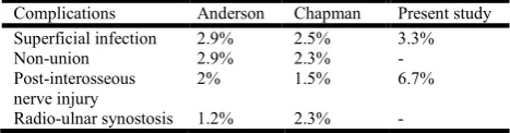
Table 9. Comparative Study and Complications
Complications Anderson Chapman Present study
Superficial infection 2.9% 2.5% 3.3%
Non-union 2.9% 2.3% -
Post-interosseous nerve injury
2% 1.5% 6.7%
Radio-ulnar synostosis 1.2% 2.3% -
Complications
In our series we had a case of superficial infection which resolved with antibiotics. We had two case of posterior interosseous nerve injury immediately following surgery. Both cases recovered spontaneously probably were neuropraxia due to retraction.
Conclusion
The incidence of fractures of both bone forearm are steadily increasing. These fractures have a poor outcome when treated conservatively and require a stable fixation and a well-planned post-operative protocol to achieve fracture union as well as mobility. In this study 30 patients underwent open reduction and internal fixation with locking compression plates for fractures of the both bones of forearm. Majority of the patients were males and most of the injuries were due to the road traffic accidents and treated with locking compression plate. Locking compression plating is effective even in osteoporotic bones and is a reliable option even in elderly patients with poor bone quality. More than 83.3% of our patients had excellent to good outcome. Hence locking compression plating is a safe and reliable technique in the fixation of fractures of the both bones of the forearm. The Radiological and clinical photography taken during the study are shown in the Annexure.
REFERENCES
Anderson. LD, Sisk.D, Tooms. R. and Park WI. 1975. Compression plate fixation in acute diaphyseal fractures of the radius and ulna. J. Bone Joint Surg. Am., 57: 287-93. Benjamin A. 2000. Injuries of the Forearm chap-22 in
Watson-Jones Fractures & Joint Injuries. Edt. J. N. Wilson, 6th Ed, 650-709.
Boussakri H, Elibrahimi A, Bachiri M, Elidrissi M, Shimi M, Elmrini A. 2016. Non-union of Fractures of the ulna And Radius Diaphyses: Clinical and Radiological results of
Surgical Treatment: Malaysian Orthopaedic Journal, 10:
2.
Chandler R N. 2015. Principles of Internal Fixation. Chapter-3, in Fractures in Adults, Vol. 1, 8th Edn., Rockwood C. A.
Jr. et al, Philadelphia; Lippincott Raven.
Chapman MW, Gordon JE, and Zissimos AG. 1989. Compression-plate fixation of acute fractures of the diaphyses of the radius and ulna. J. Bone Joint Surg. Am., 71: 159 - 169.
Colton.C. 1994. History of Osteosynthesis. Chapter-2, in AO/ASIF Instruments and implants 2nd edn, Texhammer R and C. Colton, Berlin, Springer Verlag, 3.
Crenshaw, Andrew H. 2013. Fractures of shoulder girdle, Arm and Forearm. Chapter-49, in Campbells Operative Orthopaedics, Edt. Canale, S Tery, Mosby, 3042-58 Gouda R, Patil B, Ronald I, Sharad. M.A. 2016. A Follow Up
Study To Evaluate The Management Of Both Bones Of Forearm In Adults,Using Locking Compression Plate:
IJOT, 2:119-123.
HariBabu, S., M. Rajesh, G. Suresh Babu, L. Anand, 2015. “Comparative Study of DCP versus LCP in the Management of Diaphyseal Fracture of Both Bones
Forearm”. JEBMH, 6717- 6724.
Hertle R. and Dominique A. 2006. Rothenflush. Fracture of shaft of radius and ulna. Chapter 27. Rockwood and Green’s fracture in adults. Vol. 1 Ed.6, Robert W Bucholz and James D. Heckman, Charles Court-Brown. 979-980. Hoppenfeld: The forearm. Chapter-4 in Surgical exposures
inorthopaedics, 4thEdn, Philadelphia; Lippincott Raven Publishers, 2011.
Leung F. and Chow SP. 2006. Locking compression plate in the treatment of forearm fractures a prospective study. J
Orthop Surg., 14(3):291-4.
McRae, 2002. Ronald and Max Esser,: Injuries to the forearm bones. Chapter-8, in Practical fracture treatment, 5th Edn., Edinburgh ;Churchill Livingstone, 173-186.
Michael J. and Gardner. MD. 2016. Stability of locking compression plates inosteoporotic bone. Hosp for special study, Newyork, USA (symposis);14. Gray. Henry: Osteology Chap-3 in Gray's Anatomy Edt. Williams, Peter
L et al., Norwich, Churchill Livingstone, 410-415.
Moed BR, Kellam JF, Foster RJ, Tile M. and Hansen ST. 1986. Immediate internal fixation of open fractures of the diaphysis of the Forearm. J. Bone Joint Surg. Am., 68: 1008 – 1017
Morgan, William J. and Thomas P. 1994. Breen: Complex fractures of forearm. Hand Clin., 10(3);375-390.
Schemitsch, Emil H. and. Richards. R.R. 1992. The effect of malunion on functional outcome after plate fixation of fracture of both bones of forearm in adults. J Bone & Joint Surg., 74_A (7). 1068-1078.
Sevitt Simon. 1981. Primary repair of fractures and compression fixation. Chap-10 in Bone repair and fracture healing in man. Edinburgh. Churchill Livingstone, 145-156.
Snell, Richard S. 2012. The Upper Limb. Chapter-9 in Clinical anatomy for medical students, Philadelphia; Lippincott Williams & Wilkins, 448.
Stern PJ. and Drury WJ. 1983. Complications of plate fixation of forearm fractures. Clin Orthop., 175:25-9.
Texhammer R. 1994. AO/ASIF Instrumentation. Chapter-6 in AO/ASIF Instruments and implants 2nd edn, Texhammer R and C. Colton, Berlin, Springer Verlag, 84-86.
[image:4.595.45.279.181.242.2]Annexure
Radiological and clinical photographs:
Case -1
Preoperative Post operative
3months 6 months
Case-2
Preoperative Postoperative
3months 6months
Clinical outcome- Range of movements