MR of the Head and Neck: Comparison of Fast Spin-Echo and
Conventional Spin-Echo Sequences
Robert Fulbright, David Panush, Gordon Sze, Robert C. Smith, and R. Todd Constable
PORPOSE: To compare conspicuousness of head and neck lesions on fast spin-echo sequences
and conventional spin-echo sequences. METHODS: Forty consecutive patients with 61 head and
neck lesions were evaluated. Lesion conspicuousness was qualitatively compared on conventional spin-echo and fast spin-echo sequences, using both spin-density and T2-weighted images. Thirt y-six lesions had surgical or pathologic confirmation, and 25 were assigned a presumptive diagnosis
based on clinical evaluation and imaging findings seen on conventional spin-echo T1- and T
2-weighted sequences. Forty lesions were related to neoplasms; 21 lesions consisted of infectious,
vascular, or inflammatory abnormalities. RESOL TS: Fast spin-echo sequences provided improved
lesion conspicuousness in 91% of spin-density images, in 77% of T2-weighted images, and in 84% of the combined spin-density and T2-weighted images. CONCLOSION: By providing shorter imaging times and equal or superior lesion conspicuousness, long-repetition-time fast spin-echo sequences can replace long-repetition-time conventional spin-echo sequences in evaluation of the
head and neck.
Index terms: Head, magnetic resonance; Neck, magnetic resonance; Magnetic resonance, com
-parative studies; Magnetic resonance, technique
AJNR Am J Neuroradio/15:767-773, Apr 1994
In magnetic resonance (MR) imaging of the
head and neck
,
both T1
-
weighted and
T2-weighted sequences help characterize normal and
diseased tissues. With conventional spin-echo
se-quences, the longer imaging times of T2-weighted
images and the inherent motion of the head and
neck can result in motion artifacts that degrade
image quality.
A fast spin-echo technique has been developed
that produces long repetition time (TR) images
with similar quality and tissue contrast as
long-TRconventional spin-echo sequences
,
but with
substantially shorter imaging times
,
and thus less
artifact from physiologic motion (1-4)
.
Compared
with conventional spin-echo
,
fast spin-echo has
demonstrated superior image quality in the pelvis
(5-6) and has shown promise in imaging the brain
Received March 30, 1993; accepted after revision July 6.
From the Department of Radiology, Yale University School of Medi-cine, New Haven, Conn.
Address reprint requests to Robert Fulbright, MD, Department of Radiology, Yale University School of Medicine, 333 Cedar St, New Haven, CT 06510.
AJNR 15:767-773, Apr 1994 0195-6108/94/1504-0767 © American Society of Neuroradiology
767
(7
,
8)
,
head and neck (9
,
10)
,
and spin
e
(2, 11 )
.
To determine whether fast sp
i
n-echo sequences
can replace conventional spin-echo sequen
c
es in
e
valuation of
t
he head and neck
,
we compared
the conspicuousness of 61 les
i
ons on lon
g-
TR
conventional spin-echo and long
-
TR
f
a
s
t
s
pin
-echo sequences.
Materials and Me
th
ods
The study group consisted of 40 consecutive patients
referred for MR imaging with suspected abnormalities of
the head and neck. The 27 male and 13 female patients
ranged in age from 11 months to 90 years, with a mean age of 42 years.
Imaging was performed with a 1.5-T superconductive
magnet (GE Signa, Milwaukee, Wis). All patients were
scanned using a conventional spin-echo T1-weighted (600/ 201 [TR/echo time (TE)]) sequence, followed by long-TR conventional spin-echo and long-TR fast spin-echo se-quences in the same scanning plane as the conventional
768 FULBRIGHT
superior saturation pulses were used. For long-TR fast spin-echo sequences, the parameters were 2000-2500/14, 84 (effective TE) with 2 excitations. The echo spacing was 14 msec, and the echo train length was 8; the first four echoes generated spin-density images with an effective TE of 14; the last four echoes generated T2-weighted images with an effective TE of 84. No phase wrap was used. Both se-quences used a 24-cm field of view, a 256 X 192 matrix, and a section thickness of 4 mm or 5 mm with interspace gaps of 1 mm or 2.5 mm, respectively. The section thick-ness and intersection gap were constant for conventional spin-echo and fast spin-echo sequences in each individual patient. Twenty patients were studied with a 25-cm head coil and 20 patients with an anterior neck coil. For conven-tional spin-echo sequences, the number of sections aver-aged 39; the scan time averaged 12 minutes, 4 seconds with 2 excitations (11 cases); and 8 minutes, 30 seconds with 1 excitation (29 cases). The double-echo fast spin-echo technique used 2 excitations in all cases and provided an average of 36 sections in an average time of 4 minutes, 22 seconds.
For each case, patient data and imaging parameters displayed on the films were covered from sight by opaque adhesive paper, the conventional spin-echo and fast spin-echo sequences randomly labeled A or B, and the films mounted on the viewing board side-by-side. Two experi-enced neuroradiologists, blinded to the type of pulse se-quences and clinical history, examined both sequences together at the same time. Each physician independently gave a qualitative rating of lesion conspicuousness. A grading system for each finding consisted of: 1) A superior to B; 2) A equal to B; or 3) A inferior to B. The spin density and T2-weighted images of both sequences were compared separately, and an overall grade of lesion visibility using both sequences was also given. With each lesion, therefore, there were three comparisons of conventional spin-echo and fast spin-echo lesion conspicuousness: spin-density images alone, T2-weighted images alone, and spin-density and T2-weighted images together. For each reviewer's comparison, the cases scored as A equal to B were dis-carded and the remaining cases tested for statistical signif-icance by means of a one-tailed binomial distribution. A value of P :::; .05 represented statistical significance. We used a weighted kappa statistic to evaluate interobserver variability (12).
There were 61 head and neck lesions in the 40 patients. Thirty-six lesions had pathologic or surgical confirmation,
AJNR: 15, April 1994
including neoplasms (34 lesions). The remaining 25 lesions were assigned a presumptive diagnosis based on clinical evaluation and imaging findings seen on the conventional spin-echo T 1- and T2-weighted sequences. These lesions consisted of benign adenopathy (eight lesions), benign-appearing sinus and mastoid inflammation (five lesions),
fatty atrophy of the tongue in patients with proved squa-mous cell involvement of the hypoglossal nerve (two le-sions), skin and salivary gland changes after radiation therapy (two lesions), Sjogren involvement of the parotid gland (one lesion), neck cellulitis (one lesion), neck heman-gioma (one lesion), neck arteriovenous malformation (one lesion), mucocele (one lesion), nasopalantine canal cyst (one lesion), and a neurofibroma and an optic nerve glioma in a patient with clinically proved neurofibromatosis (two lesions).
Results
Except for adipose tissue, which had high
sig-nal on fast spin-echo sequences, the sigsig-nal
char-acteristics of normal tissue such as bone
,
muscle
,
salivary glands, and fluid appeared similar on
conventional spin-echo and
fast
spin-echo
se-quences
.
In addition, the spectrum of lesions
including cystic masses, neoplasms, infections,
and inflammatory processes had the same signal
characteristics on both sequences. For example,
if the lesions appeared hyperintense on
conven-tional spin
-
echo sequences, they were also
hy-perintense on fast spin
-
echo sequences.
Table 1 summarizes the comparison of lesion
conspicuousness on fast spin-echo and
conven-tional spin
-
echo sequences. Fast spin-echo
pro-vided superior lesion conspicuousness in 48% of
the spin-density images, in 44% of the
T2-weighted images, and in 48
%
of combined
spin-density and T2-weighted images. Conventional
spin-echo sequences resulted in superior lesion
visibility in 9% of the spin-density images, in
23
%
of the T2-weighted images, and 16% of the
combined spin-density and T2-weighted images.
Lesion conspicuousness was equal on both
se-quences in
43
%
of the spin-density images,
in
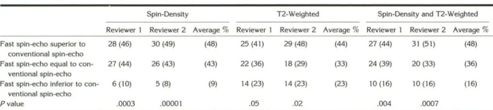
TABLE 1: Comparison of lesion conspicuousness between fast spin-echo and conventional spin-echo sequences, number (percentage) of lesions
Spin-Density T2-Weighted Spin-Density and T2-Weighted
Reviewer 1 Reviewer 2 Average% Reviewer 1 Reviewer 2 Average% Reviewer 1 Reviewer 2 Average%
Fast spin-echo superior to 28 (46) 30 (49) (48) 25 (41) 29 (48) (44) 27 (44) 31 (51) (48)
conventional spin-echo
Fast spin-echo equal to con- 27 (44) 26 (43) (43) 22 (36) 18 (29) (33) 24 (39) 20 (33) (36)
ventional spin-echo
Fast spin-echo inferior to con- 6 (10) 5 (8) (9) 14 (23) 14 (23) (23) 10 (16) 10 (16) (16) ventional spin-echo
[image:2.615.54.561.626.739.2]AJNR: 15, April 1994
33% of the T2-weighted images, and in 36% of the combined spin-density and T2-weighted im-ages. Each comparison had statistical signifi-cance (P :::; .05), with near-perfect interobserver agreement (0.81 :::; K :::; 1.0). All lesions were seen
on both sequences.
Figure 1 is an example of improved lesion visibility on fast spin-echo sequences. Because of decreased motion artifact, the margins of the paraganglioma and its abnormal vessels appear sharper.
For lesions with high signal and adjacent to fat, fast spin-echo sequences resulted in decreased conspicuousness in three cases: a parotid heman-gioma (Fig 2), an optic nerve glioma, and a peripheral neurofibroma. In each of these cases, conventional spin-echo T1-weighted sequences provided signal contrast between lesion and fat.
The cystic lesions in our study were easily identified; however, small cystic lesions adjacent to fat may be harder to see on fast spin-echo
A
B
A
B
MR OF HEAD AND NECK 769
sequences. Other lesions surrounded by fat did not have decreased conspicuousness on fast spin -echo sequences; those lesions with intermediate signals on long-TR sequences, especially the spin -density images, were easily seen next to the high signal of fat. We observed this in squamous cell carcinoma metastatic to the parotid gland (Fig 3), epidermoid carcinoma of the parotid gland, pa-rotid fibrosis, Sjogren involvement of the parotid gland, and radiation changes of the skin.
If the abnormalities contained fat and were adjacent to muscle or other soft tissue, T2-weighted fast spin-echo sequences showed them clearly as a high signal next to intermediate signal of muscle (Fig 4). Similarly, obliteration of normal fat planes by tumor was more easily detected on fast spin-echo sequences; the asymmetric loss of the normal high signal of fat indicated the pres-ence of a neoplastic or inflammatory process (Fig 4).
Fig. 1. Forty-two-year-old man with a paraganglioma.
A, Conventional spin-echo spin-density (2000/30) image.
B, Fast spin-echo spin-density (2500/14) image. The fast spin-echo image has less motion artifact; normal structures are better delineated. Although the paraganglioma is easily seen on conventional spin-echo, the fast spin-echo image provides better defini -tion of vessels (curved arrows) and lesion margins (straight arrows, A and B).
Fig. 2. Two-year-old girl with a hem an-gioma.
A, Conventional spin-echo T2-weighted (2000/80) image.
[image:3.612.56.389.338.724.2]770 FULBRIGHT
Fig. 3. Sixty-six-year-old man with squa -mous cell carcinoma metastatic to left pa-rotid.
A, Conventional spin-echo spin-density
(2000/30) image.
B, Conventional spin-echo T2-weighted (2000/80) image.
C, Fast spin-echo spin-density (2500/14) image.
D, Fast spin-echo T2-weighted (2500/ 84) image. With an intermediate signal, the lesion (straight arrow, A-D) is highlighted against the signal of fat; lesion margins ap-pear sharper on fast spin-echo images ( C
and D).
Discussion
Long-TR sequences play an integral role in MR imaging, because many pathologic processes have a long T2 relaxation time that results in a conspicuous, bright signal. Motion artifacts, how-ever, can limit the utility of MR imaging in the head and neck region. By providing shorter im-aging times, fast spin-echo represents a technique that counters image degradation caused by mo-tion yet offers long-TR images. When compared with conventional spin-echo sequences, fast spin-echo sequences provided improved or equal le-sion conspicuousness in a majority of head and neck lesions (Table 1) and did not miss any lesions seen on conventional spin-echo sequences.
Fast spin-echo sequences increase the number of lines of k-space data acquired within the TR interval by applying a series of 180° pulses after each 90° pulse. The number of 180° pulses rep-resents the echo train length; the time between pulses is the echo spacing. Each 180° pulse generates a spin-echo, with each spin-echo phase encoded by a distinct phase-encoding gradient (3, 4, 7). Those echoes collected with
low-mag-AJNR: 15, April 1994
nitude phase-encoding gradients contribute the majority of image signal and contrast (13-15). The order in which the echoes are phase encoded determines the effective TE. The application of low-magnitude phase-encoding gradients to the early echoes of each echo train results in spin-density images. If the low-magnitude phase-en-coded gradients are applied to the late echoes of the echo train, the images appear T2 weighted. Because each echo of the echo train fills a differ-ent line of k-space, fast spin-echo reduces the imaging time by a factor equal to echo train length for single-echo sequences; for double-echo sequences, imaging time is reduced by a factor of echo train length/2. In conventional spin-echo sequences, imaging time equals TR X NEX X
AJNR: 15, April 1994
A
B
Fig. 4. Fifty-eight-year-old man with spread of squamous cell
carcinoma into the masticator space and fatty atrophy of the tongue.
A, Conventional spin-echo T2-weighted (2500/80) image.
B, Fast spin-echo T2-weighted (2150/84) image. Both
se-quences depict tumor infiltration into the right pterygoid muscle
(long arrows). Because fat remains bright on long-TR fast spin-echo sequences (see text), the fast spin-spin-echo image better shows
the normal parapharyngeal space fat on the left, thus making
asymmetry more easily seen (short curved arrows). Similarly, the
fatty atrophy of the tongue (short straight arrows), a result of
tumor spread into the sublingual space, is better seen on the fast
spin-echo image. Note the large lymph node adjacent to the right masseter muscle (curved arrows).
quality: a larger matrix improves spatial
resolu-tion; longer repetition and echo times lead to
better signal intensity and tissue contrast
;
and an
increase in signal averages optimizes the
signal-to-noise ratio. In order to provide a direct
com-MR OF HEAD AND NECK 771
parison
,
however, we used similar parameters for
both fast spin-echo and conventional spin-echo
sequences.
T2-weighted fast spin-echo and conventional
spin-echo images have similar tissue contrast
except that fat has a substantially increased signal
on fast spin-echo images (16), attributable
pri-marily to the decoupling effect of multiple 180°
pulses on lipid protons (Wright GA, Lipid Signal
Enhancement in Spin-echo Trains
,
presented at
the meeting of the Society of Magnetic
Reso-nance in Medicine
,
AugU3t 1992; Rutt BK, Lipid
Signal Enhancement in CPMG MRI
:
Effect of
Field Strength, presented at the meeting of the
Society of Magnetic Resonance in Medicine
,
Au-gust 1992)
.
The protons of long-chain fatty acids
are coupled in the sense that the local magnetic
field experienced by one proton is affected by the
orientation of protons on adjacent carbon atoms.
Coupling results in splitting of frequency peaks
of lipid protons on nuclear MR spectroscopic
studies
.
On T2-weighted conventional spin-echo
sequences, coupling leads to more rapid T2 decay
of lipid protons with a relative decrease in the
signal of fat
.
In T2-weighted fast spin-echo
se-quences, however, the multiple, repetitive 180°
pulses decouple lipid protons
.
This prolongs the
T2 relaxation times of fat
,
causing it to have a
relatively increased signal on long-TR sequences
.
Increasing the echo train length and decreasing
the echo spacing contribute to a prolonged T2
value of coupled protons (17). Although imaging
parameters were concealed when the reviewers
graded lesi
o
n visibility, the increased signal of fat
seen on fast spin-echo images limited the degree
to which a strict, blinded comparison of
conven-tional spin-echo and fast spin-echo sequences
could be done.
[image:5.614.82.265.74.502.2]interme-772 FULBRIGHT
diate signal. In the parotid gland, the relative fat content increases with age; lesion conspicuous-ness may thus be affected on both conventional
spin-echo and fast spin-echo sequences,
depend-ing on the composition of the mass.
If fat surrounds a lesion, its conspicuousness is not necessarily decreased on fast spin-echo se-quences. Lesions with intermediate signal on long-TR sequences stand out against the high signal of fat, explaining in part why fast spin-echo spin-density sequences provide excellent lesion visibility. In addition, lesions that have short-T2 relaxation times and low signal on T2-weighted sequences are seen well on fast spin-echo sequences if surrounded by tissues that are hyperintense. An inverted papilloma in the nasal vault with a relatively low signal was easily visible because of surrounding high signal of sinus se-cretions. Other lesions with short T2 relaxation times and low signal on T2-weighted fast spin-echo sequences that may be highlighted by sur-rounding hyperintense tissue include cellular le-sions (sarcoidosis and lymphoma), fibrocollagen-ous stroma (orbital pseudotumor), low-spin-den-sity tissue (calcium), and paramagnetic tissue
(fungal infections, melanotic melanoma, and
blood products). T1-weighted conventional spin-echo sequences also help detect and characterize lesions that contain fat or protein or that have intermediate or dark signal on T2-weighted fast
spin-echo sequences. For lesions that have similar
signal as fat on both short- and long-TR
se-quences, fat or water chemical suppression tech
-niques may help; however, routine fat suppres-sion for all lesuppres-sions with bright signals on short-TR images is not required.
Fast spin-echo imaging has potential disadvan-tages. First, image blurring in the phase-encoding direction with loss of small lesion contrast has been observed (7, 13, 18). Certain imaging pa-rameters and tissue characteristics exacerbate blurring, including a long echo train length, a short effective TE, and small lesions with short
T2 relaxation times. Because we used a short
echo train length and a larger matrix size, blurring
did not limit the quality of fast spin-echo images.
Second, for an equal
TR
,
fast spin-echo provided an average of three fewer section locations com-pared with conventional spin-echo. In clinical use,the shorter imaging times of fast spin-echo allow
an increased TR; this results in more sections along with improved signal intensity and tissue contrast. Third, discontinuities in raw data
acqui-AJNR: 15, April 1994
sition inherent in fast spin-echo sequences cause ghosting-iike artifacts that degrade image quality (7, 13). To limit this artifact, an increased matrix size (ie, greater than 128 phase matrix) and de-creased echo spacing can be used. Finally, with short interecho spacing of fast spin-echo se-quences, diffusion-related dephasing induced by paramagnetic iron-containing molecules is de-creased, leading to fewer magnetic susceptibility artifacts (7). Although this study had no hemor-rhagic lesions, they might appear less conspicu-ous on fast spin-echo sequences.
In conclusion, compared with conventional
spin-echo sequences, fast spin-echo sequences provided superior or equal lesion conspicuous-ness in a majority of head and neck lesions. Imaging times with fast spin-echo sequences were markedly reduced. The high signal of fat on fast spin-echo images did not substantially limit lesion visibility except in a minority of lesions. In these
cases, T1-weighted conventional spin-echo and
fat suppression techniques such as inversion re-covery fast spin-echo sequences will help detect and characterize the lesions. In evaluation of the
head and neck, we advocate replacing long-TR
conventional spin-echo sequences with long-TR fast spin-echo sequences. A routine protocol therefore includes T1-weighted conventional spin-echo and spin-density and T2-weighted fast
spin-echo sequences, supplemented with
inver-sion recovery fast spin-echo and gadolinium-en-hanced conventional spin-echo T1-weighted se-quences as needed. If all four sequences and a T1-weighted or inversion recovery fast spin-echo locater scan are used, and accounting for time setting up before and during the exam, the exam time approximates 30 minutes.
References
1. Hennig J, Naureth A, Friedburg H. RARE imaging: a fast imaging
method for clinical MR. Magn Reson Med 1986;3:823-833
2. Hennig J, Friedburg H, Strobel B. Rapid nontomographic approach for MR myelography without contrast agents. J Comput Assist Tomogr 1986; 10:375-378
3. Hennig J, Friedburg H, Ott D. Fast three-dimensional imaging of
cerebrospinal fluid. Magn Reson Med 1987;5:380-383
4. Hennig J, Friedburg H. Clinical applications and methodological de
-velopments of the RARE technique. Magn Reson Imaging 1988;
6:391-395
5. Smith RC, Reinhold C, Lange RC, McCauley TR, Kier R, McCarthy S. Fast spin-echo MR imaging of the female pelvis: part 1. Use of a whole-volume coil. Radiology 1992; 184:665-669
6. Nghiem HV, Herfkens RJ, Francis IR, et al. The pelvis: T2-weighted
AJNR: 15, April1994
7. Melki PS, Mulkern RV, Panych LP, Jolesz FA. Comparing the FAlSE method with conventional dual-echo sequences. J /t1agn Reson Im
-aging 1991;1:319-326
8. Norbash AM, Glover GH, Enzmann DR. Intracerebral lesion contrast with spin-echo and fast spin-echo pulse sequences. Radiology 1992;
185:661-665
9. Yousem OM, Wang P. Comparison of standard spin-echo and fast spin-echo MR imaging of the head and neck for adenopathy. Radio
l-ogy (P) 1992; 185:270
10. Panush D. Fulbright R, Sze G. Smith RC, Constable RT. Inversio n-recovery fast spin-echo MR imaging: efficacy in the evaluation of head and neck lesions. Radiology 1993;187:421-426
11. Sze G, Merriam M, Oshio K, Jolesz FA. Fast spin-echo imaging in the evaluation of intradural disease of the spine. AJNR Am J Neuro -radiol1992; 13:1383-1392
12. Kramer MS, Feinstein AR. Clinical biostatistics LIV. The biostatistics
of concordance. C/in Pharmacal Ther 1981 ;29: 111-123
MR OF HEAD AND NECK 773
13. Mulkern RV, Wong STS, Winalski C, Jolesz FA. Contrast manipula -tion and artifact assessment of 20 and 30 RARE sequences. /t1agn
Reson Imaging 1990;8:557-566
14. Tweig DB. The k-space projectory formulation of the NMR imaging process with application in analysis and synthesis of imaging meth -ods. /t1ed Phys 1983; 10:610-621
15. Mulkern RV, Melki PS, Jakab N, Higuchi V, Jolesz FA. Phase-encode
order and its effect on contrast and artifacts in single-shot rare
sequences. /t1ed Phys 1991: 18: I 032-1037
16. Constable RT, Anderson AW, Zhong J, Gore JC. Factors influencing contrast in fast spin echo MR imaging. /t1agn Reson Imaging 1992; 10:497-511
17. Constable RT, Smith RC, Gore JC. Coupled spin FSE imaging. J
/t1agn Reson Imaging 1993;3:547-552
18. Constable RT, Gore JC. The loss of small objects in variable TE
imaging: implication for FASE, RARE. and EPI. /t1agn Reson /t1ed
1992;28:9-24
Imaging Quiz: Request for Original Submissions
This issue marks the appearance of a new feature in the AJNR, the Imaging Quiz. The quiz can be found on
page 658 and the diagnosis on page 774. We welcome other original submissions for this feature. This feature
must fit on no more than two journal pages: a quiz page and a diagnosis page. Therefore, the approximate
lengt:1 of submissions should be two to three double-spaced pages of text, including references, and up to 1 0
figures. The submissions will undergo peer review. We look forward to receiving your contributions for this