The role of mutants in the study of vertebrate limh
development: analysis of hypodactyly in the mouse
and polydactyly in the chick.
Thesis submitted for the Degree of
Doctor of Philosophy in the
Faculty of Life Sciences at the University of London
by
Katherine Ella Robertson
Department of Anatomy and Developmental Biology
University College London
ProQuest Number: 10045727
All rights reserved
INFORMATION TO ALL USERS
The quality of this reproduction is dependent upon the quality of the copy submitted. In the unlikely event that the author did not send a complete manuscript and there are missing pages, these will be noted. Also, if material had to be removed,
a note will indicate the deletion.
uest.
ProQuest 10045727
Published by ProQuest LLC(2016). Copyright of the Dissertation is held by the Author. All rights reserved.
This work is protected against unauthorized copying under Title 17, United States Code. Microform Edition © ProQuest LLC.
ProQuest LLC
789 East Eisenhower Parkway P.O. Box 1346
Abstract
Vertebrate limb mutants are a valuable source of information on how the vertebrate
limb normally develops. This thesis examines cellular and molecular aspects of limb
development in the mouse mutant hypodactyly {Hd) and the chick mutant Talpid^ {ta).
Hypodactyly heterozygotes (Hd/+) show a reduction of hindlimb digit I, while homozygotes (Hd/Hd) have only one digit on all four limbs. I have analysed Hd/Hd and
Hd/+ limb morphology in adulthood and throughout embryogenesis. Alterations in adult limb morphology are associated with reductions in size and shape of developing
limb buds and in the number of digital blastemas that develop. The polarising region
and apical ectodermal ridge (AER) are required for limb outgrowth and patterning. Hd
mutant limbs have polarising activity and a well defined AER which persists for longer
than in wildtype embryos. Sonic hedgehog {Shh) and fibroblast growth factor 4 {Fgf4),
are expressed in the polarising region and apical ectodermal ridge respectively. In
Hd/Hd mutants, expression of these genes is slightly prolonged. Hoxd genes are
downstream targets of Shh, and are important in patterning the limb. Expression of both
H oxdl3 and H o xd ll are slightly altered in Hd/Hd limbs. In addition, loss of digits in hypodactyly is associated with an increase in mesenchymal cell death in developing
limbs and an inability of limb mesenchymal cells to produce cartilage in culture.
Talpid^ {td) homozygotes have polydactylous limbs with up to eight
morphologically similar digits. I found that there is a graded distribution of polarising
activity in td limb bud mesenchyme, with t d anterior mesenchyme having very weak
ectopic polarising activity. Shh is posteriorly localised in t d limbs and therefore not
associated with weak polarising activity in anterior mesenchyme. In contrast, Bmp2 and
Contents
Abstract 2
Contents 3
List of Figures 8
List of Tables 12
Acknowledgements 14
Chapter 1
A General Introduction to Vertebrate Limb Development
L I Em bryonic Developm ent 16
L2 A Review of Vertebrate Lim b Developm ent 18
1.2.1 Limb initiation 18
1.3 Specification of limb pattern 22
1.3.1 The apical ectoderm al ridge 24
1.3.2 The progress zone 26
1.3.3 The polarising region 26
1.3.4 Limb ectoderm 32
1.4 Interspecies conservation of signalling in developing vertebrate limbs 32
1.5 Molecular basis of limb development 33
1.6 Signalling molecules in limb initiation and proximo-distal outgrowth 33
1.6.1 Fibroblast growth factors 33
1.6.2 Fibroblast growth factor receptors 36
1.7 Signalling molecules in antero-posterior patterning 36
1.7.1 Retinoic acid 36
1.7.2 Retinoid receptors and binding proteins 37
1.7.3 The hedgehog gene family of signalling molecules 37
1.7.4 Bone morphogenetic proteins 39
1.8 Dorso-ventral patterning 42
1.9 Signalling interactions along the three axes of the limb are coordinated 45
1.10 Regulatory genes expressed in the limb in response to signalling
in the limb 45
1.10.1 V ertebrate Msx genes 45
1.10.2 H oxgom s 47
1.10.3 Even-skipped hom ologues 50
1.11 Aims of this thesis 53
Chapter 2
Materials and Methods used in the analysis of the mouse mutant Hypodactyly {Hd)
2.3 Adult morphology 56
2.3.1 External morphology of adult mice 56
2.3.2 Skeletal analysis of adult mice through differential
staining of cartilage and bone 56
2.3.3 Scoring of the defects seen in the hindlimbs of
Hd/-¥ adult mice 57
2.4 Embryonic Morphology 58
2.4.1 Preparation of embryonic material 58
2.4.2 Phenotyping of embryos 58
2.4.3 External morphology of embryos 58
2.5 Analysis of limb polarising activity 58
2.5.1 Fixation and staining of manipulated embryos 60
2.6 Analysis of gene expression in developing limbs 62
2.6.1 Plasmid DNA 62
2.7 Elution of plasmid DNA from Whatmann filter paper 63
2.8 Transformation of competent cells 63
2.9 Small-scale preparation of plasmid DNA 64
2.10 Measuring the concentration of a DNA solution 65
2.11 Restriction digestion analysis of mini prep' plasmid DNA 65
2.12 Large-scale preparation of plasmid DNA 65
2.13 Purification of plasmid DNA through caesium chloride gradients 66
2.14 Removal of ethidium bromide from DNA purified by equilibrium
centrifugation in caesium chloride-ethidium bromide gradients 67
2.15 Synthesis of Digoxygenin-labelled riboprobes 67 2.16 Preparation of embryonic material for non-radioactive wholemount
in situ hybridisation 68
2.17 Non-radioactive in situ hybridisation with digoxygenin labelled probes 69
2.17.1 Protocol according to Rosen and Beddington, (1993) 69 2.17.2 Protocol according to Nieto et al, (1996) 71
2.18 Analysis of stained embryos 73
2.19 Estimation of area of distal hmb that expresses the genes analysed 73
2.20 Cellular analysis of H d mutants 73
2.20.1 Analysis of mesenchymal cell death 73
2.20.2 Skeletal analysis of the limbs of Hd mutant embryos 74 2.20.3 Cell culture of limb mesenchymal cells in high density
micromass cultures 75
2.20.4 Preparation of conditioned medium and treatment of
cultures with growth factors 75
2.20.7 Analysis of limb bud and apical ectodermal ridge
morphology with scanning electron microscopy 76
2.20.8 Histological analysis of apical ectodermal ridge
morphology in semi-thin araldite sections 77
Chapter 3
An introduction to the mouse mutant hypodactyly {Hd)
3.1 The mouse mutant hypodactyly {Hd ) 78
3.2 Appearance of normal mouse limbs 78
3.2.1 Adult limbs 78
3.2.2 Morphology of limbs during murine development 79
3.2.3 External appearance of limbs during development are
correlated with final limb morphology 80
3.3 Outgrowth and patterning of the limb 80
3.3.1 Shh m d Fgf4 83
3.3.2 Homeobox {Hox) genes 83
3.3.2.1 genes 84
3.3.2.2 Hoxa genes 85
3.3.2.3 Roles of Hox genes 86
3.4 Aims of this Chapter 86
3.5 Results 88
3.5.1 Analysis of 3 live-born Hd/Hd, Hd/+, and +/+
littermates at 28 weeks 88
3.5.2 External limb morphology of adult +/+, Hd/+,
and Hd/Hd mice 96
3.5.3 Skeletal analysis of +/+, Hd/-\-, and Hd/Hd littermates
at 28 weeks 99
3.5.4 The Hd/-\- hindlimb phenotype is variable and shows
degrees of left-right asymmetry within the same embryo 110
3.5.5 External morphology of +/+, Hd/+ and Hd/Hd mice during
embryogenesis 116
3.5.6 Polarising activity of posterior mesenchymal cells in
developing limbs of Hd mutant mice 128
3.5.7 Gene expression i n / / J mutant limb buds 130
3.5.7.1 Expression of Shh 130
3.5.7.2 Expression of Fgf4 131
3.5.7.3 Expression o f 131
3.5.7.4 Expression of H oxd ll 132
3.6 Discussion and Conclusion 142
3.6.2 Limb defects in Hd/-\- and Hd/Hd adult mice do not affect proximal structures and are more severe in the hindlimbs than in
the forelimbs 143
3.6.3 There is evidence to suggests ûvdXHd is not a null
allele of HoxalS 144
3.6.4 The hindlimbs are more susceptible to limb defects in both Hd/+
and Hd/Hd mice 145
3.6.5 The Hd/-\- hindlimb phenotype is variable 147
3.6.6 Defects induced by the Hd mutation are not restricted
to the limbs 148
3.6.7 Hd/+ and Hd/Hd embryonic phenotypes 149 3.6.8 Polarising activity in Hd/+ and Hd/Hd embryonic limbs 150
3.6.9 Gene expression in Hd/+ and Hd/Hd embryonic limbs 152
Chapter 4
Analysis of cellular effects of the Hd mutation
4.1 Introduction 154
4.1.1 Programmed cell death 155
4.1.2 Chondrogenesis during limb development 156
4.1.3 Role of the apical ectodermal ridge in shaping the limb bud 159
4.2 Aims of this Chapter 160
4.3 Results 161
4.3.1 Mesenchymal cell death in the developing limbs of H d mutants 161
4.3.2 Development of skeletal elements of the limb 165
4.3.3 In vitro chondrogenesis 174
4.3.4 The Hd mutation also affects the morphology of the
apical ectoderm al ridge 184
4.4 Discussion and Conclusion 202
4.4.1 Summary of results 202
4.4.2 Mesenchymal cell death is affected by the H d mutation 202
4.4.3 Specification of mesenchymal condensations and subsequent
chondrogenesis in vivo is affected by the H d mutation 205
4.4.4 Chondrogenic potential of undifferentiated mesenchymal cells
in vitro is affected by the H d mutation 208 4.4.5 Apical ectodermal ridge morphology is affected by the
/ / J mutation 209
Chapter 5
Distribution of polarising activity and expression of Fgf4, Shh, and Bmps in the
developing limbs of the polydactylous chicken mutant Talpid^
5.3 Materials and Methods 214
5.3.1 Chick embryos 215
5.3.2 Identification of homozygous mutants 215
5.3.3 Mapping pc4arising activity in developing limb buds of
to?tto? embryos 218
5.3.4 Grafting a normal polarising region to the anterior margin
of a stage 20/21 n^lto? limb bud 223
5.5.5 Fixation and staining, of manipulated embryos 223
5.3.6 Retinoic acid treatment of to?/ta^ limb buds 223
5.4 General molecular biology methods 224
5.5 Analysis of gene expression in limb bud of ta?lto? embryos 226
5.5.1 in situ hyWdisation 226
5.5.2 Non-radioactive, wholemount in situ hybridisation 226
5.6 Results 228
5.6.1 Distribution of polarising activity in ta^lta? limb buds 228
5.6.2 Response of tc?(to? limb mesenchyme to polarising region
grafts and retinoic acid 235
5.6.3 Expression of Shh and HoxdlS in limb buds 238
5.6.4 Expression of Fgf4 in to?!to? limb buds 238 5.6.5 Expression of Bm p genes in ta^lta? limb buds 243
5.7 Discussion and Conclusion 246
Chapter 6
General Discussion and Conclusion
6.1 Summary of the finding of this thesis 255
6.2 General conclusions 255
6.3 Future work 257
Appendix 1 260
Chapter 1 Figure 1.1
Figure 1.2
Figure 1.3
Figure 1.4
Figure 1.5
Figure 1.6
Figure 1.7
-Figure 1.8 Chapter 2 Figure 2.1
Chapter 3 Figure
3.1-List of Figures
Three dimensional, diagrammatic representation of a
vertebrate limb bud consisting of a core of undifferentiated
mesenchyme encased in an ectodennal jacket
Schematic representations of the early developing vertebrate
limb bud indicating the important signalling centres involved
in outgrowth and patterning
Final skeletal pattern of the normal chick wing and
skeletal patterns induced after grafting polarising
region cells to the anterior margin of a wing bud at stage
20 of development
Schematic representation of an early limb bud showing
localisation of Shh transcripts to posterior mesenchyme in
the same region as the polarising region
Schematic representation of a transverse section through
the limb bud in figure 1.4
Structural organisation of the Drosophila HOM-C homeotic
complex and its correspondence with genes of the four mouse
//o x clusters based on regions of homology
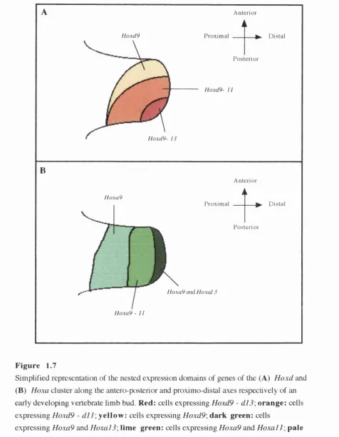
Simplified representation of the nested expression domains of
genes of the Hoxd and Hoxa cluster along the antero-posterior
and proximo-distal axes respectively of an early developing
vertebrate limb bud
A model for the role of Hox genes in limb development
Stage 20 chick wing bud showing a loop in the
anterior apical ectodermal ridge
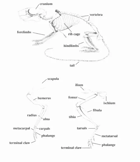
Line drawings of the mouse skeleton and the bones of the
forelimbs and hindlimbs
Figure 3.2 - Photographs of the three adult littermates
Figure 3.3 - Photographs of the hindlimbs of three adult littermates shown in figure 3.2
Figure 3.4 - Photographs of the hind-quarters of a homozygote (Hd/Hd) showing the malformed hindlimbs and the possible genital abnormalities
Figure 3.5 - External morphology of the forelimbs and hindlimbs of three +/+, Hd/+, and Hd/Hd littermates at 28 weeks
Figure 3.6 - Whole skeletons of three adult littermates at 28 weeks stained for cartilage and bone with alcian blue and alizarin red S
Figure 3.7 - Skeletal elements of forelimbs and hindlimbs of a wildtype 103 (+/+) male mouse culled at 28 weeks and stained for
cartilage and bone with alcian blue and alizarin red S 105
Figure 3.8 - Skeletal elements of forelimbs and hindlimbs of a male heterozygote (Hd/+) culled at 28 weeks and stained for
cartilage and bone with alcian blue and alizarin red S 107
Figure 3.9 - Skeletal elements of forelimbs and hindlimbs of a male homozygote (Hd/+) culled at 28 weeks and stained for
cartilage and bone with alcian blue and alizarin red S 109
Figure 3.10 - Variability of the heterozygote (Hd/+) hindlimb
phenotype in a sample population of 110 adult female mice 112
Figure 3.11 - Scanning electron micrographs of 9.5 and 10.5 day post coitum (dpc) mouse embryos from matings
of two heterozygotes (T/d/f) 120
Figure 3.12 - +/+, Hd/+, and Hd/Hd mice and limbs at 11.5 and 12.5 dpc 124
Figure 3.13 - +/+, Hd/+, and Hd/Hd mice between 13.5 and 16.5 dpc 127
Figure 3.14 - Wholemount in situ hybridisations of +/+, Hd/+, and Hd/Hd embryos between 10.5 and 12.5 dpc hybridised with species specific digoxygenin-labelled riboprobes to
ShhdinàFgf4 137
Figure 3.15 - Wholemount in situ hybridisations of +/+, Hd/+, and Hd/Hd embryos between 11.5 and 14.5 dpc hybridised with species specific digoxygenin-labelled riboprobes to
Hoxdl3 139
Figure 3.16 - Wholemount in situ hybridisations of +/+, Hd/+, and Hd/Hd embryos between 11.5 and 14.5 dpc hybridised with species specific digoxygenin-labelled riboprobes to
H o xd ll 141
Chapter 4
Figure 4.1 - Hindlimbs of +/+/Hd/+, +/+ and Hd/Hd embryos at 11.5 and 12.5 dpc showing the shape of the hindlimb buds
and the opaque patch of cells in Hd/Hd limb buds that
colocalise to regions where mesenchymal cell death
is detected 164
Figure 4.2 - +/+/Hd/+, +/+, Hd/-\- and Hd/Hd limbs at 11.5, 12.5 and 14.5 dpc showing regions of cell death as detected
Figure 4.3 - Alcian green stained limbs showing developing cartilage elements of +/+, Hd/+, and Hd/Hd embryos at 12.5 and
13.5 dpc 167
Figure 4.4 - Alcian green stained limbs showing developing cartilage
elements of +/+, Hd/+, and Hd/Hd embryos at 14.5 dpc 169
Figure 4.5 - Alcian blue and Alizarin red S stained limbs of 15.5 dpc
+/+, Hd/+, and Hd/Hd embryos 171
Figure 4.6 - Alcian blue and Alizarin red S stained Hd/Hd and +/+
embryos at 16.5 dpc 173
Figure 4.7 - Histogram showing the average number of nodules in micromass cultures set up at normal density with
mesenchyme from 11.5 dpc +/+/Hd/+ and Hd/Hd
limb buds 175
Figure 4.8 - Histogram showing the average number of nodules in micromass cultures set up at normal density with
mesenchyme from 12.5 dpc +/+, Hd/+ and Hd/Hd
limb buds 176
Figure 4.9 - Cartilage nodules in 6 day micromass cultures of 12.5 dpc +/+ and Hd/Hd]mih bud mesenchyme
stained with Alcian blue for cartilage 178
Figure 4.10 - Histogram showing average nodule number in micromass cultures of 12.5 dpc +/+, Hd/+, and Hd/Hd mesenchymal cells that were fed with
conditioned m edium 180
Figure 4.11 - Histogram showing the number of cartilage nodules in micromass cultures set up with mesenchyme from
12.5 dpc +/+ and Hd/Hd limb bud mesenchyme, and
in cultures consisting of a 50:50 mixture of +/+ and
Hd/Hd mesenchym al cells limb buds 181
Figure 4.12 - Histogram showing the average number of nodules in micromass cultures set up at double density with
mesenchyme from 11.5 dpc +/+/Hd/-\- and Hd/Hd
limb buds 182
Figure 4.13 - Histogram showing the average nodule number in micromass cultures of 12.5 dpc +/+, Hd/+ and
Hd/Hd mesenchymal cells that were fed with
different growth factors 183
Figure 4.14 - Scanning electron micrographs (SEM) of the limb
Figure 4.15 - Scanning electron micrographs (SEM) of the limb
buds of an 11.5 dpc +/+/ffû7+ embryo 188
Figure 4.16 - Scanning electron micrographs (SEM) of the limb
buds of a 12.5 dpc +/+ embryo 190
Figure 4.17 - Scanning electron micrographs (SEM) of the limb
buds of a 12.5 dpc Hd/-^ embryo 192
Figure 4.18 - Scanning electron micrographs (SEM) of the limb buds
of a 12.5 dpc Hd/Hd embryo 194
Figure 4.19 - Scanning electron micrographs (SEM) of the limb buds of 12.5 dpc +/+ and Hd/Hd embryos indicating the
apical ectoderm al ridge 196
Figure 4.20 - Scanning electron micrographs (SEM) of +/+, Hd/-\- and Hd/Hd embryos at 13.5 dpc clearly indicating the
differences in limb bud morphology between the three
phenotypes 198
Figure 4.21 - Semi-thin araldite sections of the limbs of +/+ and Hd/Hd embryos at 11.5 and 12.5 dpc showing
m orphology of the apical ectoderm al ridge 201
Chapter 5
Figure 5.1 - Photographs of a stage 20 and a stage 25 ta^/ta^
embryos in ovo 213
Figure 5.2 - Confirmation of of phenotype 217
Figure 5.3 - Schematic representation of a stage 20 chick wing bud
showing a loop in the anterior apical ectodermal ridge 220
Figure 5.4 - Diagram showing how polarising activity was mapped
in limb buds 221
Figure 5.5 - Skeletal patterns of 10-day chick wings that resulted from mesenchyme grafts from a stage 19/20 ta^/ta? limb bud
into the anterior margin of a normal stage 20/21 host 232
Figure 5.6 - Skeletal pattern of a 10-day control ta^/ta^ wing and contralateral ta^/ta^ wing after a polarising region graft
from a stage 20 normal wing had been grafted to the
anterior margin at stage 20 237
Figure 5.7 - Expression of Shh and H oxdlS in unmanipulated ta^/ta^
limb buds, and in ta^/ta^ buds treated with retinoic acid beads 240 Figure 5.8 - Expression of Fgf4 in a stage 22 normal leg bud and a
stage 22 ta^/ta? leg bud 242
Figure 5.9 - Bmp2, Bmp4, and Bm p7 expression in normal and
List of Tables
Chapter 1
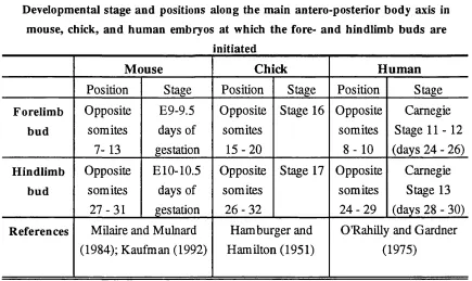
Table 1.1- Developmental stage and positions along the main antero-posterior body axis in mouse, chick, and human
embryos at which the fore- and hindlimb buds are initiated 19
Chapter 3
Table 3.1 - Number of live-born mice produced by the breeding colony
of Hd mice 89
Table 3.2 - External appearance of +/+, Hd/+, and Hd/Hd httermates
at 28 weeks 89
Table 3.3 - Regions of the adult skeleton affected by the H d mutation
in Hd/+ and Hd/Hd mice analysed at 28 weeks 101
Table 3.4 - Number of mice from a sample population of 110 adult female Hd/+ mice having different degrees of hindlimb digit I reduction, soft tissue syndactyly between digits II and ELI, and presence of an abnormal or absent terminal claw on hindlimb digit I on both
left and right hindlimbs 113
Table 3.5 - Left to right asymmetry in the Hd/+ hindlimb phenotype in a sample population of 110 adult female Hd/-^ mice
having different degrees of hindlimb digit I reduction, soft tissue syndactyly between digits II and HI, and the presence
of an abnormal or absent terminal claw on hindlimb digit I 114
Table 3.6 - Summary the number of litters and phenotypes of the embryos
in these litters from 112 Hd/+ X Hd/+ matings 117
Table 3.7 - Average crown to rump size of -i-/+, Hd/+, and Hd/Hd embryos
between 11.5 and 14.5 dpc 118
Table 3.8 - Average area of distal limb buds of +/+, Hd/+, and Hd/Hd embryos at 11.5 and 12.5 dpc, the relative increase in limb
buds size from 11.5 to 12.5 dpc, and the average difference in
size of +/+ and Hd/+ limb buds in comparison to Hd/Hd
limb buds 125
Table 3.9 - Summary of results obtained from grafting posterior mesenchyme from 4-/4-, Hd/+, and Hd/Hd limb buds to the
anterior border of a normal stage 20 chick wing 129
Chapter 5
Table 5.1 - Number of mesenchymal grafts from different regions of stage 1 9 -2 3 and stage 23 - 24 ta^/ta^ wing and leg buds that
were grafted to the anterior border of normal chick wings, and
Table 5.2 - Number of cases where skeletal alterations additional to the induction of additional digits were induced by grafted
Acknowledgements
I would like to thank my supervisors Dr. Susan Darling and Professor Cheryll Tickle for
giving me the opportunity to work on such an interesting project in their laboratories, and
for their constant support, advice and encouragement during my time at UCL. I am
indebted to them, and to many others, especially Sarah Wedden and Marty Cohn, for
helping me though. Thank you.
I ajn grateful to Dr. Donald Ede, Dr. Phillippa Francis-West, Dr. David Burt, and
Dr. Juan-Carlos Izpisùa-Behnonte fœ their collaboration on the work on Talpid^. I
would especially like to thank Anne Sheasby for her expert technical assistance, advice
and all the help she has given me over the last few years, and also to Dr. Jenny Brown,
Dr. Ketait Patel and Mary Rahman for help, advice and assistance with the molecular
biology. Three excellent graduate students have also been involved in this study; Mike
Chapman, Astrid Adams and Megan Davey. Special thanks go to Kate Lewis, Kate
Whitley, Phillippa Francis-West, and Dave Becker for letting me have access to slide
scanners, computers and printers which enabled me to set out and print my colour plates,
and to Jane Pendjiky and Chris Sym for help with the black and white photographic
plates.
The past few years here at UCL have been a lot of fun, and this is all down to
having such a lot of wonderful people around in the lab. I would like to thank them all for
their friendship, help and advice: Marty Cohn, Phillippa Bright, Liz Ensor, Jo Begby,
Jules Schofield, Annie Rowe, Kate Lewis, Han-Sung Jung, Kate Whitley, Neil
Vargesson, Lynda Erskine, Jason Hopkins, Imelda McConnell, Ronald Nittenberg,
Konstadina Kostakopoulou, Litsa Drossopoulou, Liz Bently, Deana D ’Souza, Aris
Haritos, Muriel Altabef, Ketan Patel, and Paris Ataliotis. I will miss you all.
Special thanks go to my dear friends Marty, Phillippa, Kate, Liz, Jo, Jules and
Annie, fca: making sure 1 got out and enjoyed myself once in a while^ and for fueling my
passion for great coffee. Thanks to Louise and Clare for always letting me come and stay
when I needed a break, and for putting up with my bad moods. Thanks to my friends
Leah, Paul R and the Pig, Gina, Bill and Conny who, despite the fact that they are
thousands of miles away, have k ^ t me going with their emails and the happy memories
of our times together. Thanks also go to my wonderful flatmates Alex, Caroline, and
Merrilyn for all the fun and laughs we have had over the last year.
Finally, I want to thank my parents and my sister Louise for their love, constant
To my parents and my sister Louise, and
in the memory of my dear Grandma and
Chapter 1
1. A General Introduction to Vertebrate Limb Development
1.1 Embryonic Development
Embryonic development results in the transformation of a single cell into a highly
complex, multi-cellular organism consisting of many different cell types. During
development, the structural organisation, or patterning of the embryo m ust be achieved.
Pattern formation is a term used to describe this process and the concept of positional
information provides a mechanism by which pattern formation can take place (Wolpert
1969; 1971). Cells in a developing system are instructed as to their relative positions
(positional information) within a specific region of cells with fixed boundaries (for
example the limb field, a region of the embryo from which limbs develop). Cells then
interpret this information with reference to their genetic composition and developmental
history (reviewed in Wolpert, 1994). Once cells are specified, the tissues and organs that
they form part of are moulded into shape, a process known as morphogenesis, involving
cell proliferation, cell movement, and programmed cell death.
Many different species are studied (from insects such as Drosophila melanogaster
to mammals such as Mus musculus ) in an attempt to understand the complexities of
embryonic development, and it is becoming increasingly evident that, across species,
similarities exist in the events that take place. Molecular techniques used to analyse how
genes and molecules control embryonic development are identifying genes and gene
families that are thought to be important during development, and that are conserved
across species. Analysis of molecular mechanisms of development in Drosophila has
been pivotal to the progress made in understanding molecular mechanisms of vertebrate
development Many of the genes known today to be important in development were first
identified and cloned in Drosophila, and analysis of their function during Drosophila
development has assigned putative functions to some of these genes. A significant
finding has been that a number of these genes identified in Drosophila have similar
counterparts or homologues in vertebrates, suggesting that similar molecular
Vertebrate limbs are an excellent model system in which to study pattern
formation; they develop from very simple structures (limb buds) into highly complex
functional units (limbs), and, as in the rest of the embryo, cells must differentiate at the
correct time and in the correct locations. The vertebrate limb is easily accessible to
embryonic analysis and experimental manipulation, particularly in the developing chick
embryo, and this has been the focus of much of the classical work on vertebrate limb
development (reviewed in Tickle and Eichele, 1994). A wealth of information has also
been gained from the study of murine limb development, and although experimental
manipulations of post-implantation mouse embryos in utero are technically difficult, the murine genome is accessible to analysis, and many of the genes known to be important
during vertebrate limb development have been mapped, cloned and sequenced (see The
Jackson Laboratory Mouse Genome Database and references therein, httpV/www.jax.org
). There are many genes still to be been cloned, as suggested by the large number (>
100), of spontaneous mouse mutants with limb defects (Griineberg, 1952; Lyon et al.,
1996) whose molecular basis have yet to be identified. In addition, with the development
of transgenic technology, it is now possible to artificially induce mutations in the mouse
through manipulation of the genome (Capecchi, 1994; Gatherer, 1993). Mouse mutants
(both naturally occurring and artificially induced) are an extremely useful resource in the
study of limb development as they provide an invaluable source of information about
essential gene function and an opportunity to compare and contrast normal and abnormal
development Recent advances in transgenic technology have enabled the targeting of
specific genes already known to be expressed in the developing limb (see Brandon,
1995a, 1995b, 1995c and references therein). Analysing the phenotypic outcome of such
gene targeting experiments is enhancing the understanding of the roles specific genes and
the molecules they encode play during murine limb development (discussed later in this
thesis).
The developing mouse embryo is used as a model for human embryonic
development, as many developmental mechanisms are thought to be conserved between
different mammalian species, and many of the genes known to be important during
chromosomal arrangement of these human genes is different, it has been suggested that
they may still have similar functions to those in the mouse. Already possible mouse
models of human inherited developmental defects have been identified (Belloni et al.,
1996; Clarke, 1994; Darling, 1996; Darling and Abbott, 1992; Jacenko et al., 1994;
Roessler et al., 1996; Winter, 1988). For example, very recently the targeted disruption of
a murine gene called sonic hedgehog {Shh) has been generated (Chiang et al., 1996), resulting in a phenotype closely resembling that of a human developmental defect,
holoprosencephaly (HPE) (Belloni et a l, 1996; Roessler et al., 1996). Mutations in the
human homologue of Shh have subsequently been shown to cause HPE in certain
families (Belloni et al., 1996; Roessler et al., 1996). Thus, it is becoming increasingly
evident that information gained from the study of development in both normal and
mutant mice probably has direct relevance to both normal and abnormal human
development
1.2 A Review of Vertebrate Limb Development
1.2.1 Limb initiation
Vertebrate limbs develop from small, paired, bud-like outgrowths which emerge at
specific times and at specific sites along the main antero-posterior body axis of the
developing embryo, both of which vary between species (Hamburger and Hamilton,
1951; Kaufman, 1992; Milaire and Mulnard, 1984; O'RahiUy and Gardner, 1975; Table
1.1). However, the overall appearance of limb buds in different species is somewhat
similar. Mechanisms involved m specifying limb position along the main antero
posterior body axis are not fully understood, but there is experimental evidence to suggest
that they may involve expression of genes along the developing main body axis
Table 1.1
Developmental stage and positions along the main antero-posterior body axis in mouse, chick, and human embryos at which the fore- and hindlimb buds are
initiated
M ouse Chick H um an
Position Stage Position Stage Position Stage
Forelimb bud Opposite somites 7-13 E9-9.5 days of gestation Opposite somites
1 5 -2 0
Stage 16 Opposite
somites
8 - 1 0
Carnegie
Stage 1 1 -1 2
(days 24 - 26)
Hindlimb
bud
Opposite
somites
2 7 -3 1
ElO-10.5
days of gestation
Opposite
somites 2 6 -3 2
Stage 17 Opposite somites
2 4 -2 9
Carnegie
Stage 13
(days 28 - 30)
References Milaire and Mulnard (1984); Kaufman (1992)
Hamburger and
Hamilton (1951)
O'RahiUy and Gardner
(1975)
Table 1.1
Table documenting the developmental stage and positions along the main antero-posterior
body axis in mouse, chick, and human embryos at which the fore- and hindlimb buds are
The first morphological signs of limb development can be detected
histologically as paired thickenings in the lateral plate mesoderm, the formation of which
are correlated with a decrease in levels of cell proliferation in the intervening flank
mesenchyme (Searls and Janners, 1971), Experiments have shown that prior to the
appearance of the limb buds, prospective chick limb bud mesoderm has the ability to
form hmb structures when grafted to the flank of a host embryo (Reuss and Saunders,
1965; Saunders and Reuss, 1974). This indicates that cells in the presumptive limb
regions of the embryo are specified to form limb structures even prior to the appearance
of discrete buds.
Once induced, the paired thickenings grow out and form pronounced elevations,
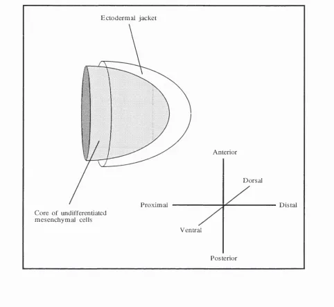
known as hmb buds, which consist of a core of undifferentiated mesenchymal cells
originating from the lateral plate mesoderm and the lateral somite, which give rise to the
connective tissues and myogenic cells of the muscles respectively (Chevalher et al., 1977;
Christ et al., 1977; Ordahl and Le Douarin, 1992), encased in a thin epithelial jacket
(Figure 1.1). The hmb buds are initiaUy fairly simple, symmetrical structures, but as
development proceeds, they grow out in a proximo-distal manner and become more
E ctod erm al jacket
Anterior
D orsal
P roxim al D ista l
C ore o f undifferentiated m e se n c h y m a l cells
V entral
P osterior
Figure 1.1
Three dimensional, diagrammatic representation of a vertebrate limb bud consisting of a
core of undifferentiated mesenchyme encased in an ectodermal jacket The three axes of
1.3 Specification of limb pattern
Limb development involves the intricate co-ordination of different developmental
processes such as tissue-specific cell differentiation, control of cell growth and cell-cell
interactions along three axes (proximo-distal, antero-posterior, and dorso-ventral; see
Figure 1.1). Specification of antero-posterior and dorso-ventral axes of the limbs occurs
prior to the appearance of limb buds (Hamburger, 1938; Saunders and Reuss, 1974).
Experimental manipulations carried out in the chick have identified three major sets of
signalling interactions in the developing limb bud that directly influence patterning and
morphogenesis (reviewed in Tickle and Eichele, 1994). Signalling centres involved in
these interactions are the specialised epithelium that runs antero-posteriorly along the
distal edge of the bud, known as the apical ectodermal ridge, a region of posterior
mesenchymal cells known as the polarising region (also known as the zone of polarising
activity), and the non-ridge limb ectoderm (Figure 1.2A and 1.2B). They play roles in the
specification of pattern along the proximo-distal, antero-posterior, and dorso-ventral axes
of the limb respectively (reviewed in Robertson and Tickle, 1997; Tickle, 1994).
Signalling interactions across the three axes are at least partly interdependent oJ)one
another, thus ensuring that patterning and morphogenesis of the limb is tightly co
Pr +
Apical ectodermal ridge
Progress zone
Polarising region
B Anterior apical ectodermal ridge
Ventral limb mesenchyme
Ventral limb ectoderm
Posterior apical ectodermal ridge
Dorsal limb mesenchyme
Dorsal limb ectoderm
Polarising region
Figure 1.2
Schematic representations of the early developing vertebrate limb bud indicating the
important signalling centres involved in outgrowth and patterning. (A) View of an early limb bud showing the localisation of the polarising region to posterior limb bud
mesenchyme, the progress zone to distal tip mesenchyme, and the specialised epithelium
that runs antero-posteriorly along the distal edge of the bud, known as the apical
1.3.1 The apical ectodermal ridge
Soon after the initial appearance of the limb buds, the ectoderm at the distal tip of the bud
undergoes morphological changes and forms a definite ridge running antero-posteriorly
along the distal edge of the bud (Saunders, 1948; Figure 1.2A). This ridge, known as the
apical ectodermal ridge, consists of tightly packed pseudostratified cells linked
extensively by gap junctions (Fallon and Kelley, 1977). Experiments in the developing
chick embryo have shown that an ectopic apical ectodermal ridge can be induced on the
flank of a host embryo by grafting presumptive limb mesenchyme to this position
(Dhouailly and Kieny, 1972; Reuss and Saunders, 1965; Saunders and Reuss, 1974;
Tanaka et al., 1997), and limb mesenchyme loses this ability once the limb buds appear,
indicating that the ridge is induced by transient signals from the underlying mesenchyme.
Recent evidence suggests that the dorso-ventral limb boundary organises the apical
ectodermal ridge structure in the limb field of early-stage chick embryos (Tanaka et al.,
1997). When new dorso-ventral boundaries are made by implanting dorsal tissue into the
ventral limb field, an ectopic apical ectodermal ridge is induced on the ventral side of the
host limb buds (Tanaka et al., 1997).
Once induced, the apical ectodermal ridge is maintained by signals from the
underlying mesenchyme (Saunders, 1948; Searls and Zwilling, 1964; Zwilling 1956b,
1961,1972). In vivo experiments have shown that the ridge degenerates within 48 hours
of grafting it over non-limb mesenchyme. This degeneration however can be prevented
by grafting small pieces of limb mesenchyme under the grafted ridge (Zwilling 1961,
1972). In addition, culturing apical ectodermal ridge dissociated from the underlying
mesenchyme in vitro results in ectodermal cell death which can be prevented by co-
culturing the apical ectodermal ridge with underlying limb mesenchymal cells (Boutin
and Fallon, 1984; Searls and Zwilling, 1964). The ability of limb mesenchymal cells to
maintain the apical ectodermal ridge is acquired at the same time as they lose their ability
to induce an apical ectodermal ridge (Saunders and Reuss, 1974). Zwilling hypothesised
that the ridge is maintained by an apical ectodermal maintenance factor (AEMF) which is
1956), and the length, morphology, and duration of the apical ectodermal ridge may be
related to the supply of this factor.
After the initial induction of the limb buds, further outgrowth and patterning
along the proximo-distal axis of the limb bud is dependent on the presence of the apical
ectodermal ridge. Removing the ridge leads to the loss of structures along the proximo-
distal axis of the limb, the extent of which is dependent on the stage of development at
which the ridge is removed, with more severely truncated limbs resulting when the ridge
is removed early (Saunders, 1948; Summerbell, 1974). Grafting an ectopic apical
ectodermal ridge to the dorsal or ventral surface of a host limb bud induces ectopic
outgrowth of host limb mesenchyme at the site of the graft, resulting in the formation of
ectopic limb structures (Saunders and Gasseling, 1968; Saunders et al., 1976).
Inductive interactions of the apical ectodermal ridge do not depend on ridge
polarity with respect to underlying mesenchyme. When the ectoderm is rotated 180°,
such that anterior mesenchyme is in contact with posterior apical ectodermal ridge, the
bud continues to grow normally and the antero-posterior axis of the final limb is not
affected (Zwilling, 1956a). Recombination experiments have also shown that the
inductive interactions of the ridge are interchangeable within the same embryo and
between embryos of different stages of development The limb type that develops
follows the mesenchymal component of the recombinant limb bud; wing mesenchyme
recombined with leg apical ectodermal ridge results in formation of a wing (Zwilling
1955) and young mesenchyme recombined with older ectoderm leads to development of
a limb related to the age of the mesenchyme (Rubin and Saunders, 1972). These
experiments show that the action of the apical ectodermal ridge on the underlying
mesenchyme is permissive and not instructive; it allows limb outgrowth to occur without
directly determining developmental fate.
There is evidence to suggest that in addition to its role during the proximo-distal
outgrowth and patterning of the limb, the apical ectodermal ridge also influences the
shape of the developing limb. It may act as a mechanical 'seam' ensuring that the bud is
of a bead soaked in retinoic acid to the anterior margin of a developing chick wing bud.
This also alters antero-posterior limb pattern and digit specification. High doses of
retinoic acid result in a decrease in the length of the apical ectodermal ridge, the antero
posterior width of the bud, and the number of digits that develop (Lee and Tickle, 1985);
low doses of retinoic acid lead to an increase in the length of the apical ectodermal ridge, a
widening of the limb buds and the development of supernumerary digits (Lee and Tickle,
1985). This suggests that the ridge may play a role in determining the antero-posterior
width of the limb bud, thus influencing the amount of mesenchyme available for digit
development Further evidence for this comes from the study of vertebrate limb
mutants in which the size of the developing limb buds is associated with changes in
length or presence of the apical ectodermal ridge (see later).
1.3.2 The progress zone
Bud outgrowth is associated with the maintenance of high levels of cell proliferation in a
zone of undifferentiated mesenchymal cells underlying the apical ridge. This region is
known as the progress zone (Summerbell et al., 1973; Figure 1.2A), and there is
evidence to suggest that the length of time cells spend in the progress zone determines
whether they will form proximal or distal limb structures. Cells that leave early, form
proximal structures, whereas those that leave later, form more distal ones (Summerbell
and Lewis, 1975). The loss of limb structures along the proximo-distal axis of the limb
that results from removal of the apical ectodermal ridge is correlated with termination of
cell proliferation in the progress zone after ridge removal (Saunders, 1948; Summerbell,
1974). Therefore, it is the interaction of the apical ectodermal ridge and the underlying
cells of the progress zone that maintains the high levels of proliferation in this region of
the limb allowing outgrowth and patterning of the limb to occur.
1.3.3 The polarising region
There are three digits in the chick wing, digits 2, 3, and 4, with 2 being most anterior and
4 m ost posterior (Figure 1.3A). When a block of posterior mesenchymal cells is grafted
chick wing bud, respecification of host anterior mesenchyme occurs such that an extra set
of digits is induced (Figure L3B). Instead of the normal 2, 3, 4 digit pattern, the
manipulated wing has a mirror-image duplication, such that the digit pattern becomes 4,
3, 2, 2, 3 ,4 , and the additional set of digits arises from the host anterior tissue (Saunders
and Gasseling, 1968; Figure L3B bottom panel). The region of posterior mesenchymal
cells capable of inducing these digits duplications is known as the polarising region, and
signals produced by this region are thought to govern patterning across the antero
posterior axis of the developing limb (Saunders and Gasseling, 1968).
The polarising region has been mapped in the developing chick wing and leg by
grafting blocks of mesenchymal cells taken from different antero-posterior locations in
the limb to the anterior margin of a host chick wing, and analysing digit patterns that
result (Hinchliffe and Sansom, 1985; Honig and Summerbell, 1985; MacCabe et al.,
1973). These experiments have shown that polarising activity is asymmetrically
distributed across the antero-posterior limb axis (Honig and Summerbell, 1985), and is at
its highest in posterior-distal mesenchyme, just proximal to the progress zone. As the
limb grows out, the polarising region continues to be located in posterior-distal
mesenchyme, and is finally lost at a stage in development when the apical ectodermal
ridge begins to regress and the hand/footplates are forming (Honig and Summerbell,
1985).
Polarising region signalling is dose-dependent; when small numbers of
polarising cells (~ 30 cells) are grafted, a partial duplication with digit patterns 2 ,2 , 3 ,4
(Figure 1.3B top panel) results whereas grafts of larger numbers of polarising cells (>
100 cells) give full duplications with digit patterns 4, 3 ,2, 2, 3 ,4 (Tickle, 1981; Figure
1.3B bottom panel). In addition, the extent of duplications induced also depends on how
long the grafted cells are left in place in the host wing; removing the graft after 15 hours
leads to the induction of an additional digit 2 whereas grafts left in for 17 - 24 hours wül
specify a digit 3 (Smith, 1980). These experiments showed that the action of grafted
polarising region cells on anterior host mesenchyme is both dose and time dependent,
and increased support for the "morphogen model" of antero-posterior limb patterning
pattern formation and morphogenesis (Brickell and Tickle, 1989), and this model
suggests that the polarising region is the source of a diffusible morphogen, that forms a
concentration gradient across the antero-posterior axis of the limb, the concentration of
which instructs cells of their positional identities (Tickle et al., 1975; Wolpert et al.,
1989). Thus, digits are specified in a concentration-dependent manner with high
concentrations of morphogen specifying posterior structures and low concentrations
specifying anterior structures (Tickle et al., 1975; Wolpert et al., 1989). Cells nearest the
polarising region would be exposed to high concentrations and those furthest away, in the
anterior limb bud, would be exposed to low concentrations giving rise to posterior and
anterior structures respectively. Grafting experiments described above are easily
explained by this model, as grafting fewer posterior mesenchymal cells or reducing
exposure time to a polarising region graft would reduce the total concentration of
morphogen anterior host cells are exposed to, leading to partial duplications in which
Apical ecUxiermal ridge clavicle
humerus
scapula M . radius
Polarising region
B
Block o f mesenchyme taken from the polarising region o f a stage 20 donor chick wing or leg bud
- 3 0 polarising region cells grafted to the anterior margin of the host wing bud
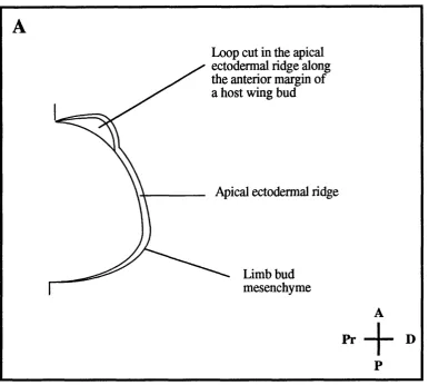
Loop in the apical ectodermal ridge along the anterior margin o f a stage 20 host wing bud
> 100 polarising region cells grafted to the anterior margin o f the host wing bud
A
Figure 1.3
Diagrams showing final skeletal pattern of the normal chick wing and skeletal patterns
induced after grafting polarising region cells to the anterior margin of a wing bud at stage
20 of development (A) Normal chick wing with digit pattern 2, 3 ,4 from anterior to
posterior. (B) Digit pattern of chick wings after grafting different amounts of polarising
region cells under a loop in the apical ectodermal ridge at the anterior margin of a stage 20
normal chick wing. The top limb has a digit pattern 2, 2, 3 ,4 which is induced by
grafting ~ 30 polarising region cells, whereas the bottom limb has a digit pattern 4, 3, 2,
2, 3, 4 which is induced by grafting more than 100 polarising region cells. A: anterior; P:
An alternative model has also been proposed to explain pattern formation during
limb development This model, known as the "polar coodinate model", suggests that cells
of the limb have an intrinsic knowledge of where they are with respect to one another
early in development; they have specific coordinates, or positional values (reviewed by
Bryant and Gardiner, 1992). When the continuity of positional information in the limb is
disrupted, for example by grafting posterior mesenchymal ceUs to the anterior margin,
the cells at the site of the disparity respond by dividing and acquiring new positional
values so as to fill in the discontinuity. This is known as intercalation.
However, there are several lines of experimental evidence suggesting that the
morphogen model can fully explain the mechanisms of pattern formation in the limb,
whereas the polar coordinate model cannot The lack of induction of digit duplications by
distal tip mesenchyme grafted anteriorly or anterior mesenchyme grafted posteriorly
(Hinchliffe and Sansom, 1985; Honig and Summerbell, 1985; MacCabe etal., 1973)
cannot be accounted for by the polar coordinate model which would predict intercalation
and production of ectopic digits. In addition, this model cannot account for the differences
in digit duplications induced by different numbers of polarising region cells, or when two
polarising regions are grafted together at the anterior margin. The polar coordinate model
would predict that no matter how many cells are grafted, or where the cells are grafted
from and to, intercalation between posterior and anterior mesenchyme would take place
leading to full digit duplications. This is clearly not the case when fewer cells are grafted
(see above), or when two polarising regions are grafted together at the anterior margin
which leads to the loss of digit 2 (Wolpert and Hombruch, 1981). The loss of digit 2 can
be explained by the morphogen model, as the anterior cells would be exposed to high
concentrations of the polarising region morphogen, leading to induction of ectopic digits
with posterior and not anterior identity (Tickle, 1981; Wolpert and Hombmch, 1981).
Finally, separation of anterior and posterior mesenchyme with an impermeable barrier
results in loss of anterior structures (Summerbell, 1979), suggesting that anterior limb
mesenchyme requires signals from the posterior mesenchyme in order to allow normal
1.3.4 Limb ectoderm
Epithelial-mesenchymal interactions control patterning across the dorso-ventral axis of
the developing limb. When limb ectoderm is rotated so that, for example, dorsal
ectoderm overlies ventral mesoderm, distal structures subsequently develop to conform
with ectoderm polarity (MacCabe et al., 1974). Both dorsal and ventral ectodermal
signals are required for patterning along the dorso-ventral axis of the limb; grafting an
ectopic apical ectodermal ridge to either the dorsal or ventral surface of a host limb bud
results in double dorsal or double ventral ectopic limb structures at the site of the graft
(Saunders et al., 1976; Shellswell and Wolpert, 1977).
1.4 Interspedes conservation of signalling in developing vertebrate limbs
Developing limb buds of different vertebrate species are alike in appearance and it is
thought that the processes of outgrowth and patterning occur via similar cellular and
molecular mechanisms. Limb buds of amphibians, birds, rats, mice, and humans consist
of a core of mesenchymal cells encased in an ectodermal jacket capped distally with an
apical ectodermal ridge (Fallon and Kelly, 1977; Jurand, 1965; O'Rahüley et al., 1956;
Saunders, 1948). The inductive interactions of limb ectoderm and apical ectodermal ridge
are conserved between different avian species and between mammals and chick
(Saunders, 1977 for references), such that the ectoderm of duck, mouse and rat limb
buds can support outgrowth and development of a normal chick limb (Joquera and
Pugin, 1971 cited in Saunders, 1977; Patou, 1968 cited in Saunders, 1977). In addition,
blocks of posterior limb mesenchyme taken from several different vertebrate species
have the ability to induce digit duplications when grafted to the anterior margin of a
normal chick wing bud (Fallon and Crosby, 1977; MacCabe and Parker, 1976; Tickle, et
a l, 1976). These digit duplications are similar to those induced by a chick polarising
region graft Thus, signalling interactions involved in outgrowth and patterning of the
vertebrate limb appear to be conserved between different species, although it is likely that
1.5 Molecular basis of limb development
Molecular analysis of gene expression in normal and experimentally manipulated
embryos has shown that outgrowth and patterning during limb development involves the
expression of genes and molecules that appear to be part of intricate signalling cascades.
Molecules involved in these cascades belong to various families, such as growth factors,
transcription factors and retinoids (see below).
1.6 Signalling molecules in limb initiation and proxim o-distal outgrow th
1.6.1 Fibroblast growth factors
The fibroblast growth factor (FGF) family consists of at least ten (FGFl - FGFIO)
structurally related peptide signalling molecules (Yamasaki et al., 1996), and FGF2,
FGF4, and FGF8 are thought to be involved in aspects of limb initiation, outgrowth and
patterning.
Recently, it has been shown in chick embryos, that beads soaked in certain
members of the FGF family can induce extra limbs when implanted into the lateral plate
mesoderm of the presumptive flank (Cohn et al., 1995; Crossley et al., 1996; Ohuchi,
1995). The type of extra limb induced is dependent on the position of bead implantation;
beads placed more anteriorly, near the prospective wing region, induce extra wings
whereas those placed more posteriorly, near the prospective leg region, tend to induce
extra legs (Cohn et al., 1995). These results suggest that the endogenous signal
responsible for the induction of paired limb buds may be an FGF. The best candidate, at
present is FgfS, which is expressed strongly in intermediate mesoderm (the region of
mesoderm that gives rise to the embryonic kidney) near the limb forming region
(Crossley et al., 1996). Recent evidence suggests that retinoic acid is also required in
conjunction with FgfS for initiation of the chick limb bud (Stratford et al., 1996; see also
section 1.7.1 in this chapter).
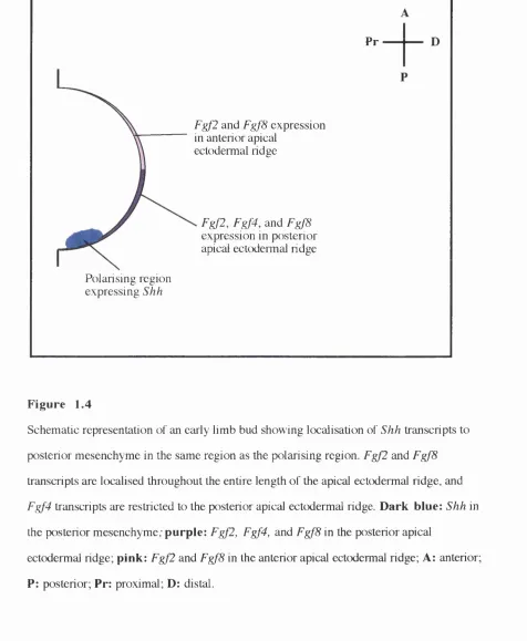
There is good evidence that FGFs also mediate signalling by the apical
ectodermal ridge. Ridge cells express at least three members of the FGF family. FGF2
Fallon et al., 1994; Savage et al., 1993; Savage et al., 1995; Figure 1.4). Fgf4 transcripts
have been found localised to the posterior apical ectodermal ridge (Niswander and
Martin, 1992; Suzuki et al., 1992; Figure 4) and FgfS transcripts are detected throughout
the entire length of the ridge prior to the appearance of Fgf4 (Crossley and Martin, 1995;
Heikinheimo et al., 1994; Ohuchi et al., 1994; Figure 1.4). Experiments in chick embryos
have shown that FGF2, FGF4 and FGF8 proteins can substitute for the ridge; limb
outgrowth and patterning after ridge removal can be maintained by grafting beads soaked
in these FGFs to the limb buds (Crossley et al., 1996; Fallon et al., 1994; Niswander et
al., 1993; Vogel et al., 1996). FGF2 and FGF4 have also been shown to maintain the
activity of the polarising region cells after removal of the posterior apical ectodermal ridge
Pr
F gjl and Fgj'8 expression in anterior apical
ectodermal ndge
Fgfl, Fgf4, and Fgf8 expression in posterior apical ectodermal ridge
Polarising region expressing Shh
Figure 1.4
Schematic representation of an early limb bud showing localisation of Shh transcripts to
posterior mesenchyme in the same region as the polarising region. Fgf2 and FgfS
transcripts are Icx^alised throughout the entire length of the apical ectcxiermal ridge, and
Fgf4 transcripts are restricted to the posterior apical ectodermal ridge. Dark blue: Shh in
the posterior mesenchyme; purple: Fgf2, Fgf4, and FgfS in the posterior apical
ectodermal ndge; pink: Fgf2 and FgfS in the anterior apical ectodermal ridge; A: anterior;
1.6.2 Fibroblast growth factor receptors
FGFs exert their effects on cells via low affinity and high affinity cell surface receptors
(Baird, 1994; Dionne et al., 1990; Klagsbrun and Baird, 1991; Partanen et al., 1991
Pasquale, 1990). Four high affinity fibroblast growth factor receptors have been
identified, FG FRl - FGFR4, three of which are expressed in the developing limb.
FG FR l is expressed in the mesenchyme of the developing limb (Orr-Urteger et al.,
1991; Patstone et al., 1993; Peters et al., 1991), FGFR2 is detected in the limb ectoderm
including the apical ectodermal ridge (Orr-Urteger et al., 1991; Patstone et al., 1993), and
FGFR3 is expressed later on, in the cartilage rudiments of developing bone and in the
cartilage growth plates during endochondral ossification (Peters et al., 1993). The exact
roles of the these receptors during limb development are not fuUy understood, but
mutations in both murine (Deng et al., 1994; Deng et al., 1996; Colvin et al., 1996;
Yamaguchi et al., 1994) and human FGFRs lead to skeletal defects (Muenke and Schell,
1995), suggesting that they are required for skeletogenesis.
1.7 Signalling molecules in antero-posterior patterning
1.7.1 Retinoic add
Retinoic acid has been detected in the developing limbs of chick and mouse embryos,
where it is enriched posteriorly, in the region of the limb where the polarising region is
found (Satre and Kochhar, 1989; Scott et al., 1994; Stratford et al., 1996; Thaller and
Eichele, 1987,1990). In the absence of an endogenous polarising region, beads soaked in
retinoic acid are capable of directing limb patterning (Eichele, 1989; Tamura, 1990).
Furthermore, application of a retinoic acid-soaked bead to the anterior border of a normal
chick wing can mimic the action of a polarising region grafted to the same position, and
lead to induction of mirror-image digit duplications (Summerbell, 1983; Tickle et al.,
1982, 1985). Effects of retinoic acid on limb pattern are dose- and position-dependent
(Eichele et al, 1985; Tickle et al., 1985), and it has been found that retinoic acid induces
an ectopic polarising region in mesenchyme distal to the bead (Wanek et al., 1991). This
suggests that retinoic acid itself is not the morphogenetic signal produced by the
to trigger the cascade of events leading to patterning of the limb, possibly by inducing the
endogenous signal/signals that then provide(s) the positional information. Treatment of
chick embryos prior to appearance of the wing buds with disulpheram, an inhibitor of
didehydroretinoic acid synthesis, abolishes limb bud outgrowth and inhibits the induction
of Shh and Fgf4 expression, but does not affect expression of FgfS (Stratford et al.,
1996). Application of a retinoic add soaked bead can rescue bud outgrowth and
expression of Shh, suggesting that retinoic acid, in conjunction with FgfS, may be
required for initiation of bud outgrowth and induction of Shh and Fgf4 expression in the
chick limb bud (Stratford et al., 1996).
1.7.2 Retinoid receptors and binding proteins
Retinoic acid exerts it's action through nuclear receptors belonging to the steroid/thyroid
hormone nuclear receptor superfamüy. These receptors act as hgand-dependent
transcription factors that have the ability to regulate the expression of other genes. Two
families of retinoid receptors have been identified, the retinoic add receptors (RA Ra, p,
and y), and the retinoid X receptors (RXRa, p, and y), and members of both families are
expressed in developing limbs suggesting that they may be involved in limb patterning,
although their exact functions remain unclear (Dollé et al., 1994; Mangelsdorf et al.,
1992; Rowe et al., 1991; Schofield et al., 1992; Smith and Eichele, 1991; ThaUer et al.,
1993). In addition to the retinoid receptors, cellular retinoic acid binding proteins
(CRA BPI and CRABP U) are expressed in developing limb buds (reviewed by
Mendelsohn et al., 1992). Their precise roles during limb outgrowth and patterning
remain unclear, but it is thought that they may play a role in controlling the concentrations
of retinoic add within the limb (DoUé, et al., 1989b; Maden, 1991).
1.7.3 The hedgehog gene family of signalling molecules
Sonic hedgehog (Shh ), a vertebrate homologue of the Drosophila hedgehog gene (hh )
(reviewed in Hammerschmidt et al., 1997; Lee et al., 1992; Mohler and Vani, 1992) is
throughout the developing embryo (Chang et al., 1994; Echelard, et al., 1993; reviewed in
Hammerschmidt et al., 1997; Johnson et al., 1994a; Riddle et al., 1993).
hh is implicated in short and long range signalling interactions at various sites during Drosophila development (reviewed in Hammerschmidt et al., 1997). During the
establishment of segment polarity, it is thought to act primarily as a short-range signal,
whereas during patterning of imaginai discs, it induces long-range effects via induction of
other signalling molecules (reviewed in Hammerschmidt et al., 1997). hh is expressed in
the posterior compartment of the imaginai discs and is involved in antero-posterior
patterning, and subsequent proximo-distal outgrowth (Easier and Struhl, 1994; Tabata et
al., 1992). Ectopic expression of hh in the anterior compartment leads to duplication of
anterior wing structures with mirror-image symmetry (Fietz et al., 1994) reminiscent of
wing duplications induced in the chick by polarising region grafts. It was suggested that
Shh in vertebrates may carry out similar functions to AA in Drosophila, thus, making Shh a good candidate for the signal produced by the polarising region that directs antero
posterior hmb patterning.
Experimental evidence supports this theory that Shh is involved in antero
posterior limb patterning; ectopic Shh expression, or the application of a bead soaked in
SHH peptide to the anterior of a normal limb bud, mimic the action of a polarising region
graft and induce a mirror-image set of digit duplications (Chang et al., 1994; Lopez-
Martinez et al., 1995; Riddle et al., 1993). Recent evidence suggests that decreasing
concentration thresholds of SHH peptide induce progressively more anterior digits
(Drossopoulou, personal communication). Reduction of Shh expression is correlated
with loss of the ulna and posterior digits in mice (Parr and McMahon, 1995) and mice
with a targeted disruption in the Shh gene have limbs that are severely malformed, with
distal structures being most affected (Chiang et al., 1996). These results suggest that
SHH is an important signal produced by the polarising region and involved in antero
posterior patterning of the limb.
(Hooper and Scott 1989; Ingham et ai., 1991). Whereas smo encodes a transmembrane
protein, SMO, with structural similarities to G-protein coupled receptors (Alcedo et al.,
1996; van den Heuvel and Ingham, 1996). A chicken patched-related gene {Ptc) has been
isolated, and is expressed in regions associated with Shh expression (Marigo et al., 1996;
Riddle et al., 1993). In the limb, it is restricted to the posterior mesenchyme overlapping
the expression domain of Shh (Riddle et al., 1993).
In Drosophila, PTC and SMO are though to function together in transducing the
HH signal. SMO is proposed to be a constitutive activator of HH target genes, and the
activity of SMO is normally repressed by PTC, and this repression is removed by HH
binding to PTC or another intermediate receptor (reviewed in Hammerschmidt et al.,
1997). Recently, direct biochemical evidence has shown that PTC binds SMO (Stone et
al., 1996) and SHH (Marigo et al., 1996; Stone et al., 1996). In contrast, SMO has no
SHH-binding activity ( Marigo et al., 1996). Thus, PTC might be the binding component
and SMO the intracellular signalling component of a receptor for SHH.
Although a vertebrate homolog of smo has yet to be isolated, the identification of a
vertebrate prc-related gene implies that as in flies, Ptc may play a role in the transduction
of the Shh signal during vertebrate limb development and suggests conservation of
signalling between flies and vertebrates.
1.7.4 Bone morphogenetic proteins
There is evidence that secondary signals are produced in response to Shh signalling and
these are likely to be involved in mediating the effects of Shh in the limb. For example,
during antero-posterior patterning of the Drosophila wing disc, reorganisation of the
anterior compartment due to overexpression of hh involves induction of decapentaplegic
{dpp ), suggesting that dpp is a target of hh. Ectopic expression of dpp can also be
induced by reducing the activity of ptc (Capdevila et al., 1994), and ectopic dpp
expression alone is sufficient to give pattern alterations similar to those caused by ectopic
hh (Basler and Struhl, 1994; Capdevila and Guerrero, 1994).
Bone morphogenetic protein 2 {Bmp2 ) is a vertebrate homologue of
superfamily of secreted signalling molecules (Kingsley, 1994), Seven Bmps have been
identified, Bmp2 - Bmp8 (Kingsley, 1994; reviewed in Rosen and Thies, 1992), which
are expressed at different times and in different locations throughout the developing
embryo, suggesting that they have multiple roles during development (Hogan, 1996;
reviewed in Kingsley, 1994). Several of the sites of expression include regions where
epithehal-mesenchymal interactions take place, and in these regions, BMPs are thought to
play key roles in the intercellular signalling that occurs between the epithehum and
underlying mesenchyme (Francis et al., 1994; Jones et al., 1991a, b; Jones et al., 1992;
Lyons et al., 1991; Vainio et al., 1993). Genetic evidence for the requirement of Bmps during development has come from recent studies on mice with targeted disruptions in
Bmp7 (Hofmann et al., 1996; Luo et al., 1995; Lyons et al., 1995). These mice exhibit a variety of defects affecting structures known to require epithelial-mesenchymal
interactions for their proper development (Hofmann et al., 1996; Luo et al., 1995; Lyons
et al., 1995).
Bmp2, Bmp4, and Bmp7 are expressed in developing limbs of chicks and mice in complex and dynamic domains (Francis et al., 1994; Jones et al., 1991b; Jones et al.,
1992; Lyons et al., 1991). During the early stages of limb development in both the chick
and the mouse, Bmp2 and Bmp4 are expressed in the mesenchyme and overlying
ectoderm; as development proceeds, Bmp2 expression becomes restricted to the posterior
mesenchyme and apical ectodermal ridge (Francis et al., 1994; Bitgood and McMahon,
1995; Bellusci et al., 1996), and Bmp4 expression becomes restricted to anterior and
posterior mesenchyme at the margins of the developing bud and the apical ectodermal
ridge (Francis et al., 1994). In the chick, Bm p7 transcripts are differentially expressed in
the wing and leg bud; in the wing Bm p7 is detected predominantly in the polarising
region and the apical ectodermal ridge, whereas in the leg, transcripts are found in
domains similar to those of Bmp4 (Francis-West et al., 1995). In the mouse, Bm p7 is expressed in the apical ectodermal ridge and diffusely throughout the limb mesenchyme
(Luo et al., 1995; Lyons et al., 1995).