R E S E A R C H A R T I C L E
Open Access
The validation study on a
three-dimensional burn estimation smart-phone
application: accurate, free and fast?
A. K. W. Cheah
1*, T. Kangkorn
2, E. H. Tan
3, M. L. Loo
3and S. J. Chong
1Abstract
Background:Accurate total body surface area burned (TBSAB) estimation is a crucial aspect of early burn management. It helps guide resuscitation and is essential in the calculation of fluid requirements. Conventional methods of estimation can often lead to large discrepancies in burn percentage estimation. We aim to compare a new method of TBSAB estimation using a three-dimensional smart-phone application named 3D Burn Resuscitation (3D Burn) against conventional methods of estimation—Rule of Palm, Rule of Nines and the Lund and Browder chart.
Methods:Three volunteer subjects were moulaged with simulated burn injuries of 25%, 30% and 35% total body surface area (TBSA), respectively. Various healthcare workers were invited to use both the 3D Burn application as well as the conventional methods stated above to estimate the volunteer subjects’burn percentages.
Results:Collective relative estimations across the groups showed that when used, the Rule of Palm, Rule of Nines and the Lund and Browder chart all over-estimated burns area by an average of 10.6%, 19.7%, and 8.3% TBSA, respectively, while the 3D Burn application under-estimated burns by an average of 1.9%. There was a statistically significant difference between the 3D Burn application estimations versus all three other modalities (p< 0.05). Time of using the application was found to be significantly longer than traditional methods of estimation.
Conclusions: The 3D Burn application, although slower, allowed more accurate TBSAB measurements when compared to conventional methods. The validation study has shown that the 3D Burn application is useful in improving the accuracy of TBSAB measurement. Further studies are warranted, and there are plans to repeat the above study in a different centre overseas as part of a multi-centre study, with a view of progressing to a prospective study that compares the accuracy of the 3D Burn application against conventional methods on actual burn patients.
Keywords:Burns, Estimation, Total body surface area burned, 3D Burn Resuscitation application
Background
Accurate total body surface area burned (TBSAB) esti-mation is a crucial aspect of early burn management. It helps guide resuscitation and is essential in the calcula-tion of fluid requirements. Convencalcula-tional methods of estimation can often lead to large discrepancies in burn percentage estimation. These inaccurate estimations can lead to a multitude of drawbacks such as unnecessary
transfers to tertiary burn centres, as well as over or under resuscitation of burn patients. TBSAB is also an extremely useful predictor of mortality outcomes. It forms a critical aspect of mortality prediction models such as the Baux Index, which has been shown to be ef-fective in predicting outcomes in 87% of patients aged 60 and above [1]. Studies have continually shown that there can be significant variability in the estimation of burn percentages depending on the assessment tool used and the assessors’level of experience in managing burns [2]. Some studies have shown that despite the ability for physicians to accurately sketch out TBSAB, there was * Correspondence:[email protected]
1Department of Plastic, Reconstructive and Aesthetic Surgery, Singapore
General Hospital, 1 Outram Road, Bukit Merah, Singapore Full list of author information is available at the end of the article
still significant over-estimation and inter-rater variability [3]. Over-estimations of up to 20% total body surface area (TBSA) have been noted in studies on the Rule of Palm [4,5], and the Rule of Nines has been shown to be inaccurate in those with high body mass indices (BMI) [6]. We aim to compare a new method of burn estimation using a smart-phone application named 3D Burn Resusci-tation (3D Burn) against conventional methods of estima-tion—Rule of Palm, Rule of Nines and the Lund and Browder chart. Conventional methods are two dimensional (2D) which can make them relatively inaccurate and labori-ous to complete.
In 2011, the free-to-use 3D Burn application was de-veloped in Thailand. It was initially dede-veloped as a TBSAB measurement tool meant to be specific to the
Asian population; quoting a difference in Asian and Caucasian physical structure, the latter of which most TBSAB measurement tools are based on. Although a few similar computerised burn estimators have been de-veloped, to our knowledge, this is the only application which has employed the use of 3D body scanning technology on Asian-specific physique to develop the models within the application. Other computerised burn estimators have different methods of burn estimation, for example, the German developed“Rapid Burn Asses-sor” [7] uses everyday objects, such as a mobile phone or credit card as a reference to objectively determine the size of burn injuries. The “PLoS One cloud-based consultation and smartphone application” similarly allows users to “paint” estimation of burn injuries albeit
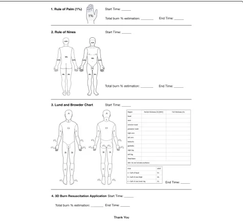
3. Lund and Browder Chart 2. Rule of Nines
1. Rule of Palm (1%)
with the help of photographs, but the subsequent TBSAB calculation is done in a sub-application which is based on the Lund and Browder chart [8].
We hope to validate the aforementioned 3D Burn ap-plication, in hopes of identifying a faster, more accurate method of estimating burn percentages. With the advent of mobile phone technology, this smart-phone applica-tion could particularly be used to great advantage in the early stages of burn management both in the emergency department and in the immediate first-responder setting.
Methods
Three volunteer subjects were moulaged with simulated burn injuries of 25%, 30% and 35% TBSA, respectively;
this was measured and marked out as accurately as pos-sible based on their estimated body surface area (BSA) [9]. The MedCalc: BSA, BMI calculator [9] was used to determine the volunteers estimated TBSA. Based on the measurements obtained from the calculator, simulated burn injuries of 25%, 30% and 35% TBSA, respectively, were then marked out on the volunteers. Flexible trans-parent plastic sheets corresponding to 1% TBSA of the subjects were measured out in square centimetre and used to trace out the simulated burn injuries.
The study was conducted in conjunction with the 2017 Singapore Annual Burns Update, with partici-pants from both local and international institutions. Various participants (Singapore Civil Service Defence
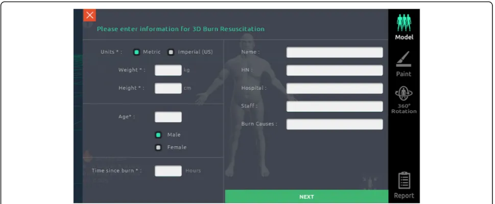
Fig. 23D Burn Resuscitation application initial data screen
Force—Paramedics/Firefighters, Singapore Armed Forces, Nurses and Physicians) were invited to use both the 3D Burn application as well as conventional methods of estimation: Rule of Palm, Rule of Nines and the Lund and Browder chart to estimate the volunteer subjects’ TBSAB (Figs.1 and2). Time to complete the estimation by each methods were recorded.
The 3D Burn application allows users to “paint” burn injuries on the on-screen models—this then automatically computes TBSAB (Fig.3).
A total of 82 participants took part in the validation study. Forty (49%) participants were male, and the remaining 42 (51%) were female. Sixty (73%) participants were between the ages of 25–40 years. Thirty-one (38%) participants had 1–5 years of experience in managing burns patients, 26 (32%) had 6–10 years of experience and 25 (30%) had beyond 10 years of experience. The participants could be broadly split into three categor-ies—first-line responders (paramedics, firefighters and emergency response teams) formed the biggest group with 46% of responses. Nurses and physicians formed the next two groups with 34% and 20% of responses, respectively (Table 1).
None of the participants had prior exposure or training with the 3D Burn application. A 15-min power-point-based tutorial was carried out on the day of the study itself. Par-ticipants were required to download and install the applica-tion on their own smart-phones.
Statistical analysis
As the three different subjects had differing burn percent-ages, the pairedttest was used to test each individual pair
of data against the 3D Burn application. For example, the pairedttest was used three times to assess the mean time to complete estimation from subject A (25% TBSA)—Rule of Palm versus (vs) 3D Burn application, Rule of Nines vs 3D Burn application and Lund and Browder chart vs 3D Burn application. The above was repeated for subject B (30% TBSA) and subject C (35% TBSA).
Results
The results from the above study were tabulated and broadly categorised into a few different aspects—mean TBSAB estimations, relative estimations to actual TBSAB and the mean time to completion of each assessment tool. In total, there are three different sets of data—one for each subject (25%, 30% and 35% TBSA, respectively) (Table2).
In order to negate the possibility of difference in time to completion due to differing burn wound sizes, the time to completion was also tested in a similar fashion (Table3).
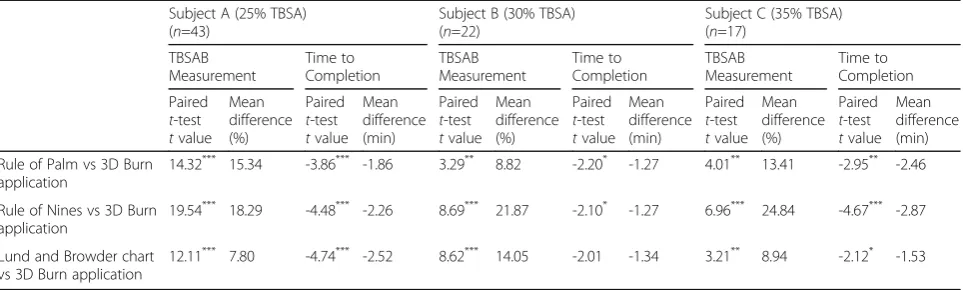
Table 4 showed that there was a statistically signifi-cant difference in both TBSAB measurements and time to completion between the 3D Burn application estimations vs all three other modalities (Rule of Palm, Rule of Nines and Lund and Browder chart) (p< 0.05 [Table 4]). There was also a statistically significant differ-ence in time to completion- usage of the application took significantly longer time compared to traditional methods of estimation.
Burn estimations relative to actual TBSAB
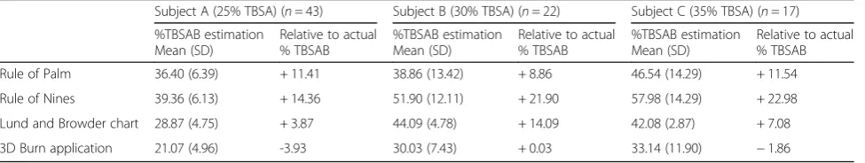
Collective relative estimations across the subjects (A, B and C) showed that when used, the Rule of Palm, Rule of Nines and Lund and Browder chart all over-estimated burns by an average of 10.6%, 19.7% and 8.3% TBSA, re-spectively. The 3D Burn application under-estimated burns area by an average of 1.9%.
Discussion
Accurate measurement of TBSAB remains a crucial, but highly subjective area in the management of burn patients. Inaccurate measurement leads to a multitude of problems, particularly in the form of unnecessary
Table 1Participants composition in the validation study of the 3D Burn Resuscitation application
Subject A (25% TBSA) n
Subject B (30% TBSA) n
Subject C (35% TBSA) n
Physicians 8 5 3
Nurses 17 4 7
First-line responders 18 13 7
Total 43 22 17
TBSATotal body surface area
Table 2Summary of TBSAB estimation by 3D Burn application and the conventional methods
Subject A (25% TBSA) (n= 43) Subject B (30% TBSA) (n= 22) Subject C (35% TBSA) (n= 17)
%TBSAB estimation Mean (SD)
Relative to actual % TBSAB
%TBSAB estimation Mean (SD)
Relative to actual % TBSAB
%TBSAB estimation Mean (SD)
Relative to actual % TBSAB
Rule of Palm 36.40 (6.39) + 11.41 38.86 (13.42) + 8.86 46.54 (14.29) + 11.54
Rule of Nines 39.36 (6.13) + 14.36 51.90 (12.11) + 21.90 57.98 (14.29) + 22.98
Lund and Browder chart 28.87 (4.75) + 3.87 44.09 (4.78) + 14.09 42.08 (2.87) + 7.08
3D Burn application 21.07 (4.96) -3.93 30.03 (7.43) + 0.03 33.14 (11.90) −1.86
transfers to tertiary burn centres and over or under resuscitation of burn patients. The ideal form of TBSAB measurement would be one that is not only 3D, but one that is entirely objective, with no subjective element. Astounding overestimations of greater than 100% have been identified in some emergency de-partments [7, 10–13]. These inaccuracies are largely due to human error, further highlighting the subject-ive element of the current methods of TBSAB measurement.
The data from our study has shown promising results. The 3D Burn application, although slower, allowed more accurate TBSAB measurements when compared to con-ventional methods. With the advent of technology and the push towards electronic data, the 3D burn applica-tion could be a step in the right direcapplica-tion to reducing the subjective element of TBSAB measurement.
We recognise that this study has its limitations. The burn models or templates in the 3D Burn appli-cation may not be an entirely accurate representation of the actual burn patient—this may ultimately affect TBSAB accuracy on the application itself. Addition-ally, the study was conducted on a relatively small
population of healthcare and frontline workers and al-though all participants had prior exposure to manage-ment of burn patients, there were varying levels of experience and not all were adept or familiar with the intricacies of TBSAB measurement. Although the dif-ference in results between the 3D Burn application and traditional methods of TBSAB measurement were statistically significant, and the clinical significance is yet to be determined.
Finally, the practical implications of using a smart-phone application for TBSAB could also be of concern. Many participants in the study found the application more challenging to use compared to the traditional pen and paper methods of measurement. Some participants also questioned patient confidentiality—although the application itself does not require pictures or patient identifiers to function. Perhaps, an encrypted dedicated smart device with a larger screen could be something that improves the user experience and assures patient confidentiality.
Conclusion
The validation study has shown that the 3D Burn appli-cation could be useful in improving the accuracy of TBSAB measurement. There are plans to address some of the limitations and repeat the study in a different centre. In addition to extending the study across sub-jects of differing body weights, we would also like to place emphasis on participants with more experience in burn TBSAB measurement to allow for a more accurate assessment. Results will be collated and analysed as part of a multi-centre study, with a view of progressing to a prospective study that compares the accuracy of the 3D Burn application against conventional methods on actual burn patients.
Table 4Comparison between 3D Burn application and other conventional methods in TBSAB estimation and time to complete the estimation
Subject A (25% TBSA) (n=43)
Subject B (30% TBSA) (n=22)
Subject C (35% TBSA) (n=17)
TBSAB Measurement Time to Completion TBSAB Measurement Time to Completion TBSAB Measurement Time to Completion Paired t-test tvalue Mean difference (%) Paired t-test tvalue Mean difference (min) Paired t-test tvalue Mean difference (%) Paired t-test tvalue Mean difference (min) Paired t-test tvalue Mean difference (%) Paired t-test tvalue Mean difference (min)
Rule of Palm vs 3D Burn application
14.32*** 15.34 -3.86*** -1.86 3.29** 8.82 -2.20* -1.27 4.01** 13.41 -2.95** -2.46
Rule of Nines vs 3D Burn application
19.54*** 18.29 -4.48*** -2.26 8.69*** 21.87 -2.10* -1.27 6.96*** 24.84 -4.67*** -2.87
Lund and Browder chart vs 3D Burn application
12.11*** 7.80 -4.74*** -2.52 8.62*** 14.05 -2.01 -1.34 3.21** 8.94 -2.12* -1.53
*P <0.05,**P
<0.01 and***P
<0.001 indicate significant differences
TBSATotal body surface area,TBSABTotal body surface area burned,3D Burn3D Burn Resuscitation
Table 3Mean time to completion by 3D Burn application and the conventional methods
Mean time to completion (min)
Subject A (25% TBSA) (n= 43)
Subject B (30% TBSA) (n= 22)
Subject C (35% TBSA) (n= 17)
Rule of Palm 2.7 3.1 3.5
Rule of Nines 2.3 3.1 3.5
Lund and Browder chart 2.0 3.1 4.9
3D Burn application 4.5 4.4 6.4
Abbreviations
BMI:Body mass index; BSA: Body surface area; SD: Standard deviation; TBSA: Total body surface area; TBSAB: Total body surface area burned; VS: Versus
Acknowledgements
We thank Mr. Raxle Ng, Mr. Louis Chia and Mr. Koh Wen Hao for volunteering as simulated patients. We also thank Ms. Chong Xin Ying and Ms. Hayley Foong for their assistance in co-ordinating the research study.
Funding Not applicable
Availability of data and materials Not applicable
Authors’contributions
AKWC led the study, analysed the results and wrote the manuscript. TK is the creator of the smart-phone application and conducted a tutorial during the study. TEH and LML played a major role in the preparation of the simulated patients, running the study and the collection of data. CSJ is the senior author and had a major role in organising and conducting the study. All authors read and approved the final manuscript.
Ethics approval and consent to participate Not applicable
Consent for publication
Formal written consent was obtained from all participants of the study.
Competing interests
Dr. Tanasit Kangkorn is the inventor of the free application. All remaining authors declare no competing interests.
Author details
1Department of Plastic, Reconstructive and Aesthetic Surgery, Singapore
General Hospital, 1 Outram Road, Bukit Merah, Singapore.2Department of Plastic and Reconstructive Surgery, Chonburi Hospital, 69 Sukhumvit Road, Muang Chonburi, Thailand.3Navy Medical Service, Republic of Singapore Navy, 210 Tanah Merah Coast Road, Singapore, Singapore.
Received: 18 November 2017 Accepted: 24 January 2018
References
1. Wibbenmeyer L, Amelon MJ, Morgan LJ, Robinson BK, Chang PX, Lewis R 2nd, et al. Predicting survival in an elderly burn patient population. Burns. 2000;27(6):583–90.
2. Wachtel TL, Berry CC, Wachtel EE, Frank HA. The inter-rater reliability of estimating the size of burns from various burn area chart drawings. Burns. 2000;26(2):156–70.
3. Nichter LS, Williams J, Bryant CA, Edlich RF. Improving the accuracy of burn-surface estimation. Plast Reconstr Surg. 1985;76(3):428–33. 4. Amirsheybani HR, Crecelius GM, Timothy NH, Peiffer M, Saggers GC,
Manders EK. The natural history of the growth of the hand: hand area as a percentage of body surface area. Plast Reconstr Surg. 2001;107:726–33. 5. Nagl TR, Schunk JE. Using the hand to estimate the surface area of burn
children. Pedatr Emerg Care. 1997;13:254–5.
6. Berry MG, Evison D, Roberts AH. The influence of body mass index on burn surface estimated from the area of the hand. Burns. 2001;27:591–4. 7. Kamolz LP, Lumenta DB, Parvizi D, Dirnberger J, Owen R, Höller J, et al.
Smartphones and burn size estimation:“rapid burn assessor”. Ann Burns Fire Disasters. 2014;27(2):101–4.
8. Wallis LA, Fleming J, Hasselberg M, Laflamme L, Lundin J. A Smartphone app and cloud-based consultation system for burn injury emergency care. PLoS One. 2016;11(2):e0147253.
9. MedCalc: body surface area, body mass index (BMI). 1999–2017 MedCalc. comhttp://www.medcalc.com/body.html. Accessed 1 Oct 2017. 10. Giretzlehner M, Dirnberger J, Owen R, Haller HL, Lumenta DB, Kamolz LP.
The determination of total burn surface area: how much difference? Burns. 2013; Epub
11. Chan QE, Barzi F, Cheney L, Harvey JG, Holland AJA. Burn size estimation in children: still a problem. Emerg Med Australas. 2012;24:181–6.
12. Jose RM, Roy DK, Vidyadharan R, Erdmann M. Burns area estimation—an error perpetuated. Burns. 2004;30:481–2.
13. Nichter LS, Bryant CA, Edlich RF. Efficacy of burned surface area estimation calculated from charts—the need for a computer based model. J Trauma. 1985;25:477–81.
• We accept pre-submission inquiries
• Our selector tool helps you to find the most relevant journal
• We provide round the clock customer support
• Convenient online submission
• Thorough peer review
• Inclusion in PubMed and all major indexing services
• Maximum visibility for your research
Submit your manuscript at www.biomedcentral.com/submit