Alaeddin Bashir Abukabda
1,2, Carroll Rolland McBride
1,2, Thomas Paul Batchelor
1,2, William Travis Goldsmith
1,2,
Elizabeth Compton Bowdridge
1,2, Krista Lee Garner
1,2, Sherri Friend
3and Timothy Robert Nurkiewicz
1,2,3*Abstract
Background:The cardiovascular effects of pulmonary exposure to engineered nanomaterials (ENM) are poorly understood, and the reproductive consequences are even less understood. Inflammation remains the most
frequently explored mechanism of ENM toxicity. However, the key mediators and steps between lung exposure and uterine health remain to be fully defined. The purpose of this study was to determine the uterine inflammatory and vascular effects of pulmonary exposure to titanium dioxide nanoparticles (nano-TiO2). We hypothesized that
pulmonary nano-TiO2exposure initiates a Th2 inflammatory response mediated by Group II innate lymphoid cells
(ILC2), which may be associated with an impairment in uterine microvascular reactivity.
Methods:Female, virgin, Sprague-Dawley rats (8–12 weeks) were exposed to 100μg of nano-TiO2via intratracheal instillation 24 h prior to microvascular assessments. Serial blood samples were obtained at 0, 1, 2 and 4 h post-exposure for multiplex cytokine analysis. ILC2 numbers in the lungs were determined. ILC2s were isolated and phosphorylated nuclear factor kappa-light-chain-enhancer of activated B cells (NF-ĸB) levels were measured. Pressure myography was used to assess vascular reactivity of isolated radial arterioles.
Results:Pulmonary nano-TiO2exposure was associated with an increase in IL-1ß, 4, 5 and 13 and TNF-α4 h post-exposure, indicative of an innate Th2 inflammatory response. ILC2 numbers were significantly increased in lungs from exposed animals (1.66 ± 0.19%) compared to controls (0.19 ± 0.22%). Phosphorylation of the transactivation domain (Ser-468) of NF-κB in isolated ILC2 and IL-33 in lung epithelial cells were significantly increased (126.8 ± 4.3% and 137 ± 11% of controls respectively) by nano-TiO2exposure. Lastly, radial endothelium-dependent arteriolar reactivity was significantly impaired (27 ± 12%), while endothelium-independent dilation (7 ± 14%) andα-adrenergic sensitivity (8 ± 2%) were not altered compared to control levels. Treatment with an anti- IL-33 antibody (1 mg/kg) 30 min prior to nano-TiO2exposure resulted in a significant improvement in endothelium-dependent dilation and a decreased level of IL-33 in both plasma and bronchoalveolar lavage fluid.
Conclusions:These results provide evidence that the uterine microvascular dysfunction that follows pulmonary ENM exposure may be initiated via activation of lung-resident ILC2 and subsequent systemic Th2-dependent inflammation.
Keywords:Engineered nanomaterials, Titanium dioxide nanoparticles, Microcirculation, Innate lymphoid cells, Inflammation
* Correspondence:[email protected]
1Department of Physiology and Pharmacology, West Virginia University
School of Medicine, 64 Medical Center Drive, Robert C. Byrd Health Sciences Center - West Virginia University, Morgantown, WV 26505-9229, USA 2Toxicology Working Group, West Virginia University School of Medicine,
Morgantown, WV, USA
Full list of author information is available at the end of the article
Introduction
Reproductive toxicity is increasingly becoming recog-nized as a critical aspect of ENM safety. However, the effects of ENM exposure on overall reproductive health have only recently been addressed and become a focus of intense study by numerous groups [1, 2]. As with other observations of systemic biologic effects after pul-monary ENM exposures, reproductive effects are equally susceptible but poorly understood. Our group has previ-ously reported that nano-TiO2 inhalation is associated
with uterine microvascular dysfunction [3], potentially deleterious epigenomic alterations [4], and cognitive def-icits in maternally exposed progeny [5]. While these studies highlight the importance of identifying the ma-ternal effects of ENM exposure, the mechanisms linking such exposures to these negative reproductive and devel-opmental outcomes have yet to be fully elucidated.
In the last decade, innate lymphoid cells (ILC) have emerged as a novel population of tissue-specific effector cells with the ability to initiate and regulate the innate and adaptive branches of the immune system [6,7]. ILCs are now divided into 3 distinct groups according to the pattern of cytokine secretion; Group 1 ILC (ILC1) are predominantly tissue-resident cells capable of secreting interferon gamma (IFN-γ) in the liver, gut, spleen, skin, peritoneum, and salivary gland [8]. The role played by ILC1 in various immunological conditions remains un-clear but is currently under investigation. Group 2 ILCs (ILC2, also known as nuocytes, natural helper cells) secrete IL-4, IL-5, IL-9, and IL-13 in response to damage-associated molecular patterns or “alarmins”and have been implicated in the immune response to parasitic infections and in allergic airway inflammation [9] to sev-eral environmental and anthropogenic agents [10, 11]. Lastly, group 3 ILCs (ILC3) produce IL-22 and or IL-17, are enriched at mucosal sites, contribute to the mainten-ance of the intestinal barriers, and may play a significant role in the promotion of the inflammatory response and etiology of inflammatory bowel diseases [12, 13]. One of the most important alarm signals secreted by cells is interleukin-33 (IL-33) [14]. IL-33 is a member of the IL-1 family of cytokines expressed by both non-immune cells such as epithelial, endothelial, smooth muscle cells, and fi-broblasts [15], as well as immune effector cells including macrophages and dendritic cells. Its role in directing the inflammatory response following pulmonary exposure to carbon nanotubes [16–18], and ozone [19] have been pre-viously reported. IL-33 has been demonstrated [20] to in-duce the release of Th2 cytokines by immune cells including ILC2s in asthmatic patients [21], and to induce airway hyperresponsiveness and increased pulmonary re-sistance [22,23].
In view of the increasing number of studies indicating a crucial role for ILCs in the initiation of the acute
inflammatory response to environmental agents, the goal of this work was to provide initial evidence of their potential involvement in the response to pulmonary ENM exposure. Therefore, the purpose of this study was threefold. First, we identified the inflammatory and uterine microvascular effects associated with acute pulmonary nano-TiO2exposure. Second, we determined
the possible role played by ILC in the immune response mounted to acute pulmonary nano-TiO2 exposure.
Lastly, the microvascular and systemic effects of treat-ment with an IL-33 antibody (a key activator of ILC2) were determined. We hypothesized that pulmonary nano-TiO2 exposure initiates a Th2 inflammatory
response mediated involving IL-33 and potentially in-volving ILC2, which may be associated with a systemic impairment in uterine microvascular reactivity.
Results
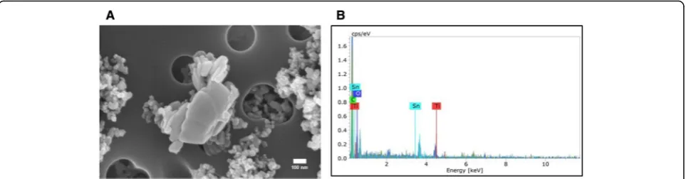
SEM images and mass spectrometry of Nano-TiO2
Figure1shows field-emission scanning electron microscope images (Hitachi S4800, Tokyo, Japan) of the nano-TiO2
sus-pension used for this study. As seen in Fig.1a, the suspended particles present significant agglomeration. Figure 1bshows the elemental composition of the nano-TiO2 suspension
(Bruker, Billerica, MA), indicating the prevalent presence of titanium.
Animal and vessel characteristics
No significant changes were observed in age, mean arterial pressure (MAP), heart rate and body weight between control and exposure groups(Table1). Additionally, radial arteriolar inner and outer diameter, tone, passive diameter, wall thick-ness, wall to lumen ratio and calculated wall tension were not affected by nano-TiO2exposure and treatment with an
anti-IL-33 antibody(Table2). From these results we can infer that acute nano-TiO2exposure does not alter vascular tone
or the balance of its contributing influences.
Acute plasma and BALF cytokine secretion patterns following pulmonary Nano-TiO2exposure
A time-course study was initially performed to identify the temporal effect of nano-TiO2on cytokine secretion. Plasma
samples were obtained at 0,1,2, and 4 h following nano-TiO2exposure by tail vein puncture. Figure2 shows
the results of the multiplex analysis. No differences existed at time 0, 1 or 2 h post-exposure. However, significant dif-ferences were detected 4 h post-exposure in plasma levels of the pro-inflammatory cytokines IL-4, IL-5, IL-13, TNF-α, IL-1β, while no alterations in the cytokines IFN-γ, KC/ GRO, TNF-α, IL-10, IL-6, MCP-1, TIMP-1, Lipocalin-2, and TSP-1 were noted(Data not shown).
also showed an almost 5-fold increase in IL-5 and a 6-fold increase in IL-4, IL-13, TNF-α, and IL-1β, while no changes were seen in the other cytokines (Data not shown). Based on the collective cytokine profile associ-ated with this exposure paradigm, the results provide evidence that acute pulmonary nano-TiO2exposure may
trigger a T-Helper cell type 2 (Th2) response beginning 4 h post-exposure.
IL-33 secretion by lung epithelial cells pre- and post- exposure and following treatment with an anti-IL-33 antibody
IL-33 plays a significant role in initiating and modulating lung inflammatory and immunological responses [24], by inducing the secretion of Th2 pro-inflammatory cytokines. It is constitutively present in mucosal epithelial cells and acts as a“danger”signal after tissue injury by activating im-mune cells. To determine the effect of nano-TiO2on
pul-monary and plasma IL-33 levels, immunohistochemistry of tracheal sections was conducted 4 h post-exposure. In a separate cohort of control and exposed animals, multiplex analysis of plasma and BALF IL-33 was also performed 4 h post-exposure. Figure 4 shows representative images of lung sections from control and exposed rats stained for IL-33 4 h post-exposure and the quantitative measurements for both groups. Relative fluorescence intensity of IL-33 was significantly increased in exposed animals by 26.8 ± 4.3%, while plasma and BALF levels indicated a 37.87 ± 2.3% and 171.26 ± 13% increase in IL-33 respectively(Fig.5). Interestingly, treatment 1 h prior to exposure to nano-TiO2
with pharmacological grade polyclonal anti-IL-33 antibody (EMD Millipore, Temecula, CA: intraperitoneal 1 mg/kg) resulted in a decrease in plasma and BALF IL-33 levels 4 h
post-exposure (61.1 ± 2.1% in plasma and 149.7 ± 7% in BALF) when compared to nano-TiO2 exposed and
un-treated animals. The results herein described provide evi-dence that reveals the critical role played by IL-33 in the initiation of the inflammatory response to ENM.
Flow cytometric analysis of ILC1 and ILC2
Polarization of T-cells requires several days [25], therefore, the early appearance of Th2 cytokines in the circulation after nano-TiO2suggests that other, more rapid immune effector
cells may be involved. Previous work has indicated that both ILC1 and ILC2 [26,27] reside in the lungs and may be in-volved in lung inflammatory responses and pathology. Therefore, we next wanted to determine the effect of nano-TiO2exposure on ILC1 and ILC2 levels in the lungs.
For this reason, multi-parametric flow cytometric analysis was conducted on lung tissue from control and nano-TiO2
exposed Sprague-Dawley rats for ILC1(Data not shown)and ILC2 (Fig. 6) 4 h post-exposure. No significant differences were noted in ILC1 levels pre- and post- exposure while ILC2 levels increased from 0.19 ± 0.22% to 1.66 ± 0.19% (Fig.7aandb).
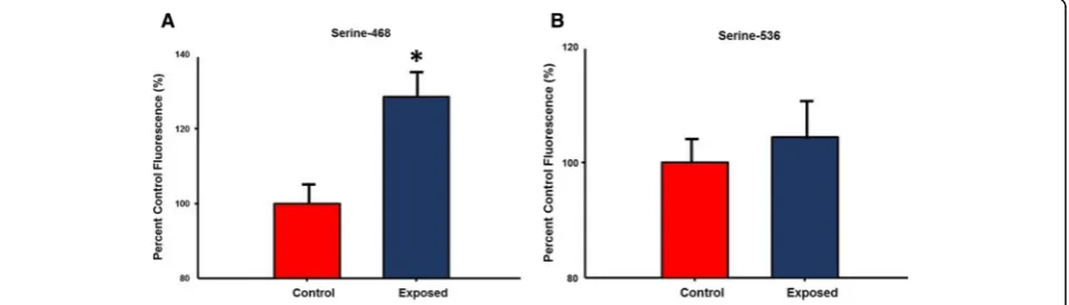
NF-ĸB phosphorylation and Th2 cytokine secretion by isolated ILC2
Activation of the NF-κB pathway has been shown to play a role in inflammation through its ability to induce the transcription of proinflammatory genes [28, 29]. Specif-ically, phosphorylation of NF—ĸB at both serine residues 468 and 536 are associated with inflammatory response [29, 30]. Therefore, levels of phospho- NF—ĸB were measured in isolated ILC2 from control and nano-TiO2
Fig. 1Characterization of Nano-TiO2. (a) SEM image and (b) Energy dispersive spectroscopy showing elemental composition of the nano-TiO2 suspension used in this study
Table 1Animal characteristics
N Age (weeks) weight (grams) Heart Rate (bpm) Map (mm Hg) Systolic Blood pressure (mm Hg) Diastolic Blood Pressure (mm Hg)
Control 13 14 ± 1 232 ± 5 319 ± 13 85 ± 3 103 ± 4 73 ± 2
Exposed 14 15 ± 1 249 ± 12 330 ± 9 86 ± 4 110 ± 6 78 ± 4
exposed animals (Fig.8). No significant differences were noted in phosphorylation of the Serine-536 residue be-tween the 2 groups, while phosphorylation of Serine-468 was increased after acute pulmonary nano-TiO2
expos-ure (128.6 ± 7.24% of control levels).
Lastly, isolated ILC2 were cultured overnight and IL-4, IL-5 and IL-13 levels in the cell-culture media were mea-sured. Nano-TiO2resulted in a significant increase in IL-4,
IL-5, and IL-13 (122 ± 7.2%, 141.4 ± 9.1%, 158.8 ± 7.6% of control levels respectively)(Fig.9).
Effect of acute pulmonary Nano-TiO2exposure and
systemic immunotherapy on uterine radial arterioles Endothelium-dependent dilation
In order to identify the acute reproductive effects asso-ciated with nano-TiO2 exposure, radial arterioles were
isolated from the uterus of control and exposed animals 24 h post-exposure and endothelium-dependent and
independent dilation along with adrenergic sensitivity were tested. Previous work by our group has shown that ENM exposure impacts vascular reactivity most severely within 24 h, a condition which has been shown to improve but not fully return to control levels after 168 h [30]. Based on these findings, all vascular assess-ments for this study were conducted 24 h post-exposure.
Endothelium-dependent dilation of radial arterioles was significantly impaired following intratracheal instillation of 100μg of nano-TiO2(Fig.10), with a mean decrease in
dila-tion of 49.23 ± 6.5% compared to controls and 24.35 ± 10% compared to anti-IL-33 antibody-treated rats. These results suggest that pulmonary nano-TiO2exposure disrupts
nor-mal physiological vascular endothelial function and that pre-treatment with an anti-IL-33 antibody partially attenu-ates the systemic acute inflammatory cascade triggered by lung-derived IL-33.
Table 2Arteriolar characteristics
n Inner diameter (μm) Outer diameter (μm) Tone(%) Passive diameter inner (μm)
Passive diameter outer (μm)
Wall Tension (Newton/meter)
Control 18 98 ± 5 152 ± 14 26 ± 7 152 ± 9 199 ± 15 0.33 ± 0.1
Exposed 16 102 ± 11 164 ± 13 21 ± 4 159 ± 15 207 ± 17 0.34 ± 0.1
Exposed + anti-L-33 12 97 ± 11 158 ± 19 21 ± 4 140 ± 14 187 ± 26 0.31 ± 0.6
Table showing characteristics of control and exposed radial arterioles (n= 12–18). All vascular assessments were performed 24 h post-exposure. Values shown are mean ± SEM. Statistics were analyzed with two-way ANOVA (P≤0.05), * Sham control group vs. nano-TiO2exposed groups
A B C
D E
In contrast to endothelium-dependent relaxation, no point to point differences were seen in α-adrenergic and endothelium-independent dilation between control, nano-TiO2exposed, and treated groups(Data not shown).
Discussion
The inflammatory paradigm tested herein has been sug-gested by previous studies [16, 31, 32]. While these studies explored pulmonary responses to exogenously
administered IL-33, carbon nanotubes and viral infec-tion, our findings provide additional evidence of a simi-lar link between acute pulmonary ENM exposure and subsequent inflammatory mechanisms with systemic microvascular consequences. Perhaps the most import-ant finding of this study is the identification of IL-33 as a potential contributor to the systemic inflammatory phenotype and microvascular dysfunction resulting from acute pulmonary ENM exposure.
Fig. 3Nano-TiO2exposure also increases T-Helper type II cytokines 4 h post-exposure in bronchoalveolar lavage fluid (BALF). Multiplex cytokine analysis showing concentrations of IL-4, IL-5, IL-13 in BALF from exposed and control animals 4 h post-exposure (N= 6). Statistics were analyzed with two-way ANOVA (P≤0.05), * Sham control group vs. nano-TiO2exposed groups
We hypothesized that the innate response to pulmonary nano-TiO2exposure is triggered by the secretion of IL-33
by lung epithelial cells. IL-33, a member of the IL-1 family, plays a significant role in initiating and modulating lung inflammatory and immunological responses [33], by indu-cing the secretion of Th2 pro-inflammatory cytokines through the Suppression of Tumorigenicity 2 (ST2) recep-tor in a variety of immune cells [32,34]. Previous studies have shown that exposure to multi-walled carbon nano-tubes triggers the IL-33/ST2 axis leading to the activation of mast-cells and macrophages [20]. Interestingly, IL-33 is also required for the maturation, activation and egress of ILC2 lineage cells from the bone marrow to specific target
organs [35]. Several previous studies by our group and others have extensively investigated the cardiovascular ef-fects of pulmonary ENM exposure [36,37]. These studies consistently reveal that nano-TiO2 exposure increases
leukocyte activation [38], proinflammatory mediators [39], and oxidative stress [40]. Collectively, this results in endothelium-dependent dysfunction. ILCs are a recently identified subpopulation of immune effector cells [33,41]. They are critical, non-cytolytic tissue-resident cells that can be activated and respond acutely to danger signals or
“alarmins” such as IL-33 from mucosal tissue by produ-cing a distinctive array of cytokines to maintain mucosal integrity. Group II ILCs specifically, secrete the cytokines Fig. 5Pre-treatment of nano-TiO2–exposed animals with an anti-IL-33 antibody lowers BALF and plasma IL-33. Interleukin-33 levels in (a) BALF and (b) plasma in control, nano-TiO2exposed groups and in rats pre-treated with an anti-IL-33 antibody. Rats were pre-treated with an anti-IL-33 antibody (1 mg/kg) 30 min prior to exposure. Plasma and BALF IL-33 levels were measured 4 h post-exposure. Statistics were analyzed with two-way ANOVA (P≤0.05). * Sham control group vs. nano-TiO2exposed groups,ŦP< 0.05 Exposed + Anti-IL-33 Antibody vs Exposed
A)
B)
IL-5, IL-13, IL-9, amphiregulin, and low quantities of IL-4 [31] and have been linked to several lung-associated con-ditions including pathogen infections (viruses and hel-minths), asthma, and pulmonary fibrosis [42]. Lung ILC2 have an important role in regulating tissue remodeling and repair during acute epithelial injury and asthma [43]. Considered together, it is reasonable to speculate that the IL-33/ILC2 axis may impact systemic microvascular func-tion after pulmonary ENM exposure.
Perhaps, the most significant finding of this study is the identification of a potential role for IL-33 in the initi-ation of an acute inflammatory response and the effects on systemic microvascular dysfunction (Fig. 10). This was achieved by interrupting the IL-33/ST2 signaling axis with an anti-IL-33 antibody treatment prior to ENM exposure. In these experiments, the control group (IT saline) did not receive the anti-IL-33 antibody. The scientific rationale for this decision was that the tech-nique (isolated, perfused and pressurized arterioles) used to assess microvascular dysfunction is a plasma free system. As such, it was impossible to assess the role of the complete IL-33/ST2 axis on arteriolar tone and/or reactivity. Whereas,
this approach directly measured the impact that IL-33 in-flammatory signaling has on microvascular function. It re-mains a distinct possibility that the IL-33/ST2 axis activated by pulmonary ENM exposure directly influences micro-vascular tone and/or reactivity. IL-33 expression has been identified in human vascular endothelial cells; whereas, is absent in murine vascular tissue [44]. This supports our pos-tulate that IL-33 is impacting microvascular function via in-flammation, rather than a direct activation of the ST2 receptor. Future in vivo studies must directly assess the im-pact of circulating IL-33 on microvascular tone, reactivity and blood flow.
While our study provides plausible mechanistic insights into both the inflammatory and microvascular effects associ-ated with ENM exposure, additional considerations as well as the limitations must be kept in mind. ENM exposure has been shown to affect other innate immune effector cells such as neutrophils [45,46], macrophages [47,48], and den-dritic cells [49]. Therefore, other mechanisms including local generation of reactive of oxygen species, secretion of chemo-kines and prostaglandins by macrophages and stimulation of the adaptive immune system by antigen-presenting cells Fig. 7Nano-TiO2exposure increases pulmonary ILC2 but not ILC1. Quantification of percentage of total pulmonary cells of (a) ILC1 and (b) ILC2 in control and nano-TiO2exposed animals (N= 6). Statistics were analyzed with two-way ANOVA (P≤0.05), * Sham control group vs. nano-TiO2 exposed groups
may play a role in the systemic inflammatory response and vascular effects seen in this study. It is also worth noting that in this study an immune response associated with pul-monary ENM exposure was observed as early as 4 h post-exposure, as evidenced not only by an increase in the proportion of lung-resident ILC2s(Fig.7), but also by aug-mented levels of the Th2 cytokines IL-4, IL-5, and IL-13 (Figs. 2 and 3). This response timeframe is in agreement
with previous studies, that have reported IL33, ILC2 num-bers as well as lung and serum Th2 cytokines increase within 6–12 h of lung injury [50,51]. Furthermore, isolated ILC2s have been shown to respond dose-dependently to transient or continuous stimulation with IL-2 and/or IL-33 within 3 h [52]. Similarly, nano-TiO2 instillation at lung
burdens of 40–160μg/rat has also been shown to increase BALF inflammatory mediators within 4 h of exposure [53]. Fig. 9Th2 cytokine secretion in isolated ILC2 is increased by nano-TiO2exposure. Multiplex cytokine analysis showing concentrations of IL-4, IL-5, IL-13 supernatant from isolated ILC2 cultured overnight from control and nano-TiO2exposed animals (N = 6). Statistics were analyzed with two-way ANOVA (P≤0.05), * Sham control group vs. nano-TiO2exposed groups
particle exposure. Rat models are primarily used in our in vivo studies as they are the most appropriate and widely used animals for microvascular research. How-ever, they are not commonly or easily adapted for trans-genic studies. As such, an ILC2 rat deletion model does not exist. If such a model were created in the future, it would then be possible to directly confirm a causative relationship between ENM exposure and ILC2 activa-tion, proliferation and recruitment.
Lastly, to allow periodic blood sampling for cytokine analysis, intratracheal instillation was chosen as the pre-ferred exposure method. The above-mentioned dose of nano-TiO2 was selected based on previous studies
con-ducted by our group which have shown that it is associ-ated with significant microvascular impairment, oxidative and nitrosative stress and alveolar macrophage recruit-ment [36,54]. Additionally, to better understand the rele-vance of the exposure paradigm used in this study to humans, alveolar surface areas must be known [55]. The rat alveolar surface area is 0.4 m2/lung. Therefore, the rat burden of 50μg/lung would result in 125 μg/m2. Given that the human alveolar surface area is 102 m2, the equivalent human burden of this exposure paradigm would be 12.75 mg. The next logical question is: How long would it take to achieve this burden in humans? In this re-gard, lung burden may be calculated as:
nano‐TiO2aerosol concentration minute ventilation
exposure duration deposition fraction;
with the following values:
25:5 mg¼nano‐TiO2aerosol concentration
7600 ml=min 8 hrhð =day60 min=hrÞ 14%;
and therefore:
25:5 mg¼nano‐TiO2aerosol concentration
0:51 m3=day:
Considering both the National Institute for Occupa-tional Safety and Health (NIOSH) and the OccupaOccupa-tional Safety and Health Administration (OSHA) Permissible Exposure Limit (0.3 mg/m3 and 5 mg/m3 respectively) [4], it would require 0.34 working years or 122 working
Materials and methods
Nanomaterial characterization
Nano-TiO2P25 powder, obtained from Evonik (Aeroxide
TiO2, Parsippany, NJ), has previously been shown to be
a mixture composed primarily of anatase (80%) and rutile (20%) TiO2, with a primary particle size of 21 nm
and a surface area of 48.08 m2/g [54], and a Zeta-poten-tial of−56.6 mV [56]. Elemental composition analysis of the nano-TiO2suspension was conducted via energy
dis-persive spectroscopy.
Scanning electron microscopy was performed by diluting the nano-TiO21:100 with filtered distilled water. 0.5 ml of
the diluted particle solution was filtered onto a 0.2μm poly-carbonate filter. A wedge-shaped portion of the polycarbon-ate filter was mounted onto carbon double stick tape which was affixed to a 13 mm aluminum stub. The sample was sputter coated with gold-palladium for two minutes. The sample was imaged using a Hitachi S4800 field-emission scanning electron microscope (Tokyo, Japan).
Experimental animals and exposure
Female (8–10 weeks) Sprague – Dawley rats were pur-chased from Hilltop Laboratories (Scottdale, PA) and housed in an AAALAC approved facility at West Virginia University (WVU) with 12:12 h light–dark cycle and reg-ulated temperature. Rats were allowed ad libitum access to food and water. All procedures were approved by the Institutional Animal Care and Use Committee of WVU.
100μg of nano-TiO2were suspended in 250μL of
ve-hicle (Normosol and 5% fetal bovine serum) for intratra-cheal instillation (IT) 24 h prior to experimentation. Nano-TiO2suspensions were sonicated for 1 min to
en-sure homogenous distribution of nanoparticles. Rats were anesthetized using 5% isoflurane and placed on a mounting stand. 250 μL of the nano-TiO2 suspension
was then instilled intratracheally. Sprague - Dawley rats instilled with 250μL of vehicle were used as controls.
Mean arterial pressure (MAP) acquisition
the right carotid artery was cannulated to acquire mean arterial pressure (MAP). The MAP was measured via a pressure transducer and recorded by PowerLab830 (AD Instruments, Colorado Springs, CO).
Multiplex cytokine panels of serum and Bronchoalveolar lavage fluid
Whole blood (1–2 ml) was collected via tail vein puncture from exposed and control animals at time 0, 1 h, 2 h, and 4 h after exposure into EDTA vacutainers and centrifuged (1000 x g) to collect plasma which was flash-frozen in liquid nitrogen and stored at−80°C until analysis. Rats were eutha-nized and tissues harvested for further analysis. Multi-spot inflammatory assays were completed per manufacturer’s directions (Meso Scale Diagnostics, Rockville, MD) for: lipocalin-2, TSP-1, TIMP-1, MCP-1, interferon (IFN-γ), inter-leukin (IL)-1β, IL-4, IL-5, IL-6, KC/GRO, IL-10, IL-13, and TNF-α.
Immunohistochemistry
Gelatin-coated cover slips of serial tissue sections were incubated with anti-rabbit (1:100) IL-33 (Cloud-Clone Corp., Katy, TX) overnight at 4°C, followed by 3 five-minute washes with cold PBS with 0.1% Triton-X. The sections were then incubated with a FITC conju-gated goat anti-rabbit (1:100, Invitrogen, Carlsbad, CA) at 37°C for one hour, followed by 3 five-minute washes with cold PBS with 0.1% Triton-X. Cover slips were then mounted on slides for visualization on a Zeiss fluores-cent microscope (Zeiss, Thornwood, NY).
Preparation of cell suspensions
Lungs were perfused with sterile PBS by injection into the right ventricle to remove remaining blood and then placed in PBS containing 20 mg/ml Collagenase A, 2.4 U/ml Dispase II solution, and 50μl/ml DNAse (Roche, Indian-apolis, IN) for 30 min.
The lung tissue was dissociated using the gentleMACS Octo Dissociator (Miltenyi Biotec, Auburn, CA) and then centrifuged (1100 x g for 10 min) to collect the sample ma-terial. The sample was resuspended with PBS and used for magnetic cell separation and flow cytometry.
Flow cytometry Antibodies and reagents
ILC1 and ILC2 numbers 4 h after exposure to nano-TiO2
from lung tissue were determined by flow cytometry as previously described [57–59]. Briefly, ILC1 were defined as Lin-CD45 + CD161 + CD335+, while ILC2 were defined as Lin-CD45 + CD90.1 + KLRG-1 + CD25 + IL1R1 + .
Monoclonal antibodies specific for CD90.1 (FITC), KLRG1 (APC EFLUOR 780), CD25 (APC), CD161 (PERCP EFLUOR 710), and CD335 (EFLUOR 450) were purchased from eBioscience (San Diego, CA). Lineage cocktail (Lin;
ALEXA FLUOR 700) was used to gate out (CD3, CD14, CD16, CD19, CD20, and CD56: Biorad, Hercules, CA) CD3 T lymphocytes, CD14 Monocytes, CD16 NK cells, granulo-cytes, CD19 B lymphogranulo-cytes, CD20 B lymphogranulo-cytes, and CD56 NK cells. Antibodies for CD45 (Cyanine 5) were from Invitrogen (Carlsbad, CA), while those for IL1R1 (PE) where from Sino Biologicals (North Wales, PA). Flow cytometry was performed on a FACSAria (BD Bioscience, Franklin Lakes, NJ). Data were analyzed with FCS Express 6 Software (De Novo Software, Glendale, CA).
Isolation of group II innate lymphoid cells
ILC2 were isolated as per manufacturer’s instructions (Miltenyi Biotec, Auburn, CA). Briefly, lineage-positive cells were indirectly labeled with a cocktail of ALEXA FLUOR 700-conjugated antibodies, as primary labeling reagent, and antibodies conjugated to MicroBeads were used as secondary labeling reagents. In the second step, lineage-negative cells were labeled with CD45, CD90.1, KLRG-1, CD25, and IL1R1, labeled with MicroBeads and isolated by positive selection from the pre-enriched lineage negative cell-fraction by separation over a MACS Column (Miltenyi Biotec, Auburn, CA), which was placed in the magnetic field of a MACS Separator. After negative selection, the cells were subsequently eluted as the positively selected cell fraction containing ILC2.
Cytokine analysis of cell culture media and measurement of Phospho-NF-ĸB
Isolated ILC2s were incubated (37 °C, 90% humidity, 5% CO2) overnight in supplemented (10% fetal bovine
serum, 1% penicillin/streptomycin, 1% sodium pyruvate, 1% L-glutamine) Dulbecco’s Modified Eagle Medium (Corning, Manassas, VA). The following day, the cell culture medium and cellular portion were separated via centrifugation (1100 x g for 10 min). IL-4, IL-5, and IL-13 levels were measured in the cell culture medium via multi-spot inflammatory assays per manufacturer’s directions (Meso Scale Diagnostics, Rockville, MD). Lastly, isolated cells were lysed and phospho-NF-κB (Ser468 and Ser536) was measured (Meso Scale Diag-nostics, Rockville, MD).
Systemic treatment with anti-Interleukin-33 antibody
Rats were treated 1 h prior to pulmonary nano-TiO2
maintained at 4 °C. Radial arterioles were isolated, transferred to a vessel chamber, cannulated between two glass pipettes, and tied with silk sutures in the chamber (Living Systems In-strumentation, Burlington, VT). The chamber was super-fused with fresh oxygenated (5% CO2/21% O2) PSS and
warmed to 37 °C. Arterioles were pressurized to 60 mmHg using a servo control system and extended to their in situ length. Internal and external arteriolar diameters were mea-sured using video calipers (Colorado Video, Boulder, CO).
Arteriolar reactivity
Previous work by our group has shown that ENM ex-posure impacts endothelium-dependent dilation most severely within 24 h, a condition which has been shown to improve but did not fully return to control levels after 168 h. Based on these findings, all vascular assessments for this study were conducted 24 h post-exposure [30].
Arterioles were allowed to develop spontaneous tone, de-fined as the degree of constriction experienced by a blood vessel relative to its maximally dilated state. Vascular tone ranges from 0% (maximally dilated) to 100% (maximal con-striction). Vessels with a spontaneous tone≥20% less than initial tone were included in this study. After equilibration, various parameters of arteriolar function were analyzed. Vessels that did not develop sufficient spontaneous tone were not included in the data analysis.
Endothelium-Dependent Dilation—arterioles were exposed to increasing concentrations of acetyl choline (ACh: 10−9- 10−4M) added to the vessel chamber. Endothelium-Independent Dilation—increasing concentrations of sodium nitroprusside (SNP: 10−9 -10−4M) were used to assess arteriolar smooth muscle responsiveness.
Arteriolar Vasoconstriction—arterioles were exposed to increasing concentrations of phenylephrine (PE: 10−9 -10−4M). The steady state diameter of the vessel was recorded for at least 2 min after each dose. After each dose curve was completed, the vessel chamber was washed to remove excess chemicals by carefully removing the superfusate and replacing it with fresh warmed oxygenated PSS. After all experimental treatments were complete, the PSS was replaced with Ca2+-free PSS until maximum passive diameter was established.
maximal diameter using the following formula:
Normalized diameter¼Dss=Dm
, where Dss is the steady state diameter recorded during each pressure change. The experimental responses to ACh, PE, and SNP are expressed using the following equation:
Diameter percent maximal diameterð Þ
¼ ðDss−DconÞ Dm−Dcon
ð Þ
x100
, where DCon is the control diameter recorded prior to the dose curve, DSS is the steady state diameter at each dose of the curve. The experimental response to PE is expressed using the following equation:
Diameter percent maximal diameterð Þ
¼ ðDcon−DssÞ Dcon
ð Þ
x100
Wall thickness (WT) was calculated from the meas-urement of both inner (ID) and outer (OD) steady state arteriolar diameters at the end of the Ca2+ free wash using the following equation:
WT ¼ðOD−IDÞ=2
Wall-to-lumen ratio (WLR) was calculated using the following equation:
WLR¼WT=ID
Statistics
Additional file
Additional file 1:Figure S1.IL-33 sequence homology. Multiple sequence alignment of the protein sequence of IL-33 in mice (query) and rats (subject) showing the significant interspecies homology of murine IL-33. (ZIP 259 kb)
Abbreviations
ACh:Acetylcholine; ANOVA: Analysis of variance; APC: Allophycocyanin; BALF: Broncho alveolar lavage fluid; CD: Cluster of differentiation; Dcon: Control diameter; Di: Initial diameter; Dm: Maximal diameter; Dss: Steady state diameter; EDTA: Ethylenediaminetetraacetic acid; ENM: Engineered nanomaterials; FITC: Fluorescein isothiocyanate; ID: Inner diameter; IFN-γ: Interferon-gamma; IL: Interleukin; ILC: Innate lymphoid cells; KC/GRO: Keratinocyte chemoattractant/human growth-regulated oncogene; KLRG-1: Killer cell lectin-like receptor subfamily G member 1; Lin: Lineage; MCP-1: Monocyte chemoattractant protein 1; Nano-TiO2: Titanium dioxide nanoparticles; NF-ĸB: Nuclear factor kappa-light-chain-enhancer of activated B cells; NIOSH: National Institute for Occupational Safety and Health; NK: Natural Killer; NO: Nitric oxide; OD: Outer diameter; OSHA: Occupational Safety and Health Administration; PBS: Phosphate buffered saline; PE: Phenylephrine; PE: Phycoerythrin; SEM: Scanning electron microscope; SEM: Standard error of the mean; Ser-468: Serine-468; SNP: Sodium nitroprusside; ST2: Suppressor of tumor tumorigenicity; Th2: T-helper type 2; TIMP-1: Tissue inhibitor of metalloproteinases 1; TNF-α: Tumor necrosis factor alpha; TSP-1: Thrombospondin 1; WLR: Wall-to-lumen ratio; WT: Wall thickness
Acknowledgments
We thank Kevin Engels from the West Virginia University (WVU) Department of Physiology and Pharmacology for his technical assistance in this study, and Dr. Kathleen Brundage from the Flow Cytometry & Single Cell Core Facility at WVU (FORTESSA S10 OD016165, FACSAria S10 RR020866). In addition, we would like to acknowledge our financial support: R01-ES015022 (TRN), and NSF-1003907 (TRN and ABA).
Availability of data and materials
All data generated or analyzed during this study are included in this published article [and its supplementary information files.
Disclaimer
The findings and conclusions in this report are those of the author(s) and do not necessarily represent the official position of the National Institute for Occupational Safety and Health, Centers for Disease Control and Prevention.
Authors’contributions
ABA conducted the vascular and immunological assessments, analyzed and interpreted the data, and was a major contributor in writing the manuscript. CRM assisted with the vascular assessments and was involved in drafting the manuscript and revising it critically for important intellectual content. TPB performed the tracheal immunohistochemistry and was involved in drafting the manuscript. WTG was involved in the animal exposures and was involved in the drafting of the manuscript. ECB and KLG were involved in the analysis and interpretation of the data and were major contributors in writing the manuscript. SF obtained the SEM images of the nano-TiO2and was involved in drafting of the manuscript. TRN made substantial contributions to the conception and design of the experiments, analysis and interpretation of the data and was a major contributor in writing of the manuscript. All the authors read and approved the final manuscript.
Ethics approval and consent to participate Not applicable.
Competing interests
The authors declare that they have no competing interests.
Publisher’s Note
Springer Nature remains neutral with regard to jurisdictional claims in published maps and institutional affiliations.
Author details
1Department of Physiology and Pharmacology, West Virginia University
School of Medicine, 64 Medical Center Drive, Robert C. Byrd Health Sciences Center - West Virginia University, Morgantown, WV 26505-9229, USA. 2Toxicology Working Group, West Virginia University School of Medicine,
Morgantown, WV, USA.3National Institute for Occupational Safety and Health, Morgantown, WV, USA.
Received: 7 June 2018 Accepted: 24 October 2018
References
1. Jackson P, et al. Prenatal exposure to carbon black (printex 90): effects on sexual development and neurofunction. Basic Clin Pharmacol Toxicol. 2011; 109(6):434–7.
2. Johansson HKL, et al. Airway exposure to multi-walled carbon nanotubes disrupts the female reproductive cycle without affecting pregnancy outcomes in mice. Part Fibre Toxicol. 2017;14(1):17.
3. Stapleton PA, et al. Maternal engineered nanomaterial exposure and fetal microvascular function: does the barker hypothesis apply? Am J Obstet Gynecol. 2013;209(3):227 e1–11.
4. Stapleton PA, et al. Maternal engineered nanomaterial inhalation during gestation alters the fetal transcriptome. Part Fibre Toxicol. 2018;15(1):3. 5. Engler-Chiurazzi EB, et al. Impacts of prenatal nanomaterial exposure on
male adult Sprague-Dawley rat behavior and cognition. J Toxicol Environ Health A. 2016;79(11):447–52.
6. McKenzie AN. Type-2 innate lymphoid cells in asthma and allergy. Ann Am Thorac Soc. 2014;11(Suppl 5):S263–70.
7. Sonnenberg GF, et al. SnapShot: innate lymphoid cells. Immunity. 2013; 39(3):622–622 e1.
8. Artis D, Spits H. The biology of innate lymphoid cells. Nature. 2015; 517(7534):293–301.
9. Turner JE, et al. IL-9-mediated survival of type 2 innate lymphoid cells promotes damage control in helminth-induced lung inflammation. J Exp Med. 2013;210(13):2951–65.
10. Vercelli D, Gozdz J, von Mutius E. Innate lymphoid cells in asthma: when innate immunity comes in a Th2 flavor. Curr Opin Allergy Clin Immunol. 2014;14(1):29–34.
11. Spits H, et al. Innate lymphoid cells--a proposal for uniform nomenclature. Nat Rev Immunol. 2013;13(2):145–9.
12. Takatori H, et al. Lymphoid tissue inducer-like cells are an innate source of IL-17 and IL-22. J Exp Med. 2009;206(1):35–41.
13. Yasuda K, et al. Contribution of IL-33-activated type II innate lymphoid cells to pulmonary eosinophilia in intestinal nematode-infected mice. Proc Natl Acad Sci U S A. 2012;109(9):3451–6.
14. Shaw JL, et al. IL-33-responsive innate lymphoid cells are an important source of IL-13 in chronic rhinosinusitis with nasal polyps. Am J Respir Crit Care Med. 2013;188(4):432–9.
15. Marsland BJ, et al. Innate signals compensate for the absence of PKC-{theta} during in vivo CD8(+) T cell effector and memory responses. Proc Natl Acad Sci U S A. 2005;102(40):14374–9.
16. Katwa P, et al. A carbon nanotube toxicity paradigm driven by mast cells and the IL-(3)(3)/ST(2) axis. Small. 2012;8(18):2904–12.
17. Wang X, et al. Intravenously delivered graphene nanosheets and multiwalled carbon nanotubes induce site-specific Th2 inflammatory responses via the IL-33/ST2 axis. Int J Nanomedicine. 2013;8:1733–48. 18. Wang X, et al. Multi-walled carbon nanotube instillation impairs pulmonary
function in C57BL/6 mice. Part Fibre Toxicol. 2011;8:24.
19. Michaudel C, et al. Inflammasome, IL-1 and inflammation in ozone-induced lung injury. Am J Clin Exp Immunol. 2016;5(1):33–40.
20. Beamer CA, et al. IL-33 mediates multi-walled carbon nanotube (MWCNT)-induced airway hyper-reactivity via the mobilization of innate helper cells in the lung. Nanotoxicology. 2013;7(6):1070–81.
21. Kabata H, et al. Group 2 innate lymphoid cells and asthma. Allergol Int. 2015;64(3):227–34.
22. Hams E, Fallon PG. Innate type 2 cells and asthma. Curr Opin Pharmacol. 2012;12(4):503–9.
23. Deckers J, Branco Madeira F, Hammad H. Innate immune cells in asthma. Trends Immunol. 2013;34(11):540–7.
course study. Int J Mol Sci. 2012;13(11):13781–803.
31. Schmitz J, et al. IL-33, an interleukin-1-like cytokine that signals via the IL-1 receptor-related protein ST2 and induces T helper type 2-associated cytokines. Immunity. 2005;23(5):479–90.
32. Zeng S, et al. IL-33 receptor (ST2) Signalling is important for regulation of Th2-mediated airway inflammation in a murine model of acute respiratory syncytial virus infection. Scand J Immunol. 2015;81(6):494–501.
33. Mjosberg JM, et al. Human IL-25- and IL-33-responsive type 2 innate lymphoid cells are defined by expression of CRTH2 and CD161. Nat Immunol. 2011;12(11):1055–62.
34. Zoltowska AM, et al. The interleukin-33 receptor ST2 is important for the development of peripheral airway hyperresponsiveness and inflammation in a house dust mite mouse model of asthma. Clin Exp Allergy. 2016;46(3):479–90. 35. Stier MT, et al. IL-33 promotes the egress of group 2 innate lymphoid cells
from the bone marrow. J Exp Med. 2018;215(1):263–81.
36. Nurkiewicz TR, et al. Nanoparticle inhalation augments particle-dependent systemic microvascular dysfunction. Part Fibre Toxicol. 2008;5:1. 37. Abukabda AB, et al. Heterogeneous vascular bed responses to
pulmonary titanium dioxide nanoparticle exposure. Front Cardiovasc Med. 2017;4:33.
38. Stapleton PA, et al. Uterine microvascular sensitivity to nanomaterial inhalation: an in vivo assessment. Toxicol Appl Pharmacol. 2015;288(3):420–8.
39. Stapleton PA, et al. Estrous cycle-dependent modulation of in vivo microvascular dysfunction after nanomaterial inhalation. Reprod Toxicol. 2018;78:20–8.
40. LeBlanc AJ, et al. Nanoparticle inhalation impairs endothelium-dependent vasodilation in subepicardial arterioles. J Toxicol Environ Health A. 2009; 72(24):1576–84.
41. Spits H, Di Santo JP. The expanding family of innate lymphoid cells: regulators and effectors of immunity and tissue remodeling. Nat Immunol. 2011;12(1):21–7.
42. Hams E, et al. IL-25 and type 2 innate lymphoid cells induce pulmonary fibrosis. Proc Natl Acad Sci U S A. 2014;111(1):367–72.
43. Monticelli LA, et al. Innate lymphoid cells promote lung-tissue homeostasis after infection with influenza virus. Nat Immunol. 2011;12(11):1045–54. 44. Pichery M, et al. Endogenous IL-33 is highly expressed in mouse epithelial
barrier tissues, lymphoid organs, brain, embryos, and inflamed tissues: in situ analysis using a novel Il-33-LacZ gene trap reporter strain. J Immunol. 2012;188(7):3488–95.
45. Sanfins E, et al. Nanoparticle effect on neutrophil produced myeloperoxidase. PLoS One. 2018;13(1):e0191445.
46. Thompson EA, et al. Innate immune responses to nanoparticle exposure in the lung. J Environ Immunol Toxicol. 2014;1(3):150–6.
47. Taylor AJ, et al. Atomic layer deposition coating of carbon nanotubes with aluminum oxide alters pro-fibrogenic cytokine expression by human mononuclear phagocytes in vitro and reduces lung fibrosis in mice in vivo. PLoS One. 2014;9(9):e106870.
48. Bonner JC, Hoffman M, Brody AR. Alpha-macroglobulin secreted by alveolar macrophages serves as a binding protein for a macrophage-derived homologue of platelet-derived growth factor. Am J Respir Cell Mol Biol. 1989;1(3):171–9.
49. Look M, et al. The nanomaterial-dependent modulation of dendritic cells and its potential influence on therapeutic immunosuppression in lupus. Biomaterials. 2014;35(3):1089–95.
50. Xu J, et al. IL33-mediated ILC2 activation and neutrophil IL5 production in the lung response after severe trauma: a reverse translation study from a human cohort to a mouse trauma model. PLoS Med.
2017;14(7):e1002365.
56. Nichols CE, et al. Reactive oxygen species damage drives cardiac and mitochondrial dysfunction following acute nano-titanium dioxide inhalation exposure. Nanotoxicology. 2018;12(1):32–48.
57. Drake LY, Kita H. Group 2 innate lymphoid cells in the lung. Adv Immunol. 2014;124:1–16.
58. Duerr CU, Fritz JH. Isolation of group 2 innate lymphoid cells from mouse lungs. Methods Mol Biol. 2017;1656:253–61.
59. Halim TY, Takei F. Isolation and characterization of mouse innate lymphoid cells. Curr Protoc Immunol. 2014;106:3 25 1–13.