Newborns With Suspected Occult Spinal Dysraphism:
A Cost-Effectiveness Analysis of Diagnostic Strategies
L. Santiago Medina, MD, MPH*; Kerry Crone, MD‡; and Karen M. Kuntz, ScD§
ABSTRACT. Objective. To assess the clinical and eco-nomic consequences of different diagnostic strategies in newborns with suspected occult spinal dysraphism.
Methods. A decision-analytic model was constructed to project the cost and health outcomes of magnetic res-onance imaging (MRI), ultrasound (US), plain radio-graphs, and no imaging in newborns with suspected occult spinal dysraphism. Morbidity and mortality rates of early versus late diagnosis of dysraphism and the sensitivity and specificity of MRI, US, and plain radio-graphs were obtained from the literature. Cost estimates were obtained from a hospital cost accounting database and from the Medicaid fee schedule.
Results. We found that the choice of imaging strategy depends on the underlying risk of occult spinal dysra-phism. In low-risk children with intergluteal dimple or newborns of diabetic mothers (pretest probability: 0.3%– 0.34%), US was the most effective strategy with an incre-mental cost-effectiveness ratio of $55 100 per quality-adjusted life year gained. For children with lumbosacral dimples, who have a higher pretest probability of 3.8%, US was less costly and more effective than the other 3 strategies considered. In intermediate-risk newborns with low anorectal malformation (pretest probability: 27%), US was more effective and less costly than radio-graphs and no imaging. However, MRI was more effec-tive than US at an incremental cost-effeceffec-tiveness of $1000 per quality-adjusted life year gained. In the high-risk group that included high anorectal malformation, cloacal malformation, and exstrophy (pretest probability: 44%– 46%), MRI was actually cost-saving when compared with the other diagnostic strategies. For the intermediate-risk group, we found our analysis to be sensitive to the costs and diagnostic performances (sensitivity and specificity) of MRI and US. Lower MRI cost or greater MRI diagnos-tic performance improved the cost-effectiveness of the MRI strategy, whereas lower US cost or greater US diag-nostic performance worsened the cost-effectiveness of the MRI strategy. Therefore, individual or institutional expertise with a specific diagnostic modality (MRI versus US) may influence the optimal diagnostic strategy.
Conclusion. In newborns with suspected occult dys-raphism, appropriate selection of patients and diagnostic strategy may increase quality-adjusted life expectancy
and decrease cost of medical work-up. Pediatrics 2001; 108(6). URL: http://www.pediatrics.org/cgi/content/full/ 108/6/e101;cost-effectiveness analysis, occult spinal dys-raphism, newborns, MRI, ultrasound.
ABBREVIATIONS. MRI, magnetic resonance imaging; QALY, quality-adjusted life year; US, ultrasound; LE, life expectancy.
T
hree percent of neonates have major central
nervous system or systemic malformations.
1Furthermore, 5% to 15% of pediatric neurology
hospital admissions are related to cerebrospinal
anomalies.
2Occult spinal dysraphism (skin-covered
lesions with no exposed neural tissue) is the most
prevalent spinal axis malformation
3and the most
common indication for spinal imaging in children.
4Occult spinal dysraphism encompasses the entire
range of skin-covered spinal column and neuraxis
anomalies resulting from the failure of primary or
secondary neurulation.
5,6Occult spinal dysraphic
le-sions include dorsal dermal sinus, tethered cord with
spinal lipoma, lipomyelomeningocele, and
diastem-atomyelia
4,6 – 8and are commonly associated with
urinary tract anomalies.
9The clinical spectrum of occult dysraphism is
broad, ranging from skin stigmata such as a dimple,
sinus tract, hair patch, or hemangioma to motor,
bladder, or bowel dysfunction.
6 – 8Approximately
50% to 80% of occult spinal dysraphic cases exhibit a
dermal lesion.
10 –13However, 3% to 5% of all children
have skin dimples.
14,15The prevalence of occult
dys-raphism has a broad range from as low as 0.34% in
children with pilonidal sinuses (intergluteal dimples)
to as high as 46% in newborns with cloacal
malfor-mation.
9,16Because of the broad range of occult
dys-raphism prevalences, controversy exists as to which
risk group may or may not benefit from imaging.
Early detection and prompt neurosurgical
correc-tion of occult spinal dysraphism may prevent upper
urinary tract deterioration, infection of dorsal dermal
sinuses, or permanent neurologic damage.
17–22Sev-eral studies have demonstrated that motor function,
urologic symptoms, and urodynamic patterns may
be improved by early surgical intervention in
pa-tients with occult spinal dysraphism.
23,24The
surgi-cal outcome may be better if intervention occurs
before the age of 3 years.
23–25Spinal neuroimaging,
therefore, has the important role of determining the
presence or absence of an occult spinal dysraphic
lesion so that appropriate surgical treatment can be
instituted in a timely manner.
From the *International Health Outcomes and Economics Center, and Di-vision of Neuroradiology, Department of Radiology, Miami Children’s Hospital, Miami, Florida; ‡Department of Neurosurgery, Children’s Hos-pital Medical Center, Cincinnati, Ohio; and §Department of Health Policy and Management, Harvard School of Public Health, Boston, Massachusetts. Received for publication Apr 9, 2001; accepted Jul 30, 2001.
Reprint requests to (L.S.M.) International Health Outcomes and Economics Center, and Division of Neuroradiology, Department of Radiology, Miami Children’s Hospital, 3100 SW 62 Ave, Miami, FL 33155. E-mail: [email protected]
There is no consensus regarding the most
appro-priate neuroimaging modality to diagnose occult
dysraphism. Some authors advocate spinal
ultra-sonography as the primary imaging modality for
lower spine congenital anomalies because of its
rea-sonable diagnostic performance at a low cost and
without sedation.
15,26Others emphasize the
impor-tance of the more expensive spinal magnetic
reso-nance imaging (MRI) examination because of its
bet-ter diagnostic performance, excellent soft tissue
characterization, and importance in presurgical
plan-ning.
6 – 8,27–29Although plain film imaging is
inex-pensive and widely available, few authors advocate
its use as a diagnostic test because of its low
speci-ficity.
30Conversely, some clinicians have
empha-sized the importance of close clinical follow-up with
no imaging in low-risk children who present with
simple intergluteal dimples, because the probability
of finding an occult dysraphic lesion is only 0.34%.
16Because occult spinal dysraphism is a relatively
common pediatric disorder and early diagnosis and
treatment are important, the role and
cost-effective-ness of the imaging modalities for different risk
groups need to be determined. Accordingly, we
con-structed a decision-analytic model to compare the
cost and health outcomes of various work-up
strate-gies in children with suspected occult spinal
dysra-phism.
METHODS The Model
Cost-effectiveness analysis is a method used to evaluate the health outcomes and costs of interventions designed to improve health.31–34Primary and secondary (medical publications) data are used to construct a comprehensive decision-analytic model. The results of an analysis are summarized in a series of cost-effective-ness ratios that show the cost of achieving 1 unit of health outcome (eg, the cost per quality-adjusted life year [QALY] gained) for different risk groups and strategies.31–34
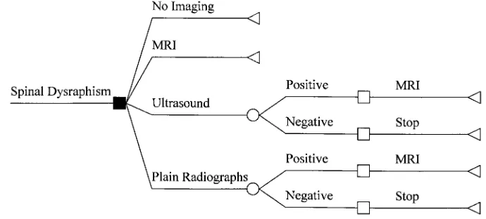
We constructed a decision-analytic Markov model to compare the costs and health outcomes of 4 diagnostic strategies in children who are suspected of having occult spinal dysraphism: 1) MRI, 2) plain radiographs followed by MRI for positive results (radio-graphs), 3) ultrasound (US) followed by MRI for positive results (US), and 4) no imaging with close clinical follow-up. We analyzed 3 risk groups based on the prevalence (pretest probability) of disease: 1) low (intergluteal dimple and infants of diabetic moth-ers; prevalence: 0.3%), 2) intermediate (low and intermediate ano-rectal malformation; prevalence: 27%–33%), and 3) high risk (high anorectal and cloacal malformation; prevalence: 44%– 46%). Figure 1 shows a simplified representation of the decision tree.
The cost-effectiveness analysis was performed from a societal perspective. Costs were expressed in 1999 US dollars. Costs and cost per QALY were discounted at 3% per year.32 Incremental cost-effectiveness ratios were calculated as the additional cost of one strategy divided by the additional effectiveness compared with the next most effective strategy.33Strategies that were both less effective and more costly than the alternative were eliminated because of dominance.33Strategies that had a higher incremental cost-effectiveness ratio and a lower effectiveness than another were eliminated because of extended dominance.33 Cost-effective-ness ratios were expressed as cost per QALY gained. The model was constructed and analyzed by using the computer program Decision Analysis (DATA; TreeAge Software, Williamstown, MA).
Data
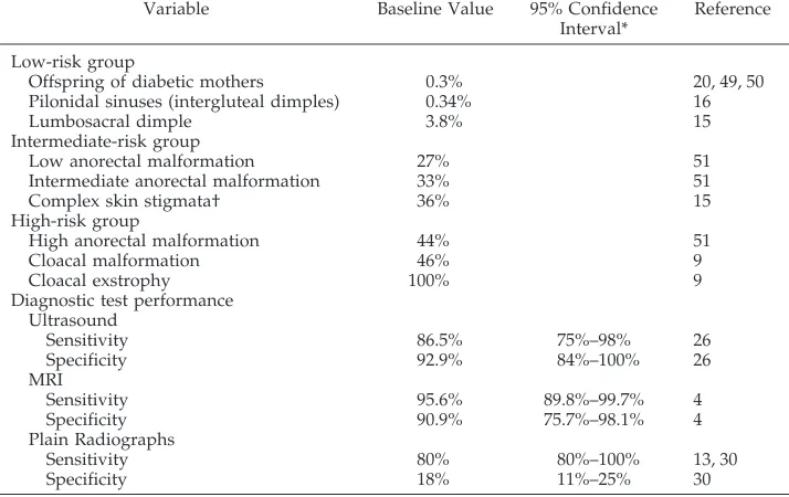
Data sources included medical publications from 1962 to 1998 reporting on occult spinal dysraphism. Model baseline values and ranges are summarized in Table 1.
Risk Groups
Children with suspected occult dysraphism were divided into low-, intermediate-, and high-risk groups. Children in the low-risk group included those with simple skin dimples as the sole mani-festation or newborns of diabetic mothers. The prevalence (pretest probability) of a dysraphic lesion among low-risk patients has been estimated at 0.3% to 3.8%. In the low range (0.3%) are children with low intergluteal dimples (pilonidal sinuses); chil-dren in the upper range (3.8%) have higher lumbosacral dim-ples.9,15,16
Children in the intermediate-risk group included those with complex skin stigmata and low and intermediate anorectal mal-formations. The prevalence (pretest probability) of a dysraphic lesion among intermediate-risk patients has been estimated at 27% to 36%. Children in the high-risk group included those with high anorectal malformations, cloacal malformation, and cloacal exstro-phy. The prevalence (pretest probability) of a dysraphic lesion among high-risk patients has been estimated at 44% to 100%.
Diagnostic Tests
Several studies have shown that MRI and US have better diag-nostic performances (ie, sensitivity and specificity) than plain radiographs4,13,26,30; these data are shown in Table 1. The sensi-tivity of spinal MRI and US has been estimated at 95.6% and 86.5%, respectively.4,26The specificity of spinal MRI and US has been estimated at 90.9% and 92.9%, respectively.4,26Conversely, the sensitivity and specificity of plain radiographs have been estimated at 80% and 18%, respectively.13,30Because there are no large series assessing the joint diagnostic performance of MRI and CT, we assumed conditional independence between these tests for the case in which a positive plain radiograph or US examination was followed by MRI (Fig 1).
Natural History and Surgical Intervention
Symptoms from occult spinal dysraphism often are not appar-ent until the child becomes older.15The most common clinical
presentations for occult dysraphic states later in infancy include delay in development of sphincter control, delay in walking, asymmetry of the legs or abnormalities of the feet (ie, pes cavus and pes equinovarus), and pain in the back or lower extremi-ties.12,13,18,24,35–39
Several studies have demonstrated improvement of the multi-ple symptoms associated with occult dysraphism if surgical inter-vention is performed.23–25 Newborns with spinal dysraphism were divided into early and delayed diagnosis. The early dysra-phism diagnosis group consisted of patients whose dysradysra-phism was diagnosed and surgically treated in the newborn period. The late dysraphism diagnosis group consisted of patients whose dys-raphism was not diagnosed in the newborn period but rather diagnosed and surgically treated by or after age 3 years as the children became symptomatic.25 Using surgical outcome data from the study by Satar et al,25in the early diagnosed and surgi-cally treated patients, 60% became asymptomatic, 30% were un-changed, and 10% worsened. Conversely, the same study data for the late diagnosed and surgically treated patients (ie, surgery after 3 years of age) demonstrated that 27% became asymptomatic, 27% improved, 27% were unchanged, and 19% worsened.25To model these surgical outcomes, 4 health states were created ranging from asymptomatic (AS) to mild (S1), moderate (S2), and severe (S3) neuromuscular, urinary, and bowel symptoms.
Dysraphic patients with a central nervous system communicat-ing dorsal dermal sinus (ie, 10% of all dysraphic patients) were considered to be at risk for infection.13The probability of becom-ing infected if diagnosed late was 50% over a 3-year period. A conservative mortality rate of 1% was used for meningitis.40 – 44 Therefore, the total probability of any dysraphic child dying of infection was estimated at .0005.
In the model, only severely symptomatic patients with dysra-phism (S3) were considered to be at risk of upper urinary tract deterioration.45In the S3 health state, only 15% had upper urinary tract deterioration45and of those with progressive renal damage, 7.5% were considered to develop end-stage renal disease over a 10-year period if undiagnosed.45
Long-Term Outcomes
The base-case patient was a newborn child without coexistent disease. The probability of dying of causes other than complica-tions from occult spinal dysraphism was obtained from the US vital statistics data at the National Center for Health Statistics. To determine the effect of imaging on outcome, the following param-eters were incorporated into the model: 1) the natural history of delayed diagnosis of occult dysraphism and associated renal ab-normalities, 2) improved prognosis from early surgical
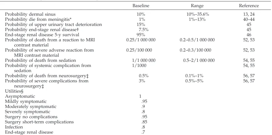
interven-tion, 3) morbidity and mortality of undiagnosed occult spinal dysraphic lesions (ie, infected dermal sinus) and associated renal abnormalities (ie, end-stage renal disease46,47), 4) morbidity and mortality of imaging sedation, and 5) quality of life (Tables 1–3). We projected long-term outcomes such as life years and QALYs gained using these parameters.
The quality-of-life weight (utility) was obtained from the treat-ing physician perspective (Table 3). A utility can vary from 0, representing death, to 1, representing perfect health. The quality weight (utility) for a year of life spent in a more favorable state (eg, mildly symptoms of dysraphism) versus a more unfavorable state (eg, severe symptoms of dysraphism) was incorporated into the Markov model.
Costs
Costs (not charges) of the radiologic and nonradiologic (ie, hospitalization, surgery, infection and complication treatment, laboratories, and pharmacy) procedures were estimated from the Cincinnati Children’s Hospital Medical Center cost accounting system. Fixed, variable, and overhead costs were analyzed. Sum-mary of cost data for the different diagnostic studies, symptomatic stage, neurosurgery, surgical complications, infection, and end-stage renal disease are shown in Table 2. The cost of the radiologic studies includes the technical and professional fee.
Sensitivity Analysis
Sensitivity analysis was performed for each parameter of the model over a clinically plausible range of values (Table 1–3). The effect of these changes on outcomes, costs, and cost-effectiveness ratios was analyzed.
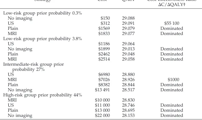
RESULTS Baseline Analysis (Table 4)
For low-risk children with intergluteal dimple or
in offspring of diabetic mothers (pretest probability:
0.3%– 0.34%), no imaging with close clinical
fol-low-up cost less and improved health outcomes
com-pared with the plain radiographs and MRI strategies.
However, the US strategy offered a higher
effective-ness at an incremental cost-effectiveeffective-ness ratio of
$55 100 per QALY gained. In low-risk children with
a lumbosacral dimple (pretest probability: 3.8%), the
TABLE 1. Summary of Data Used in the Analysis
Variable Baseline Value 95% Confidence Interval*
Reference
Low-risk group
Offspring of diabetic mothers 0.3% 20, 49, 50
Pilonidal sinuses (intergluteal dimples) 0.34% 16
Lumbosacral dimple 3.8% 15
Intermediate-risk group
Low anorectal malformation 27% 51
Intermediate anorectal malformation 33% 51
Complex skin stigmata† 36% 15
High-risk group
High anorectal malformation 44% 51
Cloacal malformation 46% 9
Cloacal exstrophy 100% 9
Diagnostic test performance Ultrasound
Sensitivity 86.5% 75%–98% 26
Specificity 92.9% 84%–100% 26
MRI
Sensitivity 95.6% 89.8%–99.7% 4
Specificity 90.9% 75.7%–98.1% 4
Plain Radiographs
Sensitivity 80% 80%–100% 13, 30
Specificity 18% 11%–25% 30
US strategy cost less and was more effective
com-pared with all other diagnostic strategies.
For children in the intermediate-risk group with
pretest probability of 27% (ie, low anorectal
malfor-mation), the US strategy cost less and was more
effective than the plain radiographs and no imaging
strategies. However, the MRI strategy was more
ef-fective than US at a marginal cost-efef-fectiveness ratio
of $1000 per QALY gained. For high-risk children
with high anorectal malformation (pretest
probabil-ity: 44%), the MRI strategy cost less and was more
effective than all other diagnostic strategies.
Sensitivity Analysis
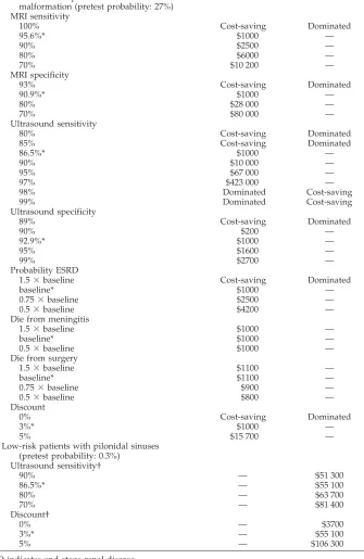
For the intermediate-risk group, the incremental
cost-effectiveness ratio of MRI decreased as the
di-agnostic performance (ie, sensitivity and specificity)
of MRI increased (Table 5). At an MRI sensitivity and
specificity at or above 100% and 93%, respectively,
MRI became cost saving in comparison with all other
strategies.
In the intermediate-risk group, the marginal
cost-effectiveness ratio of MRI increased as the diagnostic
performance (ie, sensitivity and specificity) of US
TABLE 2. Summary of Cost Data Used in the Analysis
Variable Direct Cost Total Cost* Medicaid† Sensitivity Analysis Range
Spine radiographs $21 $37 $27 20–60
Ultrasound $64 $110 $84
MRI $319 $550 $391
Sedation $70 $120 0# 0–150
Neurosurgery $12 000**
Surgical complication‡ $4000**
Infection complication $5500**
End-stage renal disease $40 000††
Symptomatic 1§ $500††
Symptomatic 2㛳 $1400††
Symptomatic 3¶ $2600††
* Medical center cost estimates, includes direct (fixed and variable) and indirect (overhead) costs. Total costs were used for the case-base study.
† Medicaid reimbursement. ‡ ie, bleeding or suture dehiscence. § Mildly symptomatic.
㛳Moderately symptomatic. ¶ Severely symptomatic.
# Sedation by nonanesthesiologist is not reimbursed by medicaid.
** Mean cost per event (including surgery, hospitalization, pharmacy, laboratory studies, and fol-low-up visits).
†† Mean cost per year.
TABLE 3. Summary of Data Used in the Analysis
Baseline Range Reference
Probability dermal sinus 10% 10%–35.6% 13, 24
Probability die from meningitis* 1% 1%–13% 40–44
Probability of upper urinary tract deterioration 15% 45
Probability end-stage renal disease† 7.5% 45
End-stage renal disease 5-y survival 95% 46
Probability of death from a reaction to MRI contrast material
0.25/1 000 000 0.2–0.5/1 000 000 52, 53
Probability of severe adverse reaction from MRI contrast material
0.25/100 000 0.2–0.3/100 000 52, 53
Probability of death from sedation 1/1 000 000 0.5–2/1 000 000 54, 55
Probability of systemic complication from sedation
1/1000 54, 55
Probability of death from neurosurgery‡ 0.5% 0.1%–1% 56, 57
Probability of severe complications from neurosurgery‡
3% 0.5%–5% 56, 57
Utilities§
Asymptomatic 1
Mildly symptomatic .95
Moderately symptomatic .9
Severely symptomatic .8
Surgery no complications .95
Surgery short-term complications .85
Infection .8
End-stage renal disease .7
* Applies to patients who have dermal sinus and get meningitis.
† Applies only to patients who have severe dysraphic findings and progressive upper urinary tract deterioration. ‡ Data from institutional statistics and available neurosurgical literature.
decreased. At an US sensitivity and specificity at or
below 85% and 89%, respectively, MRI became cost
saving in comparison with all other strategies.
Figure 2 shows a 2-way sensitivity analysis of MRI
cost and MRI sensitivity for the intermediate-risk
group. The incremental cost-effectiveness ratio of
MRI decreased as the cost of MRI decreased and MRI
sensitivity increased. MRI became a cost-saving
strategy when the following criteria were met: 1)
MRI cost of $390 and MRI sensitivity of 83% or
higher, 2) MRI cost of $500 and MRI sensitivity of
90% or higher, and 3) MRI cost of $670 and MRI
sensitivity of 100%.
In the low-risk group (pretest probability: 0.3%),
the increment cost-effectiveness ratio of US
de-creased as the sensitivity and specificity of US
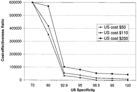
in-creased (Table 5 and Fig 3). Figure 3 shows a 2-way
sensitivity analysis of US cost and US specificity. The
incremental cost-effectiveness ratio of US decreased
as the cost of US decreased and the US specificity
increased. US became a cost-saving strategy when
the US cost was $50 and the US specificity was 98.5%
or higher.
Analysis of the data using as outcome life
expect-ancy (LE) without quality adjustment (ie, no utilities)
was done. In the low-risk group with a pretest
prob-ability of 0.3% (ie, intergluteal dimple or offspring of
diabetic mother), no imaging was cost saving
be-cause it cost less and was more effective in LE gained
compared with all other diagnostic strategies. In the
low- and intermediate-risk groups with a pretest
probability of 3.8% (ie, lumbosacral dimple) and
27%, respectively, US was cost saving because it cost
less and was more effective in LE gained compared
with all other diagnostic strategies. In the high-risk
group (pretest probability: 44%), the MRI strategy
cost less and was more effective in LE gained than
the plain radiographs and no imaging strategies.
However, the US strategy was more effective than
MRI at a high marginal cost-effectiveness ratio of
$870 000 per LE gained. In the high-risk group at a
pretest probability of 50% or higher, MRI was the
preferred strategy because it cost less and was more
effective than all other diagnostic strategies.
DISCUSSION
Controversy exists in regard to the most
appropri-ate neuroimaging modality to assess occult spinal
dysraphism in the newborn. We used the methods of
decision analysis to compare the long-term costs and
health outcomes of various imaging strategies for
children with suspected occult spinal dysraphism.
We found that the choice of imaging strategy
de-pends on the underlying risk of occult spinal
dysra-phism. In low-risk children with intergluteal dimple
or in newborns of diabetic mothers (pretest
proba-bility: 0.3%– 0.34%), the US strategy was the most
effective with an incremental cost-effectiveness ratio
of $55 100 per QALY gained. For children with
lum-bosacral dimples, who have a higher pretest
proba-bility of 3.8%, US was less costly and more effective
than the other 3 strategies considered.
In intermediate-risk newborns with low and
inter-mediate anorectal malformation or skin stigmata
(pretest probability: 27%), US was cost saving when
compared with radiographs and no imaging.
How-ever, MRI was more effective than US at an
incre-ment cost-effectiveness of $1000 per QALY gained. In
the high-risk group that included high anorectal
mal-formation, cloacal malmal-formation, and exstrophy
(pre-test probability: 44%– 46%), MRI was actually cost
saving when compared with the other diagnostic
strategies.
The cost-effectiveness ratios allow comparisons
with alternative health care programs and may assist
in resource allocation decisions.
33The low-risk
group US cost-effectiveness ratio at $55 100 per
QALY saved compared favorably to other
well-ac-TABLE 4. Cost-Effectiveness in QALYS*
Strategy Cost QALY Cost-Effectiveness Ratio
⌬C/⌬QALY† Low-risk group prior probability 0.3%
No imaging $150 29.088
US $312 29.091 $55 100
Plain $1569 29.079 Dominated
MRI $1833 29.077 Dominated
Low-risk group prior probability 3.8%
US $1186 29.064
No imaging $1899 29.013 Dominated
Plain $2462 29.048 Dominated
MRI $2514 29.058 Dominated
Intermediate-risk group prior probability 27%
US $6980 28.880
MRI $7026 28.926 $1000
Plain $8382 28.844 Dominated
No imaging $13 491 28.517 Dominated
High-risk group prior probability 44%
MRI $10 000 28.830
US $11 000 28.746 Dominated
Plain $13 000 28.695 Dominated
No imaging $22 000 28.153 Dominated
* Costs and effects are per person in the target population. † Rounded in hundreds.
cepted diagnostic strategies. For example, annual
mammography and breast examination for women
age 40 to 49 years
48costs $62 000 per life year saved,
annual cervical cancer screening for women
begin-ning at 21 years of age costs $50 000 per life year
saved,
48and colonoscopy for colorectal cancer
screening for people older than 40 years costs $90 000
per life year saved.
48For the intermediate-risk group, we found our
analysis to be sensitive to the costs and diagnostic
performances (sensitivity and specificity) of MRI and
US. Lower MRI cost or greater MRI diagnostic
per-formance improved the cost-effectiveness of the MRI
strategy, whereas lower US cost or greater diagnostic
performance of US worsened the cost-effectiveness
of the MRI strategy. Therefore, individual or
institu-tional expertise with a specific diagnostic modality
(MRI versus US) may influence the optimal
diagnos-tic strategy. For example, radiologists who are more
proficient with MRI than US may perform the
diag-TABLE 5. Cost-Effectiveness Ratios of the Sensitivity Analysis
MRI† Ultrasound
Intermediate-risk patients with low anorectal malformation (pretest probability: 27%) MRI sensitivity
100% Cost-saving Dominated
95.6%* $1000 —
90% $2500 —
80% $6000 —
70% $10 200 —
MRI specificity
93% Cost-saving Dominated
90.9%* $1000 —
80% $28 000 —
70% $80 000 —
Ultrasound sensitivity
80% Cost-saving Dominated
85% Cost-saving Dominated
86.5%* $1000 —
90% $10 000 —
95% $67 000 —
97% $423 000 —
98% Dominated Cost-saving
99% Dominated Cost-saving
Ultrasound specificity
89% Cost-saving Dominated
90% $200 —
92.9%* $1000 —
95% $1600 —
99% $2700 —
Probability ESRD
1.5⫻baseline Cost-saving Dominated
baseline* $1000 —
0.75⫻baseline $2500 —
0.5⫻baseline $4200 —
Die from meningitis
1.5⫻baseline $1000 —
baseline* $1000 —
0.5⫻baseline $1000 —
Die from surgery
1.5⫻baseline $1100 —
baseline* $1100 —
0.75⫻baseline $900 —
0.5⫻baseline $800 —
Discount
0% Cost-saving Dominated
3%* $1000 —
5% $15 700 —
Low-risk patients with pilonidal sinuses (pretest probability: 0.3%)
Ultrasound sensitivity†
90% — $51 300
86.5%* — $55 100
80% — $63 700
70% — $81 400
Discount†
0% — $3700
3%* — $55 100
5% — $106 300
ESRD indicates end-stage renal disease. * Base case.
nostic work-up of intermediate risk children with the
former, whereas radiologists who are proficient with
both may elect US because of the higher marginal
cost-effectiveness ratio of the MRI strategy. Likewise,
cost of the imaging strategies may affect the work-up
strategy of choice. For example, a developing
coun-try with low US cost but high MRI cost may elect to
recommend US to study children in the
intermedi-ate-risk group.
In the low-risk group (pretest probability: 0.3%),
our analysis was sensitive to the cost and diagnostic
performance (sensitivity and specificity) of US.
Lower US cost or higher US diagnostic performance
improved the cost-effectiveness of the US strategy.
For example, in a developing country with
well-trained radiologists performing low-cost
ultrasonog-raphy, spinal US would be the strategy of choice for
low-risk patients.
The results shown herein are summarized in Fig 4
as a suggested decision tree for use in newborns with
suspected occult spinal dysraphism. The decision
tree reinforces the primary importance of a careful
acquisition of a medical history and performance of a
thorough examination. For patients in the high-risk
group, imaging of the spine with MRI is suggested.
For patients in the intermediate-risk group, imaging
of the spine with MRI or US is suggested, whereas in
the low-risk group, the strategies of US or no
imag-ing may be indicated. Selection between these 2
strat-egies per risk group may be based on individual and
institutional diagnostic performance and cost per
test. In newborns with suspected occult dysraphism,
appropriate selection of patients for imaging based
on these risk groups may maximize health outcomes
for patients and improve health care resource
alloca-tion.
Future studies clearly are needed. Because the
spectrum of children with suspected occult
dysra-phism is so broad, other clinical risk groups need to
be studied and their prevalence (prior probability)
determined. Large prospective cohort studies in
chil-dren with other well-defined stigmata or clinical
findings of occult dysraphism are required for this
purpose. Such studies might ultimately form the
ba-sis for valuable evidence-based practice guidelines
for the large population of children evaluated for
suspected occult dysraphism each year in the United
States and other countries.
CONCLUSION
Our analysis suggested the MRI strategy in
high-risk newborns (high anorectal malformation, cloacal
malformation, or exstrophy) because it is cost saving.
In the intermediate-risk newborns (low and
interme-diate anorectal malformation or skin stigmata), MRI
or US is the preferred strategy. US or no imaging
with close clinical follow-up is the preferred strategy
in risk newborns. In the intermediate- and
low-risk newborns, choice between the 2 preferred
strat-egies is based on individual and institutional
diag-nostic performance and cost of the tests. In newborns
with suspected occult dysraphism, appropriate
selec-tion of patients and diagnostic strategy may increase
quality-adjusted LE and decrease cost of medical
work-up.
ACKNOWLEDGMENTS
This study was supported, in part, by a research grant from the American Society of Neuroradiology and General Electric-Associ-ation of University Radiologist
We are indebted to Nolan R. Altman, MD, William S. Ball, Jr,
Fig 2. Two-way sensitivity analysis of MRI according to MRI cost and MRI sensitivity in intermediate-risk patients.
Fig 3. Two-way sensitivity analysis of US according to US cost and US specificity in low-risk patients (P⫽.3%).
MD, Marguerite M. Care, MD, and Janet L. Strife, MD, for impor-tant suggestions. We are also indebted to Elsa Aguirre, RT, Maria Telarroja, and Susan DeBusk for secretarial assistance.
REFERENCES
1. Kalter H, Warkany J. Medical progress. Congenital malformations: eti-ologic factors and their role in prevention.N Engl J Med. 1983;308: 424 – 431
2. Bird TD, Hall JG. Clinical neurogenetics. A survey of the relationship of medical genetics to clinical neurology.Neurology1977;27:1057–1060 3. Egelhoff JC, Prenger EC, Coley BD. The spine. In: Ball WS Jr, ed.
Pediatric Neuroradiology. Philadelphia, PA: Lippincott-Raven Publishers; 1997:717–778
4. Medina LS, Al-Orfali M, Zurakowski D, Poussaint TY, DiCanzio J, Barnes PD. Occult lumbosacral dysraphism in children and young adults: diagnostic performance of fast-screening and conventional MRI imaging.Radiology.1999;211:767–771
5. Fitz CR, Harwood-Nash DC. The tethered conus.AJR Am J Roentgenol. 1975;125:515–523
6. Raghavan N, Barkovich AJ, Edwards M, Norman D. MR imaging in the tethered spinal cord syndrome.AJR Am J Roentgenol. 1989;152:843– 852 7. Brophy JD, Sutton LN, Zimmerman RA, Bury E, Schut L. Magnetic resonance imaging of lipomyelomeningocele and tethered cord. Neuro-surgery.1989;25:336 –340
8. Moufarrij NE, Palmer JM, Hahn JF, Weinstein MA. Correlation between magnetic resonance imaging and surgical findings in the tethered spinal cord.Neurosurgery.1989;25:341–346
9. Appignani BA, Jaramillo D, Barnes PD, Poussaint TY. Dysraphic my-elodysplasias associated with urogenital and anorectal anomalies: prev-alence and types seen with MR imaging.AJR Am J Roentgenol. 1994;163: 1199 –1203
10. Hoffman HJ, Taecholarn C, Hendrick EB, Humphreys RP. Management of lipomyelomeningoceles.J Neurosurg. 1985;62:1– 8
11. Milhorat TH, Miller JI. Neurosurgery. In: Avery GB, Fletcher MA, Mhairi GM, eds.Neonatology. 4th ed. Philadelphia, PA: JB Lippincott; 1994:1155–1163
12. Hoffman HJ, Hendrick EB, Humphreys RP. The tethered spinal cord: its protean manifestations, diagnosis and surgical correction.Childs Brain.
1976;2:145–155
13. Volpe JJ. Neural tube formation and proencephalic development. In: Volpe JJ, ed.Neurology of the Newborn. 4th ed. Philadelphia, PA: WB Saunders; 1995:3– 42
14. Haworth JC, Zachary RB. Congenital dermal sinuses in children: the relation to pilonidal sinuses in children.Lancet.1955;2:10 –14 15. Kriss VM, Desai NS. Occult spinal dysraphism in neonates: assessment
of high risk cutaneous stigmata on sonography.AJR Am J Roentgenol. 1998;171:1687–1692
16. Herman TE, Oser RF, Shackelford GD. Intergluteal dorsal dermal si-nuses. The role of neonatal spinal sonography.Clin Pediatr. 1993;32: 627– 628
17. Kaplan WE, McLone DG, Richards I. The urological manifestations of the tethered spinal cord.J Urol. 1988;140:1285–1288
18. McLone DG, Naidich TP. The tethered spinal cord. In: McLaurin RL, Schut L, Venes JL, Epstein F, eds.Surgery of the Developing Nervous System. Philadelphia, PA: WB Saunders; 1989:71–96
19. Yamada S, Zinke D, Sanders D. Pathophysiology of “tethered cord syndrome.”J Neurosurg.1981;54:494 –503
20. Mills JL. Malformations in infants of diabetic mothers.Teratology.1982; 25:385–394
21. Davis PC, Hoffman JC, Ball TI, et al. Spinal abnormalities in pediatric patients: MR imaging findings compared with clinical, myelographic and surgical findings.Radiology.1988;166:679 – 685
22. Scatliff JH, Kendall BE, Kingsley DPE, Britton J, Grant DN, Hayward RD. Closed spinal dysraphism: analysis of clinical, radiological, and surgical findings in 104 consecutive patients. AJR Am J Roentgenol.
1989;152:1049 –1057
23. Fone PD, Vapnek JM, Litwiller SE, et al. Urodynamic findings in the tethered spinal cord syndrome: does surgical release improve bladder function?J Urol. 1997;157:604 – 609
24. Pang D, Wilberger JE. Tethered cord syndrome in adults.J Neurosurg. 1982;57:32– 47
25. Satar N, Bauer SB, Shefner J, Kelly ND, Darby NM. The effects of delayed diagnosis and treatment in patients with an occult spinal dysraphism.J Urol. 1995;154:754 –758
26. Rohrschneider WK, Forsting M, Darg K, Troger J. Diagnostic value of spinal US: comparative study with MR imaging in pediatric patients.
Radiology.1996;200:383–388
27. Barnes PD, Lester PD, Yamanashi WS, Prince JR. Magnetic resonance
imaging in infants and children with spinal dysraphism.AJR Am J Roentgenol. 1986;147:339 –346
28. Altman NR, Altman DH. MR imaging of spinal dysraphism.AJNR Am J Neuroradiol. 1987;8:533–538
29. Barkovich AJ, Edwards MSB, Cogen PH. MR evaluation of spinal der-mal sinus tracts in children.AJR Am J Roentgenol. 1991;156:791–797 30. Horton D, Barnes P, Pendleton BD, Pollay M. Spina bifida occulta: early
clinical and radiographic diagnosis.J Okla State Med Assoc. 1989;82: 15–19
31. Weinstein MC, Fineberg VH.Clinical Decision Analysis. Philadelphia, PA: WB Saunders; 1980:131–265
32. Weinstein M, Stasson W. Foundations of cost effectiveness analysis for health and medical practices.N Engl J Med. 1977;296:716 –721 33. Gold MR, Siegel JE, Russell LB, Weinstein MC. Cost-effectiveness in
Health and Medicine. New York, NY: Oxford University Press; 1996:1–24 34. Drummond MF, Stoddart GL, Torrance GW. Methods for the economic evaluation of health care programmes. Oxford, England: Oxford Med-ical Publications; 1987:96 –138
35. Kaplan JO, Quencer RM. The occult tethered conus syndrome in the adult.Radiology.1980;137:387–391
36. Page LK. Occult spinal dysraphism and related disorders. In: Wilkins RH, Rengachary SS, eds.Neurosurgery. New York, NY: McGraw Hill; 1992:2053–2058
37. Westcott MA, Dynes MC, Remer EM, Donaldson JS, Dias LS. Congenital and acquired orthopedic abnormalities in patients with myelomenin-gocele.Radiographics.1992;12:1155–1173
38. Atala A, Bauer SB, Dyro FM, et al. Bladder functional changes resulting from lipomyelomeningocele repair.J Urol. 1992;148:592–594
39. Reigel DH, Tchernoukha K, Bazmi B, Kortyna R, Rotenstein D. Change in spinal curvature following release of tethered cord associated with spina bifida.Pediatr Neurosurg. 1994;20:30 – 42
40. DiTullio MV Jr. Intramedullary spinal abscess: a case report with a review of 53 previously described cases.Surg Neurol. 1977;7:351–354 41. Feigen RD, Pearlman E. Bacterial meningitis beyond the neonatal
pe-riod. In: Feign RD, Cherry JD, eds.Textbook of Pediatric Infectious Dis-eases. 4th ed. Philadelphia, PA: WB Saunders; 1998:400 – 429
42. Givner LB, Baker CJ. Anaerobic meningitis associated with a dermal sinus tract.Pediatr Infect Dis J. 1983;2:385–387
43. Law DA, Aronoff SC. Anaerobic meningitis in children: case report and review of the literature.Pediatr Infect Dis J. 1992;11:968 –971
44. Rogg JM, Benzil DL, Haas RL, Knuckey NW. Intramedullary abscess, an unusual manifestation of a dermal sinus.AJNR Am J Neuroradiol. 1993; 14:1393–1395
45. Capitanucci ML, Iacobelli BD, Silveri M, Mosiello G, De Gennaro M. Long-term urological follow-up of occult spinal dysraphism in children.
Eur J Pediatr Surg. 1996;6(suppl 1):25–26
46. US Renal Data System (USRDS).Patient Mortality and Survival. Bethesda, MD: Department of Health and Human Services; 1997;69 –90 47. US Renal Data System (USRDS).Annual Data Report. Pediatric End-Stage
Renal Disease. Bethesda, MD: Department of Health and Human Services; 1997;113–128
48. Tengs TO, Adams ME, Pliskin JS, et al. Five hundred life-saving inter-ventions and their cost-effectiveness.Risk Anal. 1995;15:369 –390 49. Rusnak SL, Discoll SG. Congenital spinal anomalies in infants of
dia-betic mothers.Pediatrics.1965;35:989 –995
50. Becerra JE, Khoury MJ, Cordero JF, Ericson JD. Diabetes mellitus during pregnancy and the risk for specific birth defects: a population-based case-control study.Pediatrics.1990;85:1–9
51. Long FR, Hunter JV, Mahboubi S, Kalmus A, Templeton JM. Tethered cord and associated vertebral anomalies in children and infants with imperforate anus: evaluation with MR imaging and plain radiography.
Radiology.1996;200:377–382
52. Carr JJ. Magnetic resonance contrast agents for neuroimaging. Neuro-imaging Clin N Am. 1994;4:43–54
53. Murphy KJ, Brunberg JA, Cohan RH. Adverse reaction to gadolinium contrast media: a review of 36 cases.AJR Am J Roentgenol. 1996;167: 847– 849
54. Cote CJ. Sedation for the pediatric patient: a review.Pediatr Clin North Am. 1994;41:31–58
55. Holzman RS. Morbidity and mortality in pediatric anesthesia.Pediatr Clin North Am. 1994;41:239 –256
56. Broggi G, Franzini A, Migliavacca F, Allegranza A. Stereotactic biopsy of deep brain tumors in infancy and childhood.Childs Brain. 1983;10: 92–98