University of Pennsylvania
ScholarlyCommons
Publicly Accessible Penn Dissertations
1-1-2013
Connectable Components for Protein Design
Gabriel B. Gonzalez
University of Pennsylvania, [email protected]
Follow this and additional works at:
http://repository.upenn.edu/edissertations
Part of the
Bioinformatics Commons
This paper is posted at ScholarlyCommons.http://repository.upenn.edu/edissertations/867
For more information, please [email protected].
Recommended Citation
Connectable Components for Protein Design
Abstract
Protein design requires reusable, trustworthy, and connectable parts in order to scale to complex challenges. The recent explosion of protein structures stored within the Protein Data Bank provides a wealth of small motifs we can harvest, but we still lack tools to combine them into larger proteins. Here I explore two approaches for connecting reusable protein components on two different length scales. On the atomic scale, I build an interactive search engine for connecting chemical fragments together. Protein fragments built using this search engine recapitulate native-like protein assemblies that can be integrated into existing protein scaffolds using backbone search engines such as MaDCaT. On the protein domain scale, I quantitatively dissect structural variations in two-component systems in order to extract general principles for engineering interfacial flexibility between modular four-helix bundles. These bundles exhibit large scissoring motions where helices move towards or away from the bundle axis and these motions propagate across domain boundaries. Together, these two approaches form the beginnings of a multiscale methodology for connecting reusable protein fragments where there is a constant interplay and feedback between design of atomic structure, secondary structure, and tertiary structure. Rapid iteration, visualization, and search glue these diverse length scales together into a cohesive whole.
Degree Type
Dissertation
Degree Name
Doctor of Philosophy (PhD)
Graduate Group
Biochemistry & Molecular Biophysics
First Advisor
William F. DeGrado
Keywords
protein design, search engine, two-component systems
Subject Categories
Bioinformatics
CONNECTABLE COMPONENTS FOR PROTEIN DESIGN
Gabriel B. Gonzalez
A DISSERTATION
in
Biochemistry and Molecular Biophysics
Presented to the Faculties of the University of Pennsylvania
in
Partial Fulfillment of the Requirements for the
Degree of Doctor of Philosophy
2013
Supervisor of Dissertation
______________________
William F. DeGrado, Professor, Pharmaceutical Chemistry
Graduate Group Chairperson
______________________
Kathryn M. Ferguson, Associate Professor, Physiology
Dissertation Committee
Roland L. Dunbrack, Adjunct Professor, Biochemistry and Biophysics
Jeffery G. Saven, Associate Professor, Chemistry
Mark Goulian, Professor, Biology
Ravi Radhakrishnan, Associate Professor, Bioengineering
Li-San Wang, Associate Professor, Pathology & Laboratory Medicine
CONNECTABLE COMPONENTS FOR PROTEIN DESIGN
COPYRIGHT
2013
Gabriel B. Gonzalez
This work is licensed under a Creative Commons Attribution-NonCommercial-ShareAlike 4.0 International License.
To view a copy of this license, visit:
iii
iv
ACKNOWLEDGMENTS
I owe my growth as a scientist to my friends in the DeGrado lab who taught me by their
wonderful example. I really want to thank Brett Hannigan: my collaboration with you has been
the most enjoyable and productive experience of my graduate study. I also am greatly indebted
to my advisor, Bill DeGrado, who patiently mentored me until I could find my own path.
The Haskell community also has my gratitude. You all educated me and molded me into a
better person. Paolo Capriotti deserves special mention: you first introduced me to the
wonderful world of computer science and gave a voice to my vague and inarticulate thoughts.
I would like to thank my parents, Gabriel Gonzalez Sr., William (Mike) Scott, and Gabriella Scott,
as well as my in-laws, Ba Phan and Khang Nguyen. You all taught me that there is no such thing
as a self-made man. Without all of your support I would never have been able to complete my
graduate studies.
To my wife Thao Phan: your love always lifted me up through difficult times. You always
supported me, listened to me, and gave me a shoulder to cry on. You even moved across the
country with me (twice!) to help me complete my degree. I will always love you and be forever
indebted to you.
Finally, my kids, Michael and Gabriella, are my greatest source of joy and perspective. No
v ABSTRACT
CONNECTABLE COMPONENTS FOR PROTEIN DESIGN
Gabriel B. Gonzalez
William F. DeGrado
Protein design requires reusable, trustworthy, and connectable parts in order to scale to
complex challenges. The recent explosion of protein structures stored within the Protein Data
Bank provides a wealth of small motifs we can harvest, but we still lack tools to combine them
into larger proteins. Here I explore two approaches for connecting reusable protein
components on two different length scales. On the atomic scale, I build an interactive search
engine for connecting chemical fragments together. Protein fragments built using this search
engine recapitulate native-like protein assemblies that can be integrated into existing protein
scaffolds using backbone search engines such as MaDCaT. On the protein domain scale, I
quantitatively dissect structural variations in two-component systems in order to extract general
principles for engineering interfacial flexibility between modular four-helix bundles. These
bundles exhibit large scissoring motions where helices move towards or away from the bundle
axis and these motions propagate across domain boundaries. Together, these two approaches
form the beginnings of a multiscale methodology for connecting reusable protein fragments
where there is a constant interplay and feedback between design of atomic structure, secondary
structure, and tertiary structure. Rapid iteration, visualization, and search glue these diverse
vi
TABLE OF CONTENTS
ACKNOWLEDGMENTS ... IV
ABSTRACT ... V
TABLE OF CONTENTS ... VI
LIST OF TABLES ... IX
LIST OF ILLUSTRATIONS ... X
CHAPTER 1 - INTRODUCTION ... 1
1.1 – Overview of protein design ... 1
1.2 – Rational and irrational protein design ... 2
1.3 – Designability ... 3
1.4 – Connecting motifs ... 4
1.5 – Project Summary ... 6
CHAPTER 2 – A REAL-TIME ALL-ATOM STRUCTURAL SEARCH ENGINE FOR
PROTEINS ... 8
2.1 – Abstract ... 8
2.2 – Introduction ... 8
2.3 – Design and Implementation ... 11
2.3.1 – Overview ... 11
2.3.2 – Forward Index ... 11
2.3.3 – Structural pages ... 14
2.3.4 – Structural Words ... 15
2.3.5 – Tokenizing Words ... 16
2.3.6 – Database ... 19
vii
2.3.8 – Streaming Results ... 21
2.3.9 – Data Set ... 21
2.4 – Results ... 22
2.4.1 – Building Motifs ... 22
2.4.2 – Assembling Larger Fragments ... 23
2.4.3 – Connecting Hot Spot Residues ... 25
2.5 – Discussion ... 26
2.5.1 – User friendliness ... 26
2.5.2 – Speed... 27
2.5.3 – Potential Applications ... 28
2.5.4 – Generalizing protein search ... 28
2.6 – External resources ... 28
2.7 – Acknowledgments ... 29
2.8 – Supporting Information ... 29
CHAPTER 3 – PHOQ, A HISTIDINE KINASE, SIGNALS ACROSS THE MEMBRANE
USING A SCISSORING MECHANISM ... 35
3.1 – Abstract ... 35
3.2 – Introduction ... 35
3.3 – Results ... 39
3.3.1 – Comparison of disulfide crosslinking efficiency to homologous crystal structures ... 41
3.3.2 – Multi-state Bayesian modeling ... 45
3.3.3 – Structural variation between signaling states ... 57
3.4 – Discussion ... 62
3.5 – Materials and Methods ... 66
3.5.1 – Plasmids ... 66
3.5.2 – Cell propagation ... 67
3.5.3 – Envelope preparations ... 67
3.5.4 – Crosslinking reactions ... 67
viii
3.5.6 – Sequence-structure threading and model manipulation ... 69
3.5.7 – Multi-State Bayesian Modeling ... 69
3.5.8 – Quantitative Structural Analysis ... 74
3.10 - Acknowledgments ... 77
CHAPTER 4 – DISCUSSION ... 78
4.1 – Connecting designable atomic substructures ... 78
4.1.1 – Mixed initiative ... 80
4.1.2 – Importance of speed and interactivity ... 81
4.2 – Connecting protein domains ... 82
4.2.1 – Signal transduction by helix bundle repacking ... 83
4.3 – Multiscale, connectable protein design ... 83
ix
LIST OF TABLES
Table 1 - Default Motif Set. ... 29
Table 2 - Search Parameters for all figures. ... 32
Table 3 – Least-squares fitting of a sinusoidal function to the crosslinking efficiency of PhoQ and the inter-residue distances of PhoQ, HtrII and Af1503 crystal structures. ... 42
Table 4 - Properties of the clusters with population greater than 3% found with 1-state, 2-state and 3-state modeling ... 49
Table 5. The largest quantified changes between pairs of correlated helices in two-component domains... 60
x
LIST OF ILLUSTRATIONS
Figure 1- Subdivision of protein structures. ... 13
Figure 2 - Incremental assembly of a motif. ... 22
Figure 3 – Building a tertiary interaction. ... 24
Figure 4- Finding backbones compatible with hot spot residues. ... 25
Figure 5 - Structural representations of PhoQ. ... 37
Figure 6 - Comparison of the crosslinking efficiency with structural models. ... 40
Figure 7 - Analysis of the fractional crosslinking of PhoQ residues. ... 44
Figure 8 - Representation and score. ... 46
Figure 9 - Analysis of the most populated cluster found in 2-state modeling. ... 47
Figure 10 Phenotypic changes in response to Cys mutations in PhoQ. ... 53
Figure 11 - Comparison of crosslinking efficiency for the periplasmic helix under different conditions. ... 56
Figure 12 - The six degrees of motion in the order they are applied to fit any given helix ... 59
Figure 13 - Cation-binding, acidic patch movements predicted by the Bayesian multi-state modeling. ... 64
Figure 14 - Scissoring motions across several two-component domains. ... 65
1
CHAPTER 1 - Introduction
1.1 – Overview of protein design
Proteins can be likened to nature’s microscopic robots, powering the majority of chemical and
mechanical processes at the molecular level [4]. Nature’s ubiquitous use of proteins testifies to
their utility, and the better we can harness their power the more precisely we can control and
orchestrate a wide variety of biological or chemical processes in exquisite detail.
There already exist several commercial applications of proteins, such as (A) transplanting
existing natural proteins to new host organisms, such as in GMO food, (B) using proteins in a
non-natural environment, such as textile processing [95], detergents [73], and biocatalysis [8],
or (C) incorporating them into medical therapeutics, such as antibodies [13]. Additionally,
several new commercial applications may emerge in the near future, including medical
diagnostics [10], bio-ethanol production [64], vaccine delivery [84], drug delivery [102], and
metabolic engineering [50].
The scientific state of the art has progressed even further. Many research groups have made
great strides in designing large-scale super-molecular protein architectures, such as protein
crystals [58] and symmetric polyhedra [53], switchable proteins [54], and highly potent catalysts
[89]. However, as these efforts grow in complexity the reliability of the design process
decreases, and the even successful and renowned research labs such as the David Baker group
can go through tens of designs on their more challenging projects [28]. This presents an
expensive and intimidating prospect for newcomers to the field who wish to break new scientific
2
Unlike software programs, proteins are difficult to “debug” when things go wrong. A
programmer can connect a software debugger and to a failed program to get a detailed portrait
of the programs’ internal state in order to diagnose problems. In contrast, a protein
biochemist’s diagnostic tools are more limited: they may run a gel and hypothesize why their
protein expression product migrates at the wrong size, assuming that it expresses at all. As
pitfalls accumulate it becomes increasingly difficult to systematically avoid them and we should
devise new ways to stem the tide of “bugs” in order to improve the quality assurance of the
protein design process.
1.2 – Rational and irrational protein design
Rational protein design is one approach that aims to solve the reliability problem that plagues
protein design. This school of thought began as an attempt to understand the first principles
underlying protein form and function [78] so that we can predict with confidence which designs
will succeed and which designs will fail with less trial and error.
The opposite of rational design is “irrational design”, which emphasizes large-scale exploration
of an enormous number of potential solutions, the great majority of which are expected to fail.
Directed evolution exemplifies this approach, where researchers generate large libraries of
protein mutants and using a high-throughput screen or selection process to discriminate which
mutants possess a functional property of interest [5]. However, I use a definition of irrational
design which is intentionally broad to also include high-throughput computational screens [55]
as well. Like directed evolution, these computational screens emphasize trial and error over
3
new first principles for rational design. Both computational and in vitro screens can produce a
large number of variations on a successful design which can provide detailed information about
what role individual mutations play [101,106].
All irrational approaches share the same disadvantage: we cannot explore complex designs
easily. For example, mutating ten positions within a protein chain to ten possible residues each
requires screening 1010 possible combinations, which pushes the limits of phage display
selections [91]. Even then, such large selections are not as desirable as lower throughput
screens which can provide more accurate measurements of fitness, but at significantly reduced
library sizes (typically at most 104 variants) [36]. Similarly, testing all of these variants
computationally within a week would require that our selection algorithm must not take longer
than 60 microseconds to run for each design we wish to test.
These limitations restrict brute-force searches to testing incremental changes to a protein rather
than designing large pieces at a time. Designing on the ten-residue scale is appropriate for
fine-grained details such as a protein’s active site or a protein binding interface, but you cannot
design a medium-sized protein of over 100 residues from scratch this way and you need an
alternative approach to fill in the remaining bulk of the protein, either by reusing natural
scaffolds [28] or by building new scaffolds de novo [57].
1.3 – Designability
My thesis explores an alternative approach to designing proteins that builds proteins by
connecting “designable” protein components together. A designable protein fragment is
4
and the concept of “designability” dates back to simple lattice models of proteins which showed
that multiple diverse sequences would independently converge on the same structure [62]. We
call such a recurring structural motif designable since we can select from many possible
variations on the motif, making it more amenable to design. A testament to the designability of
natural protein scaffolds is Dahiyat and Mayo’s redesign of a zinc finger domain scaffold to a
completely new sequence which still produced the same tertiary structure [20].
The growing size of the Protein Data Bank (PDB) and increases in computational power provide
an opportunity to harvest designable building blocks from a large repository of deposited
protein structures, currently numbering over 95,000 entries. Grigoryan et al. took this approach
of reusing natural protein components when they designed a viral-like protein coating for
nanotubes by incorporating designable helix-helix interactions using the MaDCaT search engine
[39]. Previously, the reusable unit of protein design was an entire protein domain, but MaDCaT
opened the door to reusing smaller interactions between secondary structure elements.
We can consider even smaller reusable units of design by studying conserved atomic-level
motifs. The Erebus protein search engine [87] allows one to search for conserved atomic
substructures in order to assess how abundant they are within nature, although this has not yet
been applied towards protein design.
1.4 – Connecting motifs
Identifying designable structural elements does not suffice to solve the protein design problem.
Each designable element is only a piece of the puzzle and we must provide a structured way to
5
Grigoryan et al. succeeded in designing their viral coating for carbon nanotubes by also layering
three-fold symmetry on top of two designable helix-helix interactions to generate a complete
six-helix bundle. However, this approach does not generalize to proteins that are
non-symmetric or whose anon-symmetric unit is larger than a few designable motifs.
Similarly, Erebus cannot be easily used for designability purposes because there is no way to
easily connect conserved small atomic substructures into a unified whole. This is even more
problematic on the atomic scale because one cannot apply symmetry to an atomic-level motif to
build an entire protein. Moreover, these designable atomic substructures have tighter
geometric, chemical, electrostatic requirements than designable secondary structure
interactions, which makes them more difficult to connect. A hydrogen bond distance in an
atomic-level motif may vary by tenths of an Å [93], whereas a helix-helix interaction may vary in
crossing distance by over 1 Å [97].
Additionally, we must also be able to combine multiple heterogeneous designable structural
elements in order to generate novelty. If we restrict ourselves to only incorporating one or two
designable interactions then we limit ourselves to plagiarizing existing proteins.
Knowledge-based design cannot be really considered de novo design until it can weave together many
disparate elements from unrelated protein structures.
I term this the “connectable protein design” problem: how to combine designable protein
components on multiple length scales into a unified protein without steric clashes, chemical
mismatches, or other geometric conflicts. Solving this problem would greatly generalize the
6 1.5 – Project Summary
My thesis approaches the connectable protein design problem by exploring two separate
approaches to combining reusable protein components together with as few conflicts as
possible. The first approach operates at the atomic scale and the second approach operates at
the protein domain scale.
In Chapter 2, I solve connectability at the atomic level by creating an interactive workflow for
piecing together designable atomic substructures from proteins. This workflow centers on an
all-atom search engine that I built and integrated with molecular graphics software that allows
users to interactively discover and incorporate these designable motifs into their protein
blueprints.
In Chapter 3, I study two-component signal transduction systems which frequently mix and
match a limited set of domains in diverse ways to generate novel signaling proteins. I use
quantitative structural analysis to study the interfaces between these components and tease
out the basis for their interfacial flexibility which permits such diverse inter-domain connections.
The primary novel contributions of this thesis are:
An all-atom search engine that outperforms other search engines by over two orders of
magnitude, built with technical innovations reusable by other search engines
The first integration of a protein search engine with molecular graphics software, both
7
The first interactive and connection-based protein design methodology that bridges
atomic interactions to tertiary structure
The identification of the structural basis for modularity and loose coupling for domains
8
CHAPTER 2 – A Real-Time All-Atom Structural Search Engine for Proteins
2.1 – Abstract
Protein designers use a wide variety of software tools for de novo design, yet their repertoire
still lacks a fast and interactive all-atom search engine. To solve this, we have built the Suns
program: a real-time, atomic search engine integrated into the PyMOL molecular visualization
system. Users build atomic-level structural search queries within PyMOL and receive a stream
of search results aligned to their query within milliseconds. This instant feedback cycle enables
a new “designability”-inspired approach to protein design where the designer searches for and
interactively incorporates native-like fragments from proven protein structures. We
demonstrate the use of Suns to discover protein motifs, interactively build larger protein
fragments and identify scaffolds compatible with hot-spot residues.
2.2 – Introduction
Protein structural bioinformatics rapidly approaches a big data crisis as the last decade has
witnessed a dramatic increase in protein structure depositions. In 1993 researchers had just
over 23,000 searchable structures at their disposal in the Protein Data Bank (PDB), while today
we have over 95,000. This rapid structural expansion could inform protein design, structure
determination, and structure prediction by providing numerous examples of native-like
structural interactions in exquisite detail, but researchers lack high-powered computational
tools to intelligently explore large structural data sets in detail.
One of the first popular protein structural search tools developed for this purpose was Dali by
9
distances to form a two-dimensional representation of a three-dimensional protein. Regions of
similarity between two distance maps correspond to similar substructures in their respective
proteins. Holm and Sander used Dali to create the Families of Structurally Similar Proteins
(FSSP) database [42], which aligns substructures across entries in the Protein Data Bank (PDB) to
form families and subfamilies of common folds. Researchers commonly use Dali to compare
protein folds and infer homology [23,77,81].
The more recent MaDCaT search program [105] also uses α-carbon distance maps to search for
similar protein backbone arrangements. However, where Dali uses a heuristic approach to
detect structural similarity, MaDCaT takes a query backbone structure or motif and finds
globally optimal structural matches within an entire structural database. This approach makes
MaDCaT ideal for finding the best matches to frequently occurring motifs. These “designable”
motifs promise to be excellent design scaffolds, and MaDCaT applied this approach to design a
viral-like protein coat for carbon nanotubes from designable interactions [39].
Both Dali and MaDCaT return results after a several minutes of searching. For greater speed,
Shyu et. al. developed ProteinDBS [88] in order to provide the first real-time protein backbone
search. They use image processing techniques to extract a set of features from α-carbon
distance maps and organize their structural database into a tree, allowing quick traversal and
parallelism during searches. These optimizations allow them to return search results nearly
10
We required an all-atom search engine to guide the protein design process, so that we could
search for proteins with similar active sites or binding motifs, explore protein scaffolds that can
host a specific motif, and discover atomic-scale supporting interactions.
The state of the art for all-atom search is Erebus [87], which permits all-atom rigid substructure
searches, but this is insufficient for our design purposes because we desired an interactive
search process. Several bottle-necks in the Erebus search workflow impede a fluid design
process, including time-consuming assembly of search queries, long search delays, and a web
interface for retrieving results.
A truly interactive search tool must remove every single one of these bottlenecks to bring the
feedback loop down from minutes to seconds and permit users to rapidly explore multiple
design alternatives iteratively in atomic detail. Improved speed and faster feedback lets
researchers to ask more sophisticated questions, explore structures more intelligently, and use
limited collaboration time more efficiently.
The Suns protein search engine makes it easy to search and browse a database of protein
structures at the atomic level. To our knowledge, Suns is the first real-time all-atom structural
search engine and also the first to integrate seamlessly into the popular molecular visualization
program PyMOL, so that researchers to easily click on motifs of interest, click search, and view
aligned results within a fraction of a second. We expect Suns to inform and guide protein
design, modeling, and structure determination by lowering the entry barrier to structural search
so that it becomes a staple of every structural biologist’s toolbox rather than a tool limited to
11 2.3 – Design and Implementation
2.3.1 – Overview
Our structural search engine greatly resembles a web search engine, even though these two
types of engines index different types of data: web search engines commonly index linear text
strings whereas our search engine indexes three-dimensional protein structures. Despite these
differences, we still borrow many principles from web search engines [11] to improve search
speed:
1. Divide structures into structural “pages” (3-D volumes) analogous to web pages
2. Divide these “pages” into structural “words” (chemical motifs) analogous to textual
words
3. Create a forward index that matches sets of structural words to structural pages
4. Perform slower and more accurate filters after the fast forward index lookup
5. Return only as many results as required to avoid unnecessary computation
2.3.2 – Forward Index
Web search engines derive much of their speed by preprocessing the data set using a forward
index that matches words to web pages [11]. The search engine can then tokenize each query
into words and consult the forward index to rapidly return all pages that contain every word in
the user’s search query. Protein search engines can copy this trick, but they must first decide
12
Two opposing considerations constrain the choice of page and word size. The forward index
resolves pages solely by their word counts, so larger words and smaller pages lead to more
unique word counts per page and improves the selectivity of the forward index. However, users
prefer the exact opposite: smaller words and larger page sizes increase the power and flexibility
of user search queries. Therefore, optimizing a structural search engine requires balancing user
needs against the efficiency of the forward index.
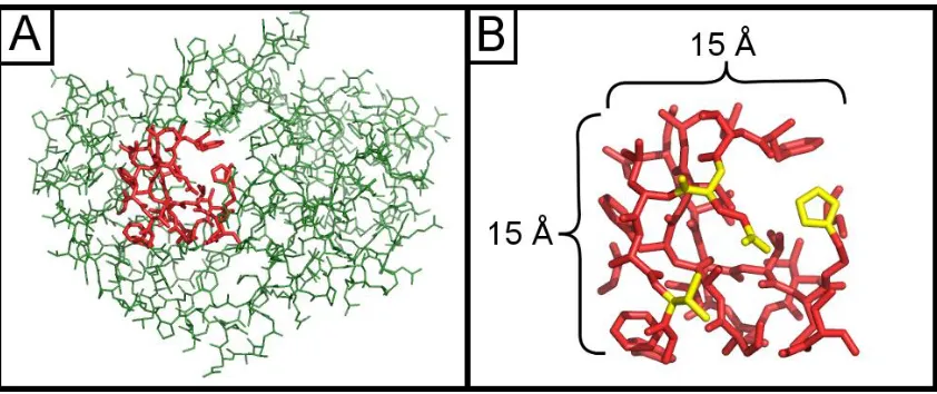
We select a compromise suitable for atomic-level search queries: we restrict structural pages to
cubes approximately 15 Å wide and we define structural words to be connected chemical
substructures ranging from 2 atoms (a hydroxyl) to 9 atoms (an indole ring) (Figure 1). Our
choice of page size assumes that larger structural patterns of interest can be reduced to a
network of bridging local interactions below the 15 Å length scale. Similarly, our choice of word
size assumes that users will accept a modest restriction on search queries to groups of chemical
motifs instead of groups of atoms. Like web search engines, we permit searches for multiple
disconnected words, allowing users to assemble complex queries from these simple chemical
13
Figure 1- Subdivision of protein structures. (A) An interior page highlighted in red from a protein of unknown function (PDB ID = 2FSQ), illustrating the maximum scale of search queries. (B) Example words (chemical motifs) within the same page highlighted in yellow. Pages are 15 Å x 15 Å x 15 Å cubes.
The forward index is a nested data structure and the outer level data structure is an array whose
elements are word indices, one for each word that suns understands:
type PrimaryIndex = Vector WordIndex
Each word index contains a list of all matches, ordered by number of matches:
type WordIndex = Vector Matches type Matches = Set PageID
The first element in the WordIndex vector consists of all pages that contain exactly one
occurrence of the given word. The second element consists of all pages that match the motif
14
Pages are grouped by number of matches so that Suns can rapidly eliminate pages that do not
have a sufficient number of matches. If the user searches for three carboxylic acids, then the
search engine can immediately skip the first two elements of the WordIndex vector for
carboxylic acids since they will be sets with fewer than three matches to carboxylic acids. Then
it folds all the remaining elements using set union to retrieve all pages with at least three
matches.
However, users can search for mixtures of diverse words, so to do this efficiently we query each
word type independently and then take the intersection of all queries. So if the user requests
two hydroxyls and two carboxylates, then the search engine will split this into two subqueries.
First, it will compute the set of all pages with at least two hydroxyls and then compute the set of
all pages with at least two carboxylates. Computing the intersection of these two sets identifies
pages with that simultaneously contain at least two hydroxyls and at least two carboxylates.
2.3.3 – Structural pages
Suns partitions protein structures spatially into pages which non-overlapping cubes
approximately 15 Å wide. Search results must fit within one of these pages, meaning that the
search engine does not return search results that span more than one page. Non-overlapping
pages were chosen for efficiency reasons, since overlapping pages would require an additional
search step to remove duplicate search results contained entirely within overlapping regions.
Protein atoms are partitioned into buckets by using a truncated Morton code [71]. First, the X,
Y, and Z coordinates are converted from floating point numbers to 21-bit integers using the
15 𝑡𝑜𝐼𝑛𝑡(𝑣) = ⌊ 2
21(𝑣 − 𝑣 𝑚𝑖𝑛)
√2(𝑣𝑚𝑎𝑥− 𝑣𝑚𝑖𝑛)
⌋
𝑣𝑚𝑎𝑥= 9999.999
𝑣𝑚𝑖𝑛= −999.999
𝑣𝑚𝑎𝑥 and 𝑣𝑚𝑖𝑛 are the upper and lower bounds, respectively, for X, Y, and Z coordinates in the
Protein Data Bank file format. 𝑡𝑜𝐼𝑛𝑡 rescales every floating point coordinate in this range to a
21 bit integer. The √2 in the denominator is a fudge factor to adjust the final page sizes to be
approximately 15 Å (the true dimension is 15.19 Å).
An atom’s X, Y, and Z coordinates are combined into a 63 bit integer using bitwise interleaving of
their binary integer representations, which corresponds to a Morton encoding of the three
coordinates. The index then assigns each atom to page by taking its Morton-encoded
coordinate (the 63 bit integer) and dropping the 33 least significant bits. The remaining 30 bits
correspond to the atom’s page ID and multiple atoms can map to the same page ID because of
truncation. Truncating the Morton encoded coordinates has the effect of portioning atoms into
cubes 15 Å wide that tile space.
2.3.4 – Structural Words
We specify structural words using PDB files, which contain the specific residue and atom types
to match. For example, one structural word consists of a single PDB file containing the Cα-Cβ-Cγ
linker of phenylalanine. When users search for the three-carbons in phenylalanine’s linker, their
16
within a phenylalanine. This allows the search index to optionally resolve motifs that are
otherwise chemically identical [15].
Structural words may also match more than one protein element, and in those cases we use
multiple PDB files to specify the structural word: one PDB file per matching chemical motif. For
example, one motif we index is a carboxylate, specified using two PDB files: one for glutamate’s
carboxylate and another for aspartate’s carboxylate. User search queries for carboxylates will
match either of these two groups.
The choice of structural words is customizable and for our public-facing server we select a
default set of substructures appropriate for general-purpose searches (Table 1). The most
important searchable substructure matches the four backbone atoms for any protein residue,
which permits geometrically exquisite backbone searches that specify all backbone atoms and
torsion angles. We partition flexible residues such as lysine and methionine into two separate
words, and also isolate important chemical moieties into their own words, such as imidazole and
guanidinium groups. Some chemical moieties are shared between residues, such as the
hydroxyl group, which matches serine, threonine, and tyrosine. However, every residue except
glycine possesses at least one unique structural word so that users can restrict searches to a
specific residue.
2.3.5 – Tokenizing Words
The search engine must tokenize protein structures into words in two separate locations. First,
the search engine must tokenizes the entire structural database into words since all search
17
every incoming search request into words before it can retrieve matching words from the
database.
Before tokenizing, Suns converts protein structures to undirected graphs, with one graph per
page in the structure. Graph nodes are atoms and vertices are bonds, so graphs are sparsely
connected since the maximum degree of any node is 4 (the maximum number of bonds per
atom). Also, since graphs are limited to atoms that fit within a single page they are small (fewer
than 100 nodes/atoms).
The tokenization algorithm is implemented as a monadic, recursive descent backtracking parser
[18], so that it can use Haskell’s do notation as syntactic sugar for assembling parsing
computations monadically. Parsers are conventionally associated with tokenizing text, but
monadic parsers in Haskell generalize to other data structures, such as chemical graphs, and
Suns takes advantage of this generalization to simplify graph tokenization code.
The monad used for parsing is a backtracking list monad enriched with state local to each
branch of the search tree [47]:
newtype ParseS a =
ParseS { unParseS :: StateT Structure [] a } deriving (Monad, Alternative)
The primitive parser takes two atom names as arguments, each of which uniquely identifies an
18
removing the matched bond from the graph and returning the indices of the nodes that
matched:
pBond :: AtomName -> AtomName -> ParseS (Int, Int)
If more than one bond matches then pBond branches one search path per potential solution. If
no bond matches then the current search path terminates and backtracks to try another
potential solution. Note that removing the matched bond does not interfere with other
branches of the backtracking search because each branch of the search maintains a separate
copy of local search state.
The higher-level pMotif function builds on top of pBond, taking a motif graph as input and
returning indices of a matching motif while simultaneously removing the matched motif from
the protein’s graph. pMotif invokes pBond as a subroutine once per bond found within the
motif graph, incrementally checking that the connectivity of each newly matched bond is
consistent with previously matched bonds:
pMotif :: Structure -> ParseS (Vector Int)
Like pBond, pMotif branches for every possible solution and backtracks if no solutions are
found.
The evalParseS function runs any parser (including bond parsers, motif parsers, or further
19
evalParseS :: ParseS a -> Structure -> [a]
This list of potential solutions is generated lazily [44], meaning that the algorithm only searches
for as many solutions as we request, performing the minimal amount of computation necessary.
Since Suns only uses the first solution the parsing algorithm usually does not explore all possible
branches and defers unnecessary evaluation.
2.3.6 – Database
Our forward index is formally a record level inverted index, meaning that it only returns matches
to pages, not to specific structural words within those pages. We supplement the forward index
with a custom in-memory database that stores two pieces of information necessary to complete
the search. First, the database stores correspondences between words in the forward index and
atoms in each structural page. Second, the database also keeps compact representations of
every structural page suitable for returning as search results.
The database is a single in memory nested data structure, where the top-level data structure is a
vector with one element per page in the data set. Each element contains the PDB ID code for
the structure the page originated from, a vector of atoms within that page, and then a nested
data structure containing all words found in that page:
20
Matches are organized by words, words are organized by incidence, and each incidence is a list
of integer indices into the page’s vector of atoms indicating which atoms that word comprises:
type Matches = Vector Words type Words = Vector Incidence type Incidence = Vector Int
When the forward index produces a matched page, the secondary index remembers which
atoms in that page correspond to the words advertised in the forward index. Sometimes the
page contains more instances of a given word than the user required, such as when the user
searches for two peptide bonds, and the page contains five. The page must try out every valid
permutation of words that match the user’s query, and the forward index minimizes the number
of permutations by prioritizing pages that most closely match or exactly match the minimum
required word count.
2.3.7 – Alignment and RMSD
Suns uses the Kabsch algorithm [48] to rapidly align each permutation to the user’s search
query. The Kabsch algorithm requires an exact atom-for-atom correspondence between the
user’s search query and a candidate motif, and Suns compiles this correspondence from
precomputed atomic correspondences for each stored motif in the custom database. After
alignment, the search engine only returns search results that match the search query within a
21
For each result below the RMSD cutoff, Suns aligns the matching page to the search query and
return the page as the search result. If a page contains multiple matches Suns aligns each match
separately and returns them as separate results. This superimposes every search result and
context on the original query for ease of visual comparison and downstream post-processing.
2.3.8 – Streaming Results
The search engine does no global ranking of results by RMSD. This means that the search
engine will immediately stream any result within the specified RMSD cutoff to the user, which
allows the user to begin visualizing results before the search has completed, improving
interactivity.
Additionally, the search protocol requires the user to specify the number of desired results up
front. While the user may request an unlimited number of results in theory, in practice the
search clients default to 100 search results, similar to how a web search engine will default to 10
search results. This allows the search engine to stop processing the request after supplying the
specified number of results, which reduces server load. Also, the search engine may also
optionally specify a search timeout to further reduce server load for users that request a large
number of search results.
2.3.9 – Data Set
The public search engine uses PISCES [99] as the non-redundant protein structure data set,
selecting a 20% sequence identity, 1.6 Å resolution, and 0.25 R-factor cutoff, which currently
22
it can index, and our stress tests on the largest PISCES data set (90% identity, 3.0 Å, 1.0 R-factor
cutoff, 24,218 chains) required 89 GB of memory or 1 GB of memory per 272 protein chains.
2.4 – Results
2.4.1 – Building Motifs
Suns lets users explore the “designable” space of protein motifs by expanding on small initial
fragments, such as building a helix N-terminal capping motif beginning from a single
guanidinium group. One might begin by searching on the guanidinium fragment from an
arginine, which recruits a cluster of nearby carboxylates forming a salt bridge with the arginine
(Figure 2A). Adding one of these carboxylates to the search query refines the motif further,
revealing a preferred rotamer for the arginine when interacting with a carboxylic acid (Figure
2B), and adding a preferred rotamer to the search query crystallizes a complete N-terminal
capping motif (Figure 2C).
23
that this salt bridge is part of an N-terminal capping motif. Search queries are represented as thick sticks and search results are shown as thin lines. Dashed lines highlight clusters in the search results, which are filtered to show the specific residue fragments of interest and neighboring water molecules within 3.0 Å as red spheres. Search parameters and fragments listed in Table 2.
The large number of close geometric matches to the final search query suggests that this is a
highly “designable” motif. Incremental searching allows users to rapidly explore and prototype
designable native-like interactions like these with very little prior knowledge in protein folding
or biophysics. Moreover, a user can discover the motif by gradually refining a specification
rather than specifying all the necessary interactions up front. This benefits people who may not
even know what designable interactions look like and simply wish to explore what options they
have available.
2.4.2 – Assembling Larger Fragments
Users can build tertiary interactions for proteins as well. To demonstrate this, we search for a
valine from glucose binding protein and grow that into three small β strands with three residues
per strand.
Beginning from an interior valine from glucose-binding protein, we seed the two adjacent β
strands with highly populated residue clusters on each side corresponding to a valine and
tyrosine (Figure 3A). To grow the three β strands in both directions, we search for pairs of
residues at a time to identify new clusters of residues within the search results that we can
insert into the sheet (Figure 3B). The PyMOL search client permits a qualitative inspection of
24
process not only provides a rough measure of residue preference, but also reveals rotameric
preference, the kind of detailed information that a sequence logo would not reveal.
Figure 3 – Building a tertiary interaction.(A) Three strands are seeded by searching on a valine, which reveals two nearby clusters of valine and tyrosine. (B) Strands are extended one residue in each direction by searching for pairs of residues (colored yellow), yielding clusters of potential inserts (colored green). (C) The final backbone fragment (green) is fed to MadCaT, which
identifies multiple compatible scaffolds. One such scaffold (PDB ID=1E54, colored light grey) possesses many exact residue/rotamer matches to the assembled fragment (blue highlights) and many close matches (yellow highlights) that differ by a related residue (threonine to serine or valine to isoleucine) or by varying the rotamer.
We repeat this process of iteratively searching for pairs of residues at a time and incorporating
clusters from the search results until we assemble a native-like fragment of a sheet where
almost every residue originates from a unique protein structure (two disconnected threonines
were inadvertently drawn from the same structure). This then provides α-carbon coordinates
that we feed into the backbone search engine MaDCaT [39], which finds suitable scaffolds to
incorporate this fragment. One MaDCaT search result greatly resembles the β sheet built using
25
complement existing coarse-grained search tools by bridging the gap between the world of
smaller atomic interactions and the world of larger secondary-structure interactions.
2.4.3 – Connecting Hot Spot Residues
Suns can also be used to find scaffolds compatible with specified residues to provide an
alternative implementation of the hotspot residue approach to design [28]. The user can select
the hotspot of interest within PyMOL, search, and find all proteins in the PDB that position the
given hot spot residues in the specified geometry.
Figure 4- Finding backbones compatible with hot spot residues. (A) A Suns search at 0.7 Å RMSD cutoff for two hotspot residues previously identified by RosettaDock [38] for a
hemagglutinin binder [28]. The majority of search results are helices that closely match the final designed protein. The search query is shown in thick green sticks, the search result matches are shown as grey carbon traces, and the designed hemagglutinin binder is shown as a purple α-carbon trace against a blue hemagglutinin surface. (B) Searching for two threonine side chains at 0.6 Å RMSD cutoff reveals two backbone clusters that can connect them, one corresponding to an α helix (green) and the other corresponding to a β sheet (yellow). The original search query is shown in thick yellow sticks.
For example, Suns recapitulates the local backbone of a designed hemagglutinin binder [28].
26
prominent cluster of α helices matching the designed protein structure, indicating that the
secondary structure of the interface could have been predicted solely from designability.
Not every hotspot search will return a single solution for the backbone. Sometimes searching
for disembodied residues will reveal multiple distinct ways to thread the backbone between
them (Figure 4B).
2.5 – Discussion
2.5.1 – User friendliness
Suns greatly improves on existing search engines in terms of ease of use. This encourages use
among a broader scientific audience, particularly people who are not programmers. Reducing
the “activation barrier” to protein search encourages users to apply protein search in novel and
previously unanticipated ways that may have never materialized had they been limited by the
availability of collaborations to computational researchers.
Ease of use also benefits non-scientists or students, who can now enjoy a private and unfiltered
learning and discovery process. Because of the low entry barrier, Suns can also be used as a
teaching tool within the classroom to present general principles of protein biophysics. An
instructor can show how electron donors cluster around the ε-nitrogen of a tryptophan, or how
water molecules form hydrogen bonds with the helical backbone on the soluble face of a helix.
After all, it is one thing to be told that a structural element is a commonly recurring motif and it
is another thing entirely to see with one’s own eyes 100 real examples of that motif from the
27 2.5.2 – Speed
The Suns search engine greatly advances the state of the art in atomic substructure search
speed. These optimizations are reusable by other search engines, such as grouping elements
into logical units so that a forward index can be employed or partitioning searchable spaces into
local volumes to prevent combinatorial explosion of atomic configurations.
This speed comes at a price: the most important optimization proved to be the coarse-graining
of atomic substructures into groups of atoms corresponding to chemical motifs. Suns differs
from the Erebus search engine by not permitting searches for arbitrary atomic configurations
and instead only allows searches for collections of motifs. This motif-based approach allows
Suns to improve the efficiency of its forward index, since motifs are more unique than atoms.
For example, the indole ring of a tryptophan ring is highly unique thanks to the rarity of
tryptophan, which allows the forward index to skip large volumes of protein structure that lack
tryptophan. On the other hand, if you view the indole ring as a nondescript bag of atoms (8
carbons and a nitrogen) then this uniqueness is lost and the forward index cannot eliminate
many structural regions.
A useful avenue for inquiry would be to combine the best features of both Suns and Erebus. It
may be possible to let the user search for atomic subsets of chemical motifs, but under the hood
the search engine supplies the entire motif to the forward index for the purpose of eliminating
potential results. If this worked, then it would combine the atomic granularity of Erebus with
28 2.5.3 – Potential Applications
Suns was originally built for protein design, but might prove useful to structural biologists. They
may be able to use Suns as a generalized PROCHECK [59] that can quickly assess if a given
structural element was modeled accurately or not.
2.5.4 – Generalizing protein search
We initially built Suns to guide the protein design process, but we are releasing it as a general
purpose search engine so that others may reuse it for applications we did not previously
anticipate.
Currently the public search engine only indexes protein structures. We also plan to add support
for ligand search queries so that Suns can be used for drug design. While this paper describes a
protein-specific application of the search engine, the underlying algorithm can be readily
generalized to ligands and other macromolecules. Such a generalized search could prove useful
for drug discovery.
2.6 – External resources
The official web site for Suns:
http://www.degradolab.org/suns/
PyMOL plugin – Master branch:
https://github.com/godotgildor/Suns
PyMOL plugin – Version referenced in manuscript:
29 Command line search tool – Master branch:
https://github.com/Gabriel439/suns-cmd
Command line search tool – Version referenced in manuscript:
https://github.com/Gabriel439/suns-cmd/commit/92c37b07b86e7e3136f732709eade5acb960adf0
Search engine – Master branch:
https://github.com/Gabriel439/suns-search
Search engine – Version referenced in manuscript:
https://github.com/Gabriel439/suns-search/commit/1100a3c12a34d1ba92f2531a4c1fdea0bb2339f5
2.7 – Acknowledgments
Brett Hannigan and William F. DeGrado are co-authors on this manuscript. Brett Hannigan
developed the PyMOL plugin for Suns. Gabriel Gonzalez built the search engine backend.
William F. DeGrado contributed to the project’s conception. All authors were involved in writing
the manuscript.
2.8 – Supporting Information
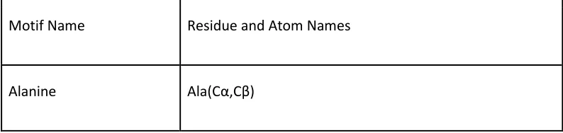
Table 1 - Default Motif Set. Default motifs indexed by the public server hosted at
suns.degradolab.org. (Motif Name): The common name for the motif. (Residue and Atom Names): The atom names used to define the motif. Some motifs may match multiple residue types, in which case all matching residues are listed with their corresponding atom names.
Motif Name Residue and Atom Names
30
Arginine Linker Arg(Cα,Cβ,Cγ,Cδ)
Asparagine Linker Asn(Cα,Cβ,Cγ)
Aspartate Linker Asp(Cα,Cβ,Cγ)
Carboxamide Asn(Cγ,Oδ,Nδ), Gln(Cδ,Oε,Nε)
Carboxyl Asp(Cγ,Oδ1,Oδ2), Glu(Cδ,Oε1,Oε2)
Cysteine Cys(Cα,Cβ,Sγ)
Glutamine Linker Gln(Cα,Cβ,Cγ,Cδ)
Glutamate Linker Glu(Cα,Cβ,Cγ,Cδ)
Guanidinium Arg(Cδ,Nε,Cζ,Nη1,Nη2)
Histidine Linker His(Cα,Cβ,Cγ)
Hydroxyl Ser(Cβ,Oγ), Thr(Cβ,Oγ), Tyr(Cζ,Oη)
Imidazole His(Cγ,Cδ,Nδ,Cε,Nε)
31
Isoleucine Ile(Cα,Cβ,Cγ1,Cγ2,δ)
Lysine End Lys(Cδ,Cε,Nζ)
Lysine Linker Lys(Cα,Cβ,Cγ,Cδ)
Methionine End Met(Cγ,Sδ,Cε)
Methionine Linker Met(Cα,Cβ,Cγ)
Peptide Bond All Residues(Cα,C,N,O)
Phenylalanine Linker Phe(Cα,Cβ,Cγ)
Phenyl Phe(Cγ,Cδ1,Cδ2,Cε1,Cε2,Cζ), Tyr(Cγ,Cδ1,Cδ2,Cε1,Cε2,Cζ)
Proline Ring Pro(Cβ,Cγ,Cδ)
Serine Linker Ser(Cα,Cβ)
Threonine Linker Thr(Cα,Cβ,Cγ)
Tryptophan Linker Trp(Cα,Cβ,Cγ)
32
Valine Val(Cα,Cβ,Cγ1,Cγ2)
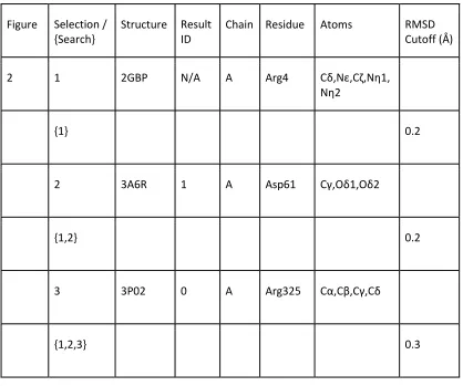
Table 2 - Search Parameters for all figures. (Figure): The figure and sub-figure the selections and searches correspond to. (Selection / {Search}): No braces indicates a saved selection referenced by searches. Braces indicate a search based in terms of previous selections of the form {sel1, sel2, …}. “sc” indicates only the side-chain was taken from the previously saved selection and “bb” indicates only the backbone atoms were used. (Structure): The PDB ID the selection originated from. (Result ID): The search result serial ID number to disambiguate selections where there are multiple results from the same PDB ID. (Chain): Chain the selection originated from. (Residue): Residue selected. (Atoms): Selected atoms. (RMSD Cutoff): Root-mean-squared deviation cutoff used for a given search. With the exception of initial selections for each figure, all selections are derived from results returned from the preceding search query in the table. †: Structure provided by the David Baker laboratory for their hot spot motif for the hemagglutinin binder [28].
Figure Selection / {Search}
Structure Result ID
Chain Residue Atoms RMSD
Cutoff (Å)
2 1 2GBP N/A A Arg4 Cδ,Nε,Cζ,Nη1,
Nη2
{1} 0.2
2 3A6R 1 A Asp61 Cγ,Oδ1,Oδ2
{1,2} 0.2
3 3P02 0 A Arg325 Cα,Cβ,Cγ,Cδ
33
3A 4 2GBP N/A A Val88 Entire Residue
{4} 0.1
5 4ASM 0 B Val353 Entire Residue
6 2WUR 0 A Tyr92 Entire Residue
3B {4bb,6bb} 0.2
7 2JCQ 1 A Thr151 Entire Residue
{4bb,7} 0.2
8 2JCQ 0 A Thr149 Entire Residue
{7sc,8bb} 0.5
9 3B34 0 A Thr37 Entire Residue
{5bb,8sc} 0.5
34
{6bb,7sc} 0.5
11 3D9A 0 H Thr482 Entire Residue
{6bb,8sc} 0.5
12 3Q1I 0 A Thr561 Entire Residue
4A 13 † N/A B Met503 Cγ,Sδ,Cε
14 † N/A B Phe504 Cγ,Cδ1,Cδ2,Cε
1,Cε2,Cζ
{13,14} 0.7
35
CHAPTER 3 – PhoQ, a histidine kinase, signals across the membrane using a scissoring mechanism
3.1 – Abstract
All organisms signal across membranes to sense and adapt to external environments. Bacteria
signal across the membrane primarily using two-component systems (TCSs), consisting of a
membrane-spanning sensor histidine kinase and a cytoplasmic response regulator. In
Salmonella enterica and other gram-negative bacteria, the PhoPQ TCS aids virulence by sensing
cations, antimicrobial peptides, and low pH, yet little is known about what structural changes
transmit the signal across the membrane. Here, we built a model of PhoQ signal transduction
using Bayesian inference, based on disulfide crosslinking data and homologous crystal
structures. We conclude that PhoQ inhabits two structurally distinct states that alternate via a
scissoring motion. These states differ in regions critical to signal transduction such as the
membrane depth of the sensor’s acidic patch and the helical packing of the dimer interface. A
comprehensive structural comparison of homologous two-component domains indicates this
scissoring transition also occurs in other TCSs, suggesting a general mechanism of signal
transduction.
3.2 – Introduction
The PhoQ sensor histidine kinase belongs to a family of two-component signal transduction
systems, which dominate signaling across prokaryotic membranes [92]. These systems respond
to diverse environmental signals, such as low pH [32], small molecules [49,60], ions [33], and
peptides [52], and regulate critical responses, such as ion transport and virulence [69]. A
36
(HK) and a cytoplasmic response regulator [65]. The periplasmic sensor responds to
environmental signals by promoting autophosphorylation of a conserved histidine, followed by
phosphotransfer to a conserved aspartate residue on its corresponding cytoplasmic response
regulator. Phosphotransfer activates the response regulator, which in turn modulates genetic
response [80].
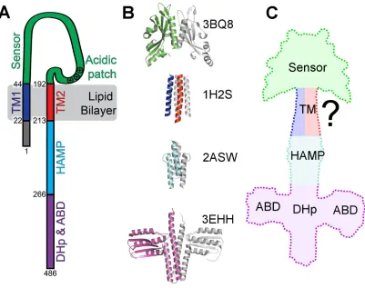
Although TCSs have been shown to be diverse [25], the topology of a canonical sensor HK
(Figure 5A) consists of a periplasmic sensing domain flanked by two transmembrane (TM)
helices, followed by one or more small domains, such as HAMP in PhoQ (named for the
domain’s presence in histidine kinases, adenylyl cyclases, methyl-accepting chemotaxis proteins,
and phosphatases) [30], and finally the kinase domain. This domain is typically known as the
dimerization and histidine phosphotransfer domain (DHp), which contains the substrate (a
conserved histidine) for autophosphorylation. The second part of this domain is a catalytic,
ATP-binding domain that mediates autophosphorylation and phosphotransfer reactions. A
functional histidine kinase is homodimeric (Figure 5B) with an extended dimer interface along
the entire length of the molecule [35]. TCSs frequently reuse these domains, so mechanistic
37
Figure 5 - Structural representations of PhoQ. (A) Schematic of the topology of a PhoQ
monomer. The numbers indicate residue numbers for E. coli PhoQ (UniProt: P23837). (B) Crystal structures used for structural comparison of each domain of PhoQ. The corresponding PDB ID is listed next to the structure. One monomer is color-coded and the other monomer is in grey. (C) A model of the first three domains of PhoQ: sensor, transmembrane (TM), and HAMP domains. The dimerization and histidine phosphotransfer domain (DHp) and ATP-binding domain (ABD) are added for clarity but were not modeled.
Structural efforts have attempted to elucidate the mechanistic details of signal transduction
spanning several domains from the periplasmic sensors to the cytoplasmic DHp domain, and
several structures have been reported. Crystal structures are now available for multiple
domains of two-component and chemotaxis systems [2,22,27,103], including a structure of the
periplasmic sensor domain of PhoQ [17]. NMR and X-ray structures have also been solved for
38
engineered, cytoplasmic two-component sensor (lacking a TM domain) was determined [22],
and the structure of the cytoplasmic region of VicK, from Streptococcus mutans, was reported
[98]. Despite these advances, there remain several competing proposals for a unified
mechanism of transmembrane signaling.
Early studies on chemotaxis systems proposed a piston-like mechanism for signal transduction
based on cysteine-scanning disulfide formation, mutagenesis, and crystallography [16]. In this
model, a transmembrane helix signals across the membrane using a rigid translation orthogonal
to the plane of the membrane [26], and later structural comparisons of the TorS TCS supported
this hypothesis [70]. However, the measured displacements in the piston model are quite small
in comparison to the length of the sensor HK protein itself or to the conformational changes
expected to power a large rearrangement of the catalytic site.
In contrast, studies on cytoplasmic signaling domains propose a gearbox model where helical
rotations within a four-helix bundle change the packing interfaces between helices [45]. Other
observed motions include inter-helical torqueing [22], helix-bending [98], or DHp domain
cracking [19]. Another study posits a combination of these models [14]. However, all proposed
mechanisms lack a crucial ingredient: structural evidence for these motions extending into the
transmembrane domain.
Critical to a membrane signal transduction model is a structural model of the TM portions of
sensor HKs. Three structures of monomeric HK transmembrane domains were recently solved
using NMR of isolated domains in micelles [66]. All three of the reported structures are limited
39
from a bona fide HK TM domain the structural starting point is not obvious. However, one
crystal structure has been solved for the dimeric TM domain of a homologous protein: the HtrII
sensory transducer [37]. A previous study utilized the HtrII X-ray structure as a model for the
transmembrane domain in HKs [37], and we have also reported similarities between the TM
domains of HtrII and PhoQ [34]. We demonstrated that the same pronounced water
hemi-channel observed in HtrII plays an important mechanistic role within PhoQ [34].
Previously, we explored local changes in the TM domain by combining molecular dynamics
simulations with disulfide crosslinking data [61]. To elucidate larger scale changes across the
membrane, we incorporate new crosslinking data in the HAMP and juxtamembrane regions of
PhoQ with previous data, and analyze it using multi-state Bayesian modeling [9,79]. This
approach provides the first investigation into the structures of the two signaling states of PhoQ,
which interconvert through a large scissoring motion. Our subsequent quantitative structural
analysis of additional TCSs also divulge large and recurring scissoring motions. Scissoring
accounts for a greater proportion of observed motions than proposed piston-shift [16] or
gearbox [24] signal transduction mechanisms.
3.3 – Results
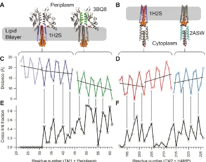
We probed the TM domain and the neighboring HAMP and periplasmic domains of PhoQ using
disulfide-scanning mutagenesis. Building upon our previous analysis of the periplasmic helix at
the dimer interface [35], new single cysteine residue mutations were introduced along the
transmembrane helices and at selected positions within the HAMP domain (Figure 6A and B).
40
formation in these mutants required the presence of an oxidative catalyst,
Cu(II)(1,10-phenanthroline)3 (CuPhen). For each residue in the predicted TM domain, we calculated the
fraction of crosslinking from the measured intensities of covalent dimer and monomer bands on
a Western blot.
Figure 6 - Comparison of the crosslinking efficiency with structural models. (A) PhoQ TM1-periplasm-TM2 model in a lipid bilayer. The color-coded helical regions (blue-green-red, respectively) indicate where cysteine mutations were made. An orange envelope marks the water hemichannel. (B) PhoQ TM1-TM2-HAMP model in a lipid bilayer. Color-coding (blue-red-cyan, respectively) is applied to the regions probed by cysteine mutations. The water
41
to each helical segment. (D) Inter-monomer distances between dimeric structures of structural models for TM2-HAMP. The second TM helix is from HtrII, and the HAMP helix is from
Archaeoglobus fulgidus (PDB ID: 2ASW). Distances and fits were done as in (C). (E) Crosslinking data from the full length PhoQ protein in a native membrane for cysteine mutants 22 through 61. (F) Crosslinking data from the full-length PhoQ protein in a native membrane for cysteine mutants 192 to 226.
3.3.1 – Comparison of disulfide crosslinking efficiency to homologous crystal structures
The crosslinking efficiency should depend inversely on the distance between the reacting thiol
groups [67], so in an initial modeling approach, we compared the measured crosslinking
efficiency for all three domains against their individual structures or homologous structures. To
model a full-length PhoQ, we mapped the periplasmic crosslinking data on the crystal structure
of the PhoQ periplasmic sensor, the transmembrane crosslinking data on the transmembrane
structure of HtrII, and the cytoplasmic crosslinking data on the HAMP structure of Af1503 from
Archaeoglobus fulgidus (Figure 5B). These comparisons test how faithfully these individual
domains represent the full-length structure of PhoQ (Figure 5C). Importantly, the crosslinking
data also adds new structural insight by spanning the intact juxtamembrane regions, which were
not present in previous single domain structures.
We compared the inter-residue distances in the transmembrane helical bundles of HtrII with the
corresponding experimental crosslinking data (Figure 6). The transmembrane four-helix bundle
of HtrII and other HKs are oriented with the N-terminus of TM1 and the C-terminus of TM2
directed towards cytoplasm. The core of the HtrII bundle is well packed near the periplasm, but
its helices kink and diverge slightly near the cytoplasm. The crosslinking fractions agree
qualitatively with this bipartite structure. Near the periplasmic end of the bundle, we observe a
42
with a period of 3.6 residues. Fitting a sinusoidal function to the data resulted in a period of 3.5
residues for TM2 and 3.7 residues for TM1 (Table 3). We computed a phase offset to determine
if there was relationship between variation in crosslinking efficiency and the expected distance
variation for an alpha helix.
Table 3 – Least-squares fitting of a sinusoidal function to the crosslinking efficiency of PhoQ and the inter-residue distances of PhoQ, HtrII and Af1503 crystal structures.
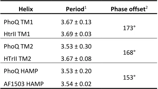
Helix Period1 Phase offset2
PhoQ TM1 3.67 ± 0.13
173°
HtrII TM1 3.69 ± 0.03
PhoQ TM2 3.53 ± 0.30
168°
HTrII TM2 3.67 ± 0.08
PhoQ HAMP 3.53 ± 0.20
153°
AF1503 HAMP 3.54 ± 0.02
1 Number of residues per repeat
2 Differences in phase for the fitted sinusoidal waves between the
experimental crosslinking data and the inter-monomer distance data (Cβ
-Cβ’distance or Cα-Cα’ for Gly) taken from corresponding crystal structure
There was little, if any, crosslinking observed in the cytoplasmic end of the bundle along the
polar cavity of PhoQ (Figure 6E and F). Thus, the low degree of crosslinking near the cytoplasmic
side of the bundle agrees with the presence of a water hemichannel, shown as solvent
accessible surface in Figure 6A and B. However, the complete lack of crosslinking on the
cytoplasmic side of PhoQ TM1 helices cannot be explained by the HtrII structure. The lack of
cross-linking suggests a larger separation in the PhoQ hemi-channel compared to that in HtrII.
At the periplasmic side of the TM bundle, we observed that the TM1 helices crosslink as strongly


![Figure 4 - Finding backbones compatible with hot spot residues. (A) A Suns search at 0.7 Å RMSD cutoff for two hotspot residues previously identified by RosettaDock [38] for a hemagglutinin binder [28]](https://thumb-us.123doks.com/thumbv2/123dok_us/9342223.1468585/37.612.108.533.292.466/finding-backbones-compatible-residues-previously-identified-rosettadock-hemagglutinin.webp)