R E S E A R C H
Open Access
Brain network involved in visual processing of
movement stimuli used in upper limb robotic
training: an fMRI study
Federico Nocchi
1,2*†, Simone Gazzellini
3,4†, Carmela Grisolia
3,4, Maurizio Petrarca
3,4, Vittorio Cannatà
1,
Paolo Cappa
4,5, Tommaso D
’
Alessio
2and Enrico Castelli
3,4Abstract
Background:The potential of robot-mediated therapy and virtual reality in neurorehabilitation is becoming of increasing importance. However, there is limited information, using neuroimaging, on the neural networks involved in training with these technologies. This study was intended to detect the brain network involved in the visual processing of movement during robotic training. The main aim was to investigate the existence of a common cerebral network able to assimilate biological (human upper limb) and non-biological (abstract object) movements, hence testing the suitability of the visual non-biological feedback provided by the InMotion2 Robot.
Methods:A visual functional Magnetic Resonance Imaging (fMRI) task was administered to 22 healthy subjects. The task required observation and retrieval of motor gestures and of the visual feedback used in robotic training. Functional activations of both biological and non-biological movements were examined to identify areas activated in both conditions, along with differential activity in upper limb vs. abstract object trials. Control of response was also tested by administering trials with congruent and incongruent reaching movements.
Results:The observation of upper limb and abstract object movements elicited similar patterns of activations according to a caudo-rostral pathway for the visual processing of movements (including specific areas of the occipital, temporal, parietal, and frontal lobes). Similarly, overlapping activations were found for the subsequent retrieval of the observed movement. Furthermore, activations of frontal cortical areas were associated with congruent trials more than with the incongruent ones.
Conclusions:This study identified the neural pathway associated with visual processing of movement stimuli used in upper limb robot-mediated training and investigated the brain’s ability to assimilate abstract object movements with human motor gestures. In both conditions, activations were elicited in cerebral areas involved in visual perception, sensory integration, recognition of movement, re-mapping on the somatosensory and motor cortex, storage in memory, and response control. Results from the congruent vs. incongruent trials revealed greater activity for the former condition than the latter in a network including cingulate cortex, right inferior and middle frontal gyrus that are involved in the go-signal and in decision control. Results on healthy subjects would suggest the appropriateness of an abstract visual feedback provided during motor training. The task contributes to highlight the potential of fMRI in improving the understanding of visual motor processes and may also be useful in detecting brain reorganisation during training.
Keywords:Neurorehabilitation, Robot-mediated therapy, fMRI, Motor processing, Upper limb rehabilitation
* Correspondence:[email protected] †Equal contributors
1
Clinical Technology Innovations Research Area, Bambino Gesù Children’s Hospital, IRCCS, Piazza S. Onofrio 4, Rome, Italy
2
Department of Applied Electronics, University Roma Tre, Via della Vasca Navale 84, Rome, Italy
Full list of author information is available at the end of the article
Background
New technologies such as robotics and virtual reality are increasingly being used alongside traditional rehabilita-tion treatments in order to assist, enhance and assess motor training. The role of these technologies within therapeutic treatment programs and their effectiveness are currently under debate [1-6]. In particular, their as-sociation with traditional therapy [7] and the differences between improvements in motor control and in activities of daily living are discussed in the literature [8]. Recent evidence suggests that the contribution of new technolo-gies to clinical practice is currently limited to providing intensive and repetitive movements and that the poten-tial of robotic devices in neurorehabilitation has not been completely investigated [9]. Furthermore, there is limited description of the neural networks involved and of the brain reorganisation related to training. In this re-gard, neuroimaging techniques represent a powerful tool. Simple and complex movement and motor imagery tasks were used to investigate motor processing in healthy and neurologically impaired subjects in experi-ments with functional Magnetic Resonance Imaging (fMRI) [10-12], Single Photon Emission Computerised Tomography [13], Positron Emission Tomography [14-16], Magnetoencephalography [17], and Direct Current potentials [18,19]. Moreover, changes have been described in neuronal motor networks as a consequence of a reha-bilitation training by means of functional and morpho-metric Magnetic Resonance techniques [20-23].
In studies with fMRI involving patients suffering from motor deficits, the selection of an appropriate task is a particularly relevant issue [24] and visual tasks may be
preferred to motor ones in order to reduce involuntary head movements during scanning. With regard to this aspect, it has been shown that the neural network acti-vated in humans following the observation of an action overlaps with that involved during the execution of the same action [25-27]. Furthermore, recent clinical trials have proved that action observation in chronic stroke patients improves functional abilities measured by clin-ical scales [28,29] and induces cortclin-ical reorganisation as revealed by enlarged fMRI activation in the bilateral ven-tral premotor cortex, bilateral superior temporal gyrus, supplementary motor area (SMA) and contralateral supramarginal gyrus [28].
However, there is an open question regarding whe-ther or not the processing of non-biological movements relies on the same structures as those involved in bio-logical motion processing. In case of light-points portray-ing human biological motion significant fMRI activations were reported, compared with non-rigid random points motion, in the lingual and fusiform gyri, superior temporal sulcus (STS), kinetic occipital area (KO, lateral occipital cortex sensitive to kinetic contours), and lateral cerebel-lum [30]. Servos et al. [31] confirmed the role of the lingual gyrus during the recognition of light-points simu-lating human biological motion. The specific involvement in the perception of biological movements of the right posterior STS has also been highlighted by previous stud-ies [32,33]. Further data support the need of observing biological plausible movements to elicit a corresponding pre-motor cortex activation [34,35]. On the contrary, other works reported that the observation of upper limb movements carried out by humanoid robotic devices may
generate the same fMRI pattern activated by the observa-tion of the human arm [36-38]. Sayginet al.[39] reported no difference in the fMRI activation pattern during obser-vation of body movements executed by humans or by robots. However, they found that the anterior part of the intraparietal sulcus (IPS) was significantly active in detect-ing the mismatch between appearance and motion, as in the case of androids with biological appearance perform-ing mechanical movements.
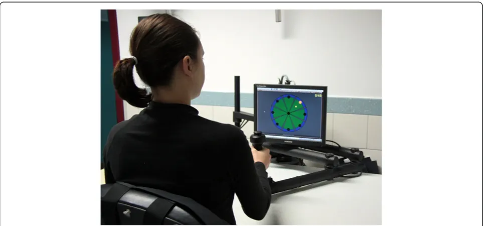
In this study, we present a visual fMRI task able to in-directly estimate, by measuring brain haemodynamics [40,41], the neuronal activation patterns related to the visual stimulation during upper limb robotic training. The task simulates the human and abstract object move-ments observed during a planar point-to-point reaching task with the InMotion2 Robot (Interactive Motion Technologies Inc., Boston, Massachusetts, USA), a de-vice that exploits both visual and haptic feedback [42] (Figure 1). Human movements are presented as an arm performing a reaching gesture similar to that carried out during robotic training, whereas abstract object movements consist in a dot moving along straight trajec-tories that represent the visual feedback provided by the InMotion2 Robot. The experimental task was designed to identify the cerebral activity associated with visual processing (observation, analysis, and representation) of movements similar to those executed during robotic training. The main aim was to verify commonalities and differences in the brain networks able to process bio-logical (human upper limb) and non-biobio-logical (abstract object) movements. A group of healthy subjects partici-pated in this study. Comparing neuronal activity before and after rehabilitative training in impaired subjects is out of the scope of the present study. However, this
analysis could be useful in testing the biological effect-iveness of the visual feedback associated with robotic training. Furthermore, this study could contribute to the current debate about the use of action observation train-ing in the treatment of stroke and cerebral palsy [43,44].
Methods
Subjects
The study was carried out on a sample of 22 healthy volunteers (8 males, age 25.6 ± 4.3 years, min = 19.2, max = 36.0). The inclusion criteria were: 18 to 39 years of age; right-handedness; no current or previous motor and neurological disorders; no current or previous re-habilitation protocols or robot-mediated training. A clin-ical evaluation was performed in each participant by a skilled physician prior to scanning. Informed consent was obtained from all subjects and the study was approved by the hospital’s Ethical Committee.
Experimental design
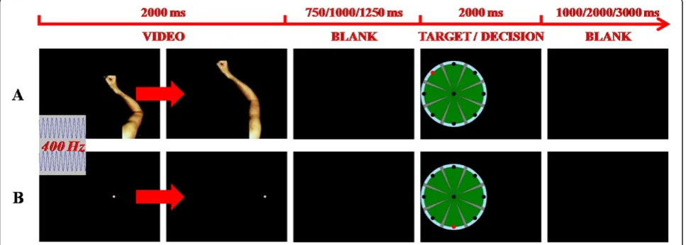
An fMRI visual task was set up. The task consisted in administering a video sequence of both human arm planar movements and straight trajectories of a dot, presented in a random order, along eight possible direc-tions starting from the centre of the screen. The motion stimuli were followed by the image of the video screen presented during the rehabilitation training with the InMotion2 Robot, with a randomly positioned target point. The subjects were required to compare the direc-tion of the observed modirec-tion stimulus (arm planar move-ment or dot trajectory) with the target position (Figure 2) and they were asked to mentally count the number of congruencies of the target position with any kind of motion stimulus. We hypothesised that, in each trial, the
subject should process the observed stimulus (arm movement or dot trajectory), retrieve the arm movement or the dot trajectory, assess the congruence between dir-ection of the stimulus and position of the target, and make a decision. According to this hypothesis, arm and dot trials only differ in the perceptual analysis of the an-thropomorphic movement, which is absent in the case of the dot trajectory presentation. The use of a visual task allowed reducing involuntary head movements during the fMRI session. With the same aim, any kind of motor response from the subjects (e.g.: pushing a response but-ton in a keypad) was avoided.
Both right and left arm movements from a healthy adult subject were video recorded during the execution of an InMotion2 training session (planar point-to-point reaching task) and adapted for the stimulation sequence. An acoustic signal was given to the subjects at the be-ginning of each trial (i.e., when an arm movement or a dot trajectory started). A time interval of random dur-ation (750, 1,000, or 1,250 ms), during which subjects saw a blank screen, followed the motion stimulus and preceded the presentation of the target. A second vari-able time interval (1,000, 2,000, or 3,000 ms) was intro-duced following the target and before the onset of the successive motion stimulus (Figure 2). Four runs were performed by each subject. Each run consisted of 54 trials with randomised presentation of both biological and non-biological stimuli and all stimuli (right arm movements, left arm movements and dot trajectories) occurring with the same frequency at each run. Taking the 4 runs together, each movement direction was pre-sented the same number of times and an equal number of congruent and incongruent trials occurred. To avoid possible ambiguities related to the interpretation of the human arm movements, the angular mismatch between the movement direction and the position of the target was greater than or equal to 90° both for arm move-ments and dot trajectories.
A 15’ practice session with the InMotion2 Robot was administered to each subject one hour before the fMRI exam. This very short session, aimed at familiarising with the task used in fMRI, was part of the instructions given to subjects prior to scanning and was followed by administering a set of trials of the fMRI task to verify their ability to perform it.
MRI acquisition
All scans were performed on the same MRI scanner (1.5 T Achieva, Philips Medical Systems, Best, the Neth-erlands) equipped with an 8-channels Sense head coil. The imaging protocol for each subject consisted of a 3D T1-weighted anatomical scan acquired as a structural reference and of 4 T2*-weighted Echo-Planar Imaging (EPI) sequences. The following parameters were used in
EPI scans: repetition time (TR) = 2,500 ms, echo time (TE) = 46 ms, flip angle = 90°, field of view (FOV) = 256 x140x256 mm, 28 axial slices (slice thickness = 5 mm, without gap), reconstruction matrix = 64x64 (pixel size = 4x4 mm), 6 dummy scans and 153 dynamic scans for each sequence (duration: 6’22”). The total scan time for the protocol was 31’45”. The stimuli sequences were implemented in the STIM2 software (Compumedics Neuroscan, El Paso, Texas, USA) and delivered by an MR-compatible stimulation system (NordicNeuroLab, Bergen, Norway).
FMRI data analysis
Analysis of fMRI data was performed with SPM8 (The Wellcome Department of Imaging Neuroscience, Univer-sity College, London, UK) running in MATLAB (version 7.9.0.529, R2009b) (The Mathworks, Natick, Massachu-setts, USA). For each subject, after motion correction, the anatomical MRI was coregistered to the mean func-tional image. Parameters for normalisation to the stand-ard Montréal Neurological Institute (MNI) space were subsequently estimated and applied to the functional images. Finally, spatial smoothing was performed by con-volving with an 8-mm full-width at half-maximum iso-tropic Gaussian kernel.
Statistical analysis was performed in a 2-stage mixed-effect procedure using the general linear model ap-proach for event-related fMRI designs. In the 1st level, individual subject analysis, 6 regressors were used in each run to model the blood oxygenation level dependent (BOLD) response for each of 6 event types. The events were defined as observation of an arm move-ment (AM), observation of a dot trajectory (DT), pre-sentation of a congruent target point following an AM (AMCT), presentation of a congruent target point following a DT (DTCT) and presentation of incong-ruent target points following AMs or DTs (AMIT and DTIT, respectively). Estimates of the 6 head movement parameters obtained from the realignment stage of pre-processing were included as additional regressors. Con-trasts between regressors were then obtained for each subject.
thresholded, binarised, and multiplied voxel-wise with each other [47]. The analysis of congruence between the observed arm movement or dot trajectory and the position of the target point implies retrieval from memory of the previously observed movement and imagery of the movement toward the target, which were referred to as “representation of arm movements” (AMT) and “representation of dot trajectories” (DTT). A second conjunction analysis was performed for these
“representations”.
Results
Subjects’performance
At the end of each fMRI run, subjects reported the num-ber of congruent trials. Errors were less than 12 % for each run in each subject. This low percentage error was assumed to be a sufficient proof of subjects’continued participation in the task. Therefore, no subject nor run was excluded from the group analysis due to low task performance.
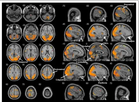
Observation of arm movements and dot trajectories When the observation of AMs was compared to the IB, a significant bilateral activation was found in the occipi-tal lobe (primary, secondary and associative visual cor-tex, Brodmann Areas (BA) 17, 18, 19), parietal lobe (superior parietal lobules, BA 5, 7; supramarginal gyrus, BA 40; angular gyrus, BA 39; postcentral gyrus, BA 3, 1, 2; precuneus, BA 7 medial; paracentral lobule, BA 3, 1, 2, 5 medial), temporal lobe (middle and inferior tem-poral gyrus, fusiform gyrus, BA 20, 21, 22, 37), several areas of the frontal lobe (precentral gyrus-primary motor area, BA 4; SMA, BA 6; medial frontal gyrus, BA 8, 9; superior frontal gyrus, BA 4, 6, 8; middle frontal gyrus, BA 9; inferior frontal gyrus, BA 44, 46, 47), limbic lobe (cingulate gyrus, BA 31; hippocampus and parahippo-campal gyrus, BA 27, 30), and insula (BA 13). Further-more, activations were found in a large portion of the cerebellum (vermis, anterior and posterior cortex) and in the thalamus.
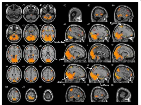
The comparison of DTs observation with the IB revealed a large pattern of activations overlapping with the above described network. Consequently, the con-junction analysis identified significant activations in all the regions mentioned above, with medial frontal gyrus activated only in the left hemisphere and a right lateral-isation of the cingulate gyrus and parahippocampal gyrus (Figure 3).
The differential analysis between AM and DT condi-tions (AM>DT) showed significant differences in oc-cipital lobe, temporal lobe and cerebellum (Table 1). Finally, the reverse contrast (DT>AM) highlighted only small clusters of voxels in the occipital and temporal regions.
Representation of arm movements and dot trajectories and congruence analysis
A network of activations was observed when the repre-sentation of arm movements (AMT) was compared to the IB. Regions with significant activations included the occipital lobe (primary, secondary and associative visual cortex, BA 17, 18, 19), the parietal lobe (superior parietal lobules, BA 5, 7; right supramarginal gyrus, BA 40; an-gular gyrus, BA 39; postcentral gyrus, BA 3, 1, 2; precu-neus, BA 7 medial; paracentral lobule, BA 3, 1, 2, 5 medial), the temporal lobe (superior, middle and inferior temporal gyrus, fusiform gyrus, BA 20, 21, 22, 37, 38), the frontal lobe (left precentral gyrus-primary motor area, BA 4; right SMA, BA 6; medial frontal gyrus, BA 8, 9; superior frontal gyrus, BA 4, 6, 8; middle frontal gyrus, BA 9; inferior frontal gyrus, BA 46, 47), the limbic lobe (hippocampus, parahippocampal gyrus, BA 27, 30), insula, cerebellum, basal ganglia (caudate, left putamen) and the thalamus.
Similar to the case of movement observation, an over-lapping pattern of activations was found when compar-ing the representation of dot trajectories (DTT) to the IB, so that all the above cited regions were present in the conjunction analysis of the two contrasts (Figure 4).
The presentation of the target following arm move-ments was compared with the presentation of the target following dot trajectories, showing significant differences in the superior parietal cortex and in the precuneus (AMT>DTT, Table 1). The reverse contrast (DTT> AMT) revealed a large cluster of voxels in the occipital lobe, temporal lobe and cerebellum, with a similar pat-tern to that obtained with the AM>DT contrast.
reverse contrast (IT>CT) no cluster survived the cor-rected threshold. However, when component contrasts were analysed, while no cluster survived a p<0.10 FWE threshold in the AMIT>AMCT contrast, the DTIT> DTCT contrast revealed significant activations in the left occipital cortex (primary, secondary and associative vis-ual cortex, BA 17, 18, 19).
Discussion
In the present study, an fMRI experiment was used to investigate the brain regions involved in a visual task adapted from the training with the InMotion2 Robot. The task required upper limb motor gesture recognition and activation of the cognitive representations of arm movements and dot trajectories, both presented on a screen. The results showed a caudo-rostral neuronal pathway. Common activations between upper limb
gestures and dot trajectories were found with regard to both movement observation and retrieval. Furthermore, the activation of areas involved in higher level cognitive functions was associated with the processing of congru-ent trials more than with the incongrucongru-ent ones.
Observation of arm movements and dot trajectories The pathway of activations found is consistent with pre-vious studies [49-53] and points out that the neural cor-relates of movement are a property of the brain that emerges from a complex cortical-subcortical network. A significant bilateral activation was elicited during percep-tion and recognipercep-tion of arm movements, depicting a caudo-rostral pathway: visual perception (primary, sec-ondary and associative visual cortex [54]), sensory inte-gration [55], subsequent recognition of arm movement [56] and attention shifting [57,58] (posterior parietal
Table 1 Differential activations between conditions
Contrast Significant clusters Brodmann areas Size
(voxels)
p T MNI coordinates
x y z
AM>DT Occipital_Mid_L, Fusiform_R, Calcarine_L, Fusiform_L, Occipital_Inf_R, Cuneus_R, Occipital_Inf_L, Occipital_Mid_R, Cuneus_L, Occipital_Sup_L, Lingual_L, Cerebelum_6_R, Occipital_Sup_R, Temporal_Mid_R, Cerebelum_6_L, Cerebelum_Crus1_L, Lingual_R, Temporal_Mid_L, Calcarine_R, Temporal_Inf_R, Cerebelum_Crus1_R, Temporal_Inf_L, Cerebelum_4_5_R, Parietal_Sup_R
19, 18, 37, 17, 39, 36, 20, 23 4008 0.000 14.65 −42 −88 4
DT>AM Temporal_Mid_R, Temporal_Sup_R, Rolandic_Oper_R, SupraMarginal_R, Postcentral_R, Heschl_R
21, 22, 42, 43, 41, 40 167 0.000 6.21 57 −16 13
Temporal_Inf_L, Temporal_Mid_L 37, 21, 22 54 0.061* 5.53 −54 −55 −11
Angular_R, Parietal_Inf_R, Occipital_Mid_R, Occipital_Sup_R, Parietal_Sup_R
7, 40, 19, 39 88 0.009 4.84 36 −64 43
AMT>DTT Precuneus_R, Precuneus_L, Parietal_Sup_R, Parietal_Sup_L
7 114 0.003 5.19 −6 −70 55
DTT>AMT Occipital_Mid_L, Fusiform_R, Fusiform_L, Occipital_Mid_R, Occipital_Inf_R, Occipital_Inf_L, Occipital_Sup_R, Lingual_L, Cerebelum_6_R, Occipital_Sup_L, Lingual_R, Cerebelum_6_L, Cerebelum_Crus1_L, Cuneus_L, Cuneus_R, Calcarine_L,
Temporal_Mid_R, Temporal_Mid_L, Temporal_Inf_R, Cerebelum_4_5_R, Cerebelum_Crus1_R
19, 18, 37, 39, 17, 7 1830 0.000 10.72 −42 −88 1
CT>IT Caudate_R, Thalamus_L, Thalamus_R — 184 0.000 6.93 12 5 16
Postcentral_L, Precentral_L 4, 3, 6 57 0.069* 6.41 −51 −10 46
Cerebelum_6_R, Fusiform_R, Lingual_R, Cerebelum_Crus1_R, Temporal_Inf_R
18, 19, 37 252 0.000 5.59 33 −58 −23
Putamen_R, Amygdala_R, Olfactory_R, ParaHippocampal_R, Rectus_R, Frontal_Sup_Orb_R
34, 25, 47 80 0.021 5.35 15 8 −23
AMCT>AMIT Frontal_Mid_R, Frontal_Inf_Tri_R 46, 10 76 0.021 6.67 45 47 4
Postcentral_L, Precentral_L 4, 3, 6 55 0.069* 6.20 −51 −10 46
Cerebelum_6_R, Fusiform_R, Cerebelum_Crus1_R, Temporal_Inf_R
37, 19 91 0.010 4.52 33 −58 −23
Cingulum_Mid_L, Supp_Motor_Area_R, Supp_Motor_Area_L, Cingulum_Mid_R
24, 6, 31 74 0.024 4.00 0 −4 64
DTCT>DTIT — — — — — — — —
IT>CT — — — — — — — —
AMIT>AMCT — — — — — — — —
DTIT>DTCT Occipital_Sup_L, Calcarine_L, Cuneus_L, Occipital_Mid_L, Lingual_L
18, 17, 19 189 0.000 7.24 −12 −99 10
cortex), spatial input processing [59] (inferior parietal cortex: supramarginal gyrus), re-mapping on the som-atosensory and motor cortex (postcentral gyrus [60], precentral gyrus [61,62] and SMA [63]), storage in mem-ory (hippocampus and parahippocampal gyrus, temporal areas [64-66] and dorsolateral pre-frontal cortex, see [67] for a review), motor control and motor learning (cerebellum [68], hippocampus and parahippocampal gyrus [69]). As far as the dorsolateral fronto-parietal activations are concerned, a similar pattern was observed when dot trajectories were presented, showing a wide overlapping of areas significantly activated during arm movement and dot trajectory processing, as revealed by a conjunction analysis. However, the larger activations in the lateral and medial occipital and temporal lobes in arm movement presentation with respect to dot trajec-tory trials, revealed by the AM>DT subtractive analysis,
may only in part be explained by the visuo-perceptual differences among the two conditions. Actually, the more intense BOLD signal from lingual and fusiform gyri during the AM condition supports the specific role of theselociin biological movement processing and con-firms previous fMRI data [30,31]. The wider cerebellar involvement in AM reveals the contribution of such a structure to motor activities (see also [30,32]). Interest-ingly, no difference was found in the motor and pre-motor areas.
Representation of arm movements and dot trajectories In each trial, following the presentation of the target, subjects were required to analyse the congruence be-tween the observed arm movement or dot trajectory and the position of the target point. This implies retrieval from memory of the observed movement and imagery of
Figure 4Functional activations for the representation of arm movements and dot trajectories.Cerebral regions involved in both the representation of arm movements and dot trajectories (orange) as revealed by conjunction analysis. The t-maps of AMT>IB and DTT>IB contrasts at the group level were thresholded (p<0.05 at cluster-level, FWE corrected), binarised and multiplied voxel-wise with each other to identify common areas of activation. Areas activated only for arm movements’representation are shown in red, while areas activated only for dot trajectories’representation are shown in yellow. Activations are superimposed on the MNI single subject T1 template. The coordinates
the movement toward the target (both for human arm motor gestures, AMTs, and dot trajectories, DTTs). Results showed activation patterns consistent with data from previous studies [13-18]. The active loci corre-sponded to those found during movement observation, with the minor differences described in the Results sec-tion. Similar to the case of movement observation, a wide overlapping of activated areas for biological and non-biological trials was also found with regard to movement retrieval and imagery. This result was again confirmed by a conjunction analysis. However, more in-tense parietal cortical activity (superior parietal lobule) was found for AMT than for DTT. Together with pre-motor and prefrontal cortex, the superior parietal lobule is part of the cerebral network for working memory [70]. In the reverse contrast (DTT>AMT), the differential activations involved occipital and temporal lobes and the cerebellum, which were the same areas that were more activated during AM than DT observation.
While there is convincing evidence about the cerebral structures involved in biological motion processing, it is still not clear whether or not the processing of non-biological movements relies on the same structures. Pre-vious studies have described the specific anatomical net-works for the perception of biological movements, compared with non-biological ones [30-33]. However, other works have reported that observing upper limb movements carried out by humanoid robotic devices may generate the same fMRI pattern activated by the observation of a human arm [36-39]. Therefore, a pos-sible explanation of the results provided by the present study would be that dot trajectories (i.e., abstract move-ments corresponding to the visual feedback in the InMotion2 Robot training) activate the same brain areas involved in the processing (recognition, retrieval and im-agery) of the human arm motor gesture. In that case, our data would support the hypothesis that the non-biological feedback is processed in a manner similar to that for the natural motor gesture. As far as the dorso-lateral fronto-parietal regions are concerned (in particu-lar motor, pre-motor and sensorimotor areas), these results would enforce the idea that a biological input is not necessarily required [36-39], whereas significant dif-ferences mainly involve the lingual and fusiform gyri [30,31].
The fact that significant areas in the DTT>AMT comparison overlapped (with smaller extension and lower intensity) with those in which activations were stronger during arm movement presentation than during dot trajectory observation (AM>DT; Table 1), would suggest that when participants were seeing the target, and hence had to make the decision on the congruity of the dot trajectory previously seen, they mentally recon-structed the motor gesture that subtends such a
trajectory. Therefore, in trials presenting dot trajectories, a more intense effort was required to reconstruct the human gesture generating the corresponding non-biological motion. On the contrary, in case of previous presentation of human arm movements, subjects did not need to actively reconstruct the gesture, since a stored trace was still available in working memory. However, a possible objection could be that such a result may also be related to the characteristics of our experimental de-sign. In the event-related task that was implemented in this study, the presentations of AM and DT trials were randomised, which could have facilitated a strategy based on the recall of the corresponding motor gesture even in case of dot trajectory presentation. In summary, from a rehabilitative point of view, we may speculate that inducing patients with hemiplegia to mentally re-construct the gestures by requiring an active analysis of the goal of the action, might increase the cerebral haemodynamic response from the network involved in movement processing, with respect to passive stimula-tion only. This argument implies that the mostimula-tion ana-lysis of inanimate objects could be more effective than just observing a gesture or requiring the immediate ac-tion repetiac-tion.
Processing of congruence
activation, as far as silent speech production is con-cerned. With the exclusion of these two areas, there is no reason to ascribe significant cortical activations to a counting process.
The role played by the knowledge of results provided by the feedback during robotic training deserves further consideration. In the fMRI task, the target point was shown after the end of movement observation and it was not superimposed on the motor gesture or dot tra-jectory. Literature on motor learning suggests that a delayed feedback (i.e., a feedback provided at the end of the action), similar to the one used in our study, increases the processing of the features of performance and promotes a more stable learning than an instan-taneous feedback, like the one provided during the InMotion2 Robot training [78]. While this paper addresses the issue of the biological or non-biological nature of the visual feedback, the different effectiveness of continuous vs. delayed feedback during robotic ther-apy remains unclear.
Conclusions
A visual fMRI task was used to identify the neural pathway associated with the visual processes involved in upper limb motor training performed with the InMotion2 Robot. This study investigated the suitability of non-biological movement presentation, with respect to human movement observation, in activating brain networks for motor processing. Results from healthy adult subjects would support the appropriateness of the visual feedback (movement of a dot) during robotic treatment, while the task does not address whether a continuous feedback is more or less efficient than a feed-back based on delayed knowledge of results. However, due to the nature of the task, this study does not take into account some relevant contributions that affect motor control such as motor learning related to the hap-tic feedback and the aspects of motor execution pro-vided by robotic training. Moreover, the dissociation between the processing of arm movements and abstract object trajectories cannot be fully discarded since its absence in the results could be due to the characteristics of our task, which, similar to what happens during robotic training, may induce the assimilation of strategies for upper limb movement and dot trajectory processing.
Abbreviations
AM: Observation of an arm movement; AMCT: Presentation of a congruent target point following an arm movement; AMIT: Presentation of an incongruent target point following an arm movement; AMT: Presentation of (congruent or incongruent) target points following arm movements (also referred to as“representation of arm movements”); BA: Brodmann area; CT: Presentation of congruent targets (following arm movements or dot trajectories); DT: Observation of a dot trajectory; DTCT: Presentation of a congruent target point following a dot trajectory; DTIT: Presentation of an incongruent target point following a dot trajectory; DTT: Presentation of (congruent or incongruent) target points following dot trajectories (also
referred to as“representation of dot trajectories”); EPI: Echo-planar imaging; fMRI: Functional magnetic resonance imaging; FWE: Family-wise error; IB: Implicit baseline; IPS: Intraparietal sulcus; IT: Presentation of incongruent targets (following arm movements or dot trajectories); KO: Kinetic occipital area; MNI: Montréal neurological institute; SMA: Supplementary motor area; SPM: Statistical parametric map; STS: Superior temporal sulcus.
Competing interests
The authors have not competing interests as defined by the BioMed Central Publishing Group, or other interests that may influence results and discussion reported in this study.
Authors’contributions
FN contributed to conception and design, test conduction, data acquisition, data analysis and writing. SG contributed to conception and design, test conduction, data acquisition, interpretation of data and writing. CG contributed to test conduction, interpretation of data and writing. MP contributed to conception and design, test conduction, interpretation of data and writing. VC, PC, TD, EC participated in design, acquisition of founding and coordination of the research. All authors read and approved the final manuscript.
Acknowledgements
The authors wish to thank Dr. Marcello Costantini for his precious suggestions and comments.
The work was supported by the Italian Ministry of Health (Grant 2010), and co-funded by the Italian 5x1000 contribution 2008.
P. Cappa acknowledges the financial support of the Italian Ministry of Education, University, and Scientific Research (Grant 2006 International University Cooperation“Robot-mediated therapy”) and the Italian Ministry of Health (Grant 2007“Pilot study on a novel typology of medical devices: robotic systems for rehabilitation and tele-rehabilitation”).
Author details
1Clinical Technology Innovations Research Area, Bambino Gesù Children’s
Hospital, IRCCS, Piazza S. Onofrio 4, Rome, Italy.2Department of Applied
Electronics, University Roma Tre, Via della Vasca Navale 84, Rome, Italy.
3
Department of Neuroscience and Neurorehabilitation, Bambino Gesù Children’s Hospital, IRCCS, Via Torre di Palidoro, Passoscuro, Rome, Italy.
4MARlab (Movement Analysis and Robotics Laboratory), Neurorehabilitation
Division of Bambino Gesù Children’s Hospital, IRCCS, Via Torre di Palidoro, Passoscuro, Rome, Italy.5Department of Mechanical and Aerospace Engineering, Sapienza University of Rome, Via Eudossiana 4, Rome, Italy.
Received: 8 August 2011 Accepted: 24 July 2012 Published: 24 July 2012
References
1. Adamovich SV, Fluet GG, Tunik E, Merians AS:Sensorimotor training in virtual reality: a review.Neuro Rehabilitation2009,25:29–44. 2. Castelli E:Robotic movement therapy in cerebral palsy.Dev Med Child
Neurol2011,53:481.
3. Henderson A, Korner-Bitensky N, Levin M:Virtual reality in stroke rehabilitation: a systematic review of its effectiveness for upper limb motor recovery.Top Stroke Rehabil2007,14:52–61.
4. Huang VS, Krakauer JW:Robotic neurorehabilitation: a computational
motor learning perspective.J Neuroeng Rehabil2009,6:5.
5. Masia L, Frascarelli F, Morasso P, Di Rosa G, Petrarca M, Castelli E, Cappa P:
Reduced short term adaptation to robot generated dynamic
environment in children affected by Cerebral Palsy.J Neuroeng Rehabil
2011,8:28.
6. Prange GB, Jannink MJ, Groothuis-Oudshoorn CG, Hermens HJ, Ijzerman MJ:
Systematic review of the effect of robot-aided therapy on recovery of the hemiparetic arm after stroke.J Rehabil Res Dev2006,43:171–184. 7. Waldner A, Tomelleri C, Hesse S:Transfer of scientific concepts to
clinical practice: recent robot-assisted training studies.Funct Neurol
2009,24:173–177.
8. Kwakkel G, Kollen BJ, Krebs HI:Effects of robot-assisted therapy on upper
limb recovery after stroke: a systematic review.Neurorehabil Neural Repair
9. Brochard S, Robertson J, Médée B, Rémy-Néris O:What’s new in new
technologies for upper extremity rehabilitation?Curr Opin Neurol2010,
23:683–687.
10. Binkofski F, Buccino G, Posse S, Seitz RJ, Rizzolatti G, Freund H:A fronto-parietal circuit for object manipulation in man: evidence from an fMRI-study.Eur J Neurosci1999,11:3276–3286.
11. Kollias SS, Alkadhi H, Jaermann T, Crelier G, Hepp-Reymond MC:
Identification of multiple nonprimary motor cortical areas with simple movements.Brain Res Rev2001,36:185–195.
12. Lotze M, Markert J, Sauseng P, Hoppe J, Plewnia C, Gerloff C:The role of multiple contralesional motor areas for complex hand movements after internal capsular lesion.J Neurosci2006,26:6096–6102.
13. Ryding E, Decety J, Sjöholm H, Stenberg G, Ingvar DH:Motor imagery activates the cerebellum regionally. A SPECT rCBF study with 99mTc-HMPAO.Brain Res Cogn Brain Res1993,1:94–9.
14. Boecker H, Ceballos-Baumann AO, Bartenstein P, Dagher A, Forster K, Haslinger B, Brooks DJ, Schwaiger M, Conrad B:A H(2)(15)O positron emission tomography study on mental imagery of movement
sequences–the effect of modulating sequence length and direction.
NeuroImage2002,17:999–1009.
15. Grèzes J, Decety J:Does visual perception of object afford action?
Evidence from a neuroimaging study.Neuropsychologia2002,40:212–222.
16. Samuel M, Ceballos-Baumann AO, Boecker H, Brooks DJ:Motor imagery in
normal subjects and Parkinson’s disease patients: an H215O PET study.
Neuroreport2001,12:821–828.
17. Lang W, Cheyne D, Höllinger P, Gerschlager W, Lindinger G:Electric and magnetic fields of the brain accompanying internal simulation of
movement.Cogn Brain Res1996,3:125–129.
18. Beisteiner R, Höllinger P, Lindinger G, Lang W, Berthoz A:Mental representations of movements. Brain potentials associated with
imagination of hand movements.Electroencephalogr Clin Neurophysiol
1995,96:183–193.
19. Hwang HJ, Kwon K, Im CH:Neurofeedback-based motor imagery training for brain-computer interface (BCI).J Neurosci Methods2009,179:150–156. 20. Cope SM, Liu XC, Verber MD, Cayo C, Rao S, Tassone JC:Upper limb
function and brain reorganization after constraint-induced movement
therapy in children with hemiplegia.Dev Neurorehabil2010,13:19–30.
21. Gauthier LV, Taub E, Perkins C, Ortmann M, Mark VW, Uswatte G:
Remodeling the brain: plastic structural brain changes produced by different motor therapies after stroke.Stroke2008,39:1520–1525. 22. Sutcliffe TL, Gaetz WC, Logan WJ, Cheyne DO, Fehlings DL:Cortical
reorganization after modified constraint-induced movement therapy in pediatric hemiplegic cerebral palsy.J Child Neurol2007,22:1281–1287. 23. Takahashi CD, Der-Yeghiaian L, Le V, Motiwala RR, Cramer SC:Robot-based
hand motor therapy after stroke.Brain2008,131:425–437.
24. Price CJ, Crinion J, Friston KJ:Design and analysis of fMRI studies with
neurologically impaired patients.J Magn Reson Imaging2006,23:816–826.
25. Buccino G, Binkofski F, Fink GR, Fadiga L, Fogassi L, Gallese V, Seitz RJ, Zilles K, Rizzolatti G, Freund HJ:Action observation activates premotor and
parietal areas in a somatotopic manner: an fMRI study.Eur J Neurosci
2001,13:400–404.
26. Jeannerod M:Neural simulation of action: a unifying mechanism for
motor cognition.NeuroImage2001,14:S103–S109.
27. Rizzolatti G, Craighero L:The mirror-neuron system.Annu Rev Neurosci 2004,27:169–192.
28. Ertelt D, Small S, Solodkin A, Dettmers C, McNamara A, Binkofski F, Buccino
G:Action observation has a positive impact on rehabilitation of motor
deficits after stroke.NeuroImage2007,36(Suppl 2):164–173. 29. Franceschini M, Agosti M, Cantagallo A, Sale P, Mancuso M, Buccino G:
Mirror neurons: action observation treatment as a tool in stroke rehabilitation.Eur J Phys Rehabil Med2010,46:517–523.
30. Vaina LM, Solomon J, Chowdhury S, Sinha P, Belliveau JW:Functional
neuroanatomy of biological motion perception in humans.Proc Natl
Acad Sci USA2001,98:11656–11661.
31. Servos P, Osu R, Santi A, Kawato M:The neural substrates of biological
motion perception: an fMRI study.Cereb Cortex2002,12:772–782.
32. Grossman E, Donnelly M, Price R, Pickens D, Morgan V, Neighbor G, Blake R:
Brain areas involved in perception of biological motion.J Cogn Neurosci
2000,12:711–720.
33. Morris JP, Pelphrey KA, McCarthy G:Perceived causality influences brain activity evoked by biological motion.Soc Neurosci2008,3:16–25.
34. Buccino G, Lui F, Canessa N, Patteri I, Lagravinese G, Benuzzi F, Porro CA, Rizzolatti G:Neural circuits involved in the recognition of actions
performed by nonconspecifics: an fMRI study.J Cogn Neurosc2004,
16:114–126.
35. Costantini M, Galati G, Ferretti A, Caulo M, Tartaro A, Romani GL, Aglioti SM:
Neural systems underlying observation of humanly impossible
movements: an FMRI study.Cereb Cortex2005,15:1761–1767.
36. Engel A, Burke M, Fiehler K, Bien S, Rosler F:How moving objects become animated: the human mirror neuron system assimilates non-biological
movement patterns.Soc Neurosci2008,3:368–387.
37. Gazzola V, Rizzolatti G, Wicker B, Keysers C:The anthropomorphic brain:
the mirror neuron system responds to human and robotic actions.Neuro
Image2007,35:1674–1684.
38. Press C, Bird G, Flach R, Heyes C:Robotic movement elicits automatic imitation.Cogn Brain Res2005,25:632–640.
39. Saygin AP, Chaminade T, Ishiguro H, Driver J, Frith C:The thing that should not be: predictive coding and the uncanny valley in perceiving human
and humanoid robot actions.Soc Cogn Affect Neurosci2011. doi:. Epub 22
Apr 2011.
40. Buckner RL:The hemodynamic inverse problem: making inferences
about neural activity from measured MRI signals.Proc Natl Acad Sci USA
2003,100:2177–2179.
41. Shibasaki H:Human brain mapping: hemodynamic response and electrophysiology.Clin Neurophysiol2008,119:731–743.
42. Krebs HI, Hogan N, Aisen ML, Volpe BT:Robot-aided neurorehabilitation.
IEEE Trans Rehabil Eng1998,6:75–87.
43. Sgandurra G, Ferrari A, Cossu G, Guzzetta A, Biagi L, Tosetti M, Fogassi L, Cioni G:Upper limb children action-observation training (UP-CAT): a
randomised controlled trial in Hemiplegic Cerebral Palsy.BMC Neurol
2011,11:80.
44. Small SL, Buccino G, Solodkin A:The mirror neuron system and treatment of stroke.Dev Psychobiol. doi:. Epub 24 Nov 2010.
45. Friston KJ, Holmes AP, Worsley KJ:How many subjects constitute a study?
NeuroImage1999,10:1–5.
46. Forman SD, Cohen JD, Fitzgerald M, Eddy WF, Mintun MA, Noll DC:
Improved assessment of significant activation in functional magnetic
resonance imaging (fMRI): use of a cluster-size threshold.Magn Reson
Med1995,33:636–647.
47. Nichols T, Brett M, Andersson J, Wagner T, Poline JB:Valid conjunction
inference with the minimum statistic.NeuroImage2005,25:653–660.
48. Tzourio-Mazoyer N, Landeau B, Papathanassiou D, Crivello F, Etard O, Delcroix N, Mazoyer B, Joliot M:Automated anatomical labeling of activations in SPM using a macroscopic anatomical parcellation of the
MNI MRI single-subject brain.NeuroImage2002,15:273–289.
49. Boyd LA, Vidoni ED, Siengsukon CF, Wessel BD:Manipulating time-to-plan alters patterns of brain activation during the Fitts’task.Exp Brain Res 2009,194:527–539.
50. Cunnington R, Windischberger C, Deecke L, Moser E:The preparation and readiness for voluntary movement: a high-field event-related fMRI study
of the Bereitschafts-BOLD response.NeuroImage2003,20:404–412.
51. Goodyear BG, Douglas EA:Decreasing task-related brain activity over repeated functional MRI scans and sessions with no change in
performance: implications for serial investigations.Exp Brain Res2009,
192:231–239.
52. Hanakawa T, Dimyan MA, Hallett M:Motor planning, imagery, and execution in the distributed motor network: a time-course study with functional MRI.Cereb Cortex2008,18:2775–2788.
53. Jäncke L, Lutz K, Koeneke S:Converging evidence of ERD/ERS and BOLD
responses in motor control research.Prog Brain Res2006,159:261–271.
54. Waberski TD, Gobbelé R, Lamberty K, Buchner H, Marshall JC, Fink GR:
Timing of visuo-spatial information processing: electrical source
imaging related to line bisection judgements.Neuropsychologia2008,
46:1201–1210.
55. Fink GR, Marshall JC, Halligan PW, Frith CD, Driver J, Frackowiak RS, Dolan RJ:The neural consequences of conflict between intention and the senses.Brain1999,122:497–512.
56. Ohgami Y, Matsuo K, Uchida N, Nakai T:An fMRI study of tool-use gestures:
body part as object and pantomime.Neuroreport2004,15:1903–1906.
58. Corbetta M, Kincade JM, Ollinger JM, McAvoy MP, Shulman GL:Voluntary orienting is dissociated from target detection in human posterior parietal cortex.Nat Neurosci2000,3:292–7.Erratum in: Nat Neurosci2000, 3:521.
59. Amorapanth PX, Widick P, Chatterjee A:The neural basis for spatial relations.J Cogn Neurosci2010,22:1739–1753.
60. Ebisch SJ, Perrucci MG, Ferretti A, Del Gratta C, Romani GL, Gallese V:The sense of touch: embodied simulation in a visuotactile mirroring
mechanism for observed animate or inanimate touch.J Cogn Neurosci
2008,20:1611–1623.
61. Binkofski F, Fink GR, Geyer S, Buccino G, Gruber O, Shah NJ, Taylor JG, Seitz RJ, Zilles K, Freund HJ:Neural activity in human primary motor cortex
areas 4a and 4p is modulated differentially by attention to action.J
Neurophysiol2002,88:514–519.
62. Naito E:Sensing limb movements in the motor cortex: how humans sense limb movement.Neuroscientist2004,10:73–82.
63. Rumiati RI, Weiss PH, Tessari A, Assmus A, Zilles K, Herzog H, Fink GR:
Common and differential neural mechanisms supporting imitation of
meaningful and meaningless actions.J Cogn Neurosci2005,17:1420–1431.
64. Powell HW, Koepp MJ, Symms MR, Boulby PA, Salek-Haddadi A, Thompson PJ, Duncan JS, Richardson MP:Material-specific lateralization of memory encoding in the medial temporal lobe: blocked versus event-related design.NeuroImage2005,27:231–239.
65. Machielsen WC, Rombouts SA, Barkhof F, Scheltens P, Witter MP:FMRI of
visual encoding: reproducibility of activation.Hum Brain Mapp2000,
9:156–164.
66. Maguire EA, Mummery CJ, Büchel C:Patterns of hippocampal-cortical
interaction dissociate temporal lobe memory subsystems.Hippocampus
2000,10:475–482.
67. Fletcher PC, Henson RN:Frontal lobes and human memory: insights from functional neuroimaging.Brain2001,124:849–881.
68. Manto M, Bower JM, Conforto AB, Delgado-García JM, da Guarda SN, Gerwig M, Habas C, Hagura N, Ivry RB, Mariën P, Molinari M, Naito E, Nowak DA, Oulad Ben Taib N, Pelisson D, Tesche CD, Tilikete C, Timmann D:
Consensus paper: roles of the cerebellum in motor control-the diversity
of ideas on cerebellar involvement in movement.Cerebellum. doi:. Epub
13 Dec 2011.
69. Albouy G, Sterpenich V, Balteau E, Vandewalle G, Desseilles M, Dang-Vu T, Darsaud A, Ruby P, Luppi PH, Degueldre C, Peigneux P, Luxen A, Maquet P:
Both the hippocampus and striatum are involved in consolidation of
motor sequence memory.Neuron2008,58:261–272.
70. Clayton EC, D’Esposito M:Functional neuroimaging of working memory.
InHandbook of Functional Neuroimaging of Cognition. 2nd edition. Edited
by Cabeza R, Kingstone A. Cambridge MA: MIT Press; 2006:269–306. 71. Chevrier AD, Noseworthy MD, Schachar R:Dissociation of response
inhibition and performance monitoring in the stop signal task using
event-related fMRI.Hum Brain Mapp2007,28:1347–1358.
72. Kemmotsu N, Villalobos ME, Gaffrey MS, Courchesne E, Müller RA:Activity and functional connectivity of inferior frontal cortex associated with response conflict.Brain Res Cogn Brain Res2005,24:335–342. 73. Kübler A, Dixon V, Garavan H:Automaticity and reestablishment of
executive control-an fMRI study.J Cogn Neurosci2006,18:1331–1342.
74. Dehaene S:The organization of brain activations in number comparison:
event-related potentials and the additive-factors methods.J Cogn
Neurosc1996,8:47–68.
75. Fias W, Lammertyn J, Reynvoet B, Dupont P, Orban GA:Parietal
representation of symbolic and nonsymbolic magnitude.J Cogn Neurosc
2003,15:47–56.
76. Nieder A, Freedman DJ, Miller EK:Representation of the quantity of visual items in the primate prefrontal cortex.Science2002,297:1708–1711. 77. Zorzi M, Di Bono MG, Fias W:Distinct representations of numerical and
non-numerical order in the human intraparietal sulcus revealed by
multivariate pattern recognition.NeuroImage2011,56:674–680.
78. Swinnen SP, Schmidt RA, Nicholson DE, Shapiro DC:Information feedback for skill acquisition: Instantaneous knowledge of results degrades learning.J Exp Psychol Learn Mem Cogn1990,16:706–716.
doi:10.1186/1743-0003-9-49
Cite this article as:Nocchiet al.:Brain network involved in visual processing of movement stimuli used in upper limb robotic training: an fMRI study.Journal of NeuroEngineering and Rehabilitation20129:49.
Submit your next manuscript to BioMed Central and take full advantage of:
• Convenient online submission
• Thorough peer review
• No space constraints or color figure charges
• Immediate publication on acceptance
• Inclusion in PubMed, CAS, Scopus and Google Scholar
• Research which is freely available for redistribution