Cases Administrated of CBCT by Dentists of Kerman: A
Questionnaire Study
Molok Torabi 1,2,3, Jahangir Haghani 1,2,3, Majid Asadi-Shekaari 4, Parviz Amini 1,2,5, Simin Esmaeli 1,2, Maryam Alsadat Hashemipour 1,2,6*
1. Kerman Dental and Oral Diseases Research Center, Kerman University of Medical Sciences, Kerman, Iran. 2. Kerman Social Determinants on Oral Health Research Center, Kerman University of Medical Sciences, Kerman, Iran. 3. Department of Oral and Maxillofacial Pathology, Dental School, Kerman University of Medical Science, Kerman, Iran. 4. Department of Basic Neuroscience, Neuroscience Research Center, Kerman University of Medical Sciences, Kerman, Iran. 5. Department of Prosthodontics, Dental School, Kerman University of Medical Science, Kerman, Iran.
6. Department of Oral Medicine, Dental School, Kerman University of Medical Science, Kerman, Iran.
* Corresponding Author: Maryam Alsadat Hashemipour, PhD
Address: Department of Oral Medicine, Dental School, Kerman University of Medical Science, Kerman, Iran. Tel: +98 (913) 2996183 Tel: +98 (341) 2118074 Fax: +98 (341) 21180713
Introduction: Cone beam computed tomography (CBCT) is a new method that is able to provide three-dimensional images in radiology and is useful for dentomaxillofacial imaging. This study aimed to investigate the cases administrated CBCT by dentists of Kerman (2015 year).
Methods: This cross-sectional study was conducted on dentists in Kerman. The data were gathered using standard research questionnaires containing 20 questions about the CBCT prescription and 10-question on diagnostic value of CBCT with intraoral radiographs and other information regarding the participants (age, sex, job, participate in the retraining of radiology, retraining courses, radiation protection). Data was analyzed by T test, regression and SPSS 18. Results: Among 182 participants, 107 (52.26%) were male and the rest were female with an average age of 37.16±8.93 years. The mean score of the questions of CBCT was 75.75±3.43, and the diagnostic value of the study was 16.17±2.15. The value of questions was statistically significant between general practitioners and specialists (P=0.01). There was no difference between men and women in the administration of CBCT. The difference between male and female dentists for diagnostic value of CBCT and other X-ray was statistically significant (P=0.029). Between continuing medical education courses, retraining radiology, radiation protection programs, CBCT and diagnostic value with other radiological statistically significant correlation was observed. Conclusion: The results of this study showed that knowledge of dentists in the administration of CBCT and its diagnostic value with other X-rays was satisfactory.
A B S T R A C T
Article info:Received: 11 Mar. 2014 Accepted: 09 Jul. 2014
Key Words:
CBC, Dentists, Administration
1. Introduction
edicated cone beam computed tomography (CBCT) scanners for the oral and maxillo-facial region were pioneered in late 1990s independently by Arai et al. [1] in Japan and Mozzo et al. [2] in Italy. Therefore, CBCT is a new technology that has been recently useful for dento-maxillofacial imaging [3].
When compared with conventional CT scanners, CBCT unit cost less and require less space. They have rapid scan time and they reduce the radiation doses [3, 4, 5]. Also, the beams rays are confined to head and neck only. Lower dos-age of X-rays and the ability to take different imdos-ages from a certain structure and also the possibility of reconstructing sagittal and coronal views, all make CBCT a convenient technology [6-8].
Common indications for CBCT in dentistry include as-sessment of the jaws for placement of implants, examina-tion of teeth and facial structures for orthodontic treatment
planning, evaluation of TMJ for osseous degenerative changes, evaluation of mandibular third molar root prox-imity to mandibular canal prior to extraction, evaluation of teeth and bone for cysts and tumors [5, 6]. The disad-vantages of CBCT are low resolution of its soft tissue and scattering beams from tooth tissue [9]. Usual indications of CBCT in dentistry are implantation, orthodontic treat-ments, assessment of temporomandibular joint, relation-ship of third mandibular molar with inferior alveolar nerve block and presence of tumors and cysts [10].
Researchers have investigated the use of digital radiologi-cal imaging in various countries. In 2000, it was estimated that 5% of dental practitioners in North America used digi-tal radiography in their practice [11]. In studies conducted in Norway, the usage rate was estimated to be 11-14% [12, 13] and in the Netherlands it was estimated as 12% [14]. Considering the significance of CBCT in dental treatments, it seems that assessment of dentists’ awareness for indica-tions of this system is important. Therefore, the aim of this study is to investigate the cases in which the CBCT was administrated by dentists of Kerman.
D
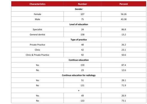
Percent Number
Characteristics
Gender
56.26 107
Female
43.38 75
Male
Level of education
86.8 24
Specialist
13.2 158
General dentist
Type of practice
26.2 48
Private Practice
23.1 42
Clinic
50.6 92
Clinic & Private Practice
Continue education
87.4 159
Yes
12.6 23
No
Continue education for radiology
28.1 51
Yes
71.9 131
No
+
26.9 49
Yes
73.1 133
No
2. Materials & Methods
This cross-sectional descriptive study was performed on 182 dentists in Kerman. First a questionnaire was designed according to previous studies [15-17]. The questionnaire comprised of: a demographic part [age, sex, years of em-ployment, educational degree], as well as general questions and questions regarding the CBCT technology. Validity of the questionnaire was evaluated by 10 specialists in Ker-man. They were asked to express their opinions on each question by the words: totally appropriate, appropriate, no idea, inappropriate and totally inappropriate.
After evaluation by the specialists, level of the questions and their comprehensibility was evaluated. Validity of the questionnaire was good. Its overall validity was 79% and the validity co-efficient for each question was between 77-89%. The reliability of the questionnaire was assessed using Cronbach’s alpha. Reliability of the questionnaire was assessed by gathering the responses provided by 15 dentists to the same questionnaire within a 15-day interval.
Cronbach’s coefficient for the reliability was 0.87, which was suitable for the study. The questionnaire was distribut-ed by a senior student. Confidentiality was guarantedistribut-ed and subjects were asked to freely express their opinions. Ques-tionnaires were designed un-identified and un-addressed and dentists were ensured that the results of this study will be used only for educational purposes of the dental soci-ety and will not be used for evaluating the dentists. T-test, Chi-square and regression test was used to analyze the data using SPSS 18.
3. Results
A total number of 182 questionnaires were completed by specialists and general dentists. 107 male (56.66%) and 75 female (43.38%) subjects with the average age of 37.16±8.93 years participated in this study. Most of the den-tists had 1-10 years of professional experience (10.92±7.89 years; range:1-40 years). The demographic characteristics of the dentists are shown in Table 1.
Percent Number
Question
3.3 6
Patients with systemic disease
58 107
Patients with malignant disease
66.5 121
Endo-perio lesions
25.3 46
Patients with xerostomia
17.6 32
Maxillofacial fractures
0.5 1
Check the patient’s sinuses
80.2 146
Root fractures
53.8 98
Temporomandibular joint pain and limited movement of jaw
32.4 59
Trauma to the face
18.1 33
Before and after maxillofacial surgery
79.1 144
The Relationship between impacted tooth with normal structures
3.3 6
Salivary gland stones
89 162
Area search implants
63.2 115
Check the type of fixture
72 131
Check diameter implant fixture
71.4 130
Check the length of the implant fixture
1.1 2
Before and after orthodontic treatment
1.6 3
Assessment of growth and development after 6 years
3.8 7
Assessment of growth and development before age 6
32.5 60
Dental anomalies
Table 2 shows the reasons for using CBCT. Most of the dentists (89%) prefer CBCT due to the implant, 80.2% for root fracture and 79.1 for maxillary sinus.
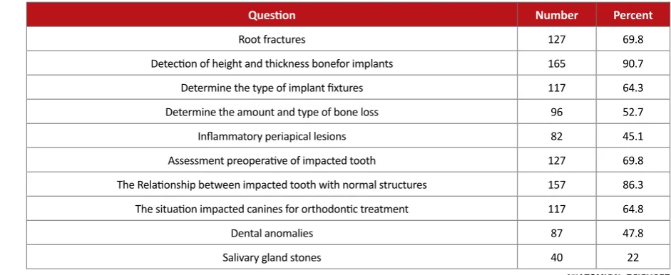
Table 3 shows the diagnostic value of CBCT. Thus, study shows that thickness of bone height for implant (90.7%) and impacted teeth (86.3%) were the most correct respons-es.
The mean score for CBCT indications was 75.76±3.41 out of 100 score (74.76±3.41 and 75.85±3.48 for specialist
and general dentist, respectively), with no significant dif-ferences between specialist and general dentist (P=0.28).
The mean score for diagnostic value of CBCT was 16.17±2.15 out of 20 score (17.02±1.52 and 15.28±2.79 for specialist and general dentist, respectively), with sig-nificant differences between specialist and general dentist (P=0.01) (Table 4).
The mean score for CBCT indication for male and fe-male was 75.90±3.28 and 75.53±3.67 respectively, with no
Percent Number
Question
69.8 127
Root fractures
90.7 165
Detection of height and thickness bonefor implants
64.3 117
Determine the type of implant fixtures
52.7 96
Determine the amount and type of bone loss
45.1 82
Inflammatory periapical lesions
69.8 127
Assessment preoperative of impacted tooth
86.3 157
The Relationship between impacted tooth with normal structures
64.8 117
The situation impacted canines for orthodontic treatment
47.8 87
Dental anomalies
22 40
Salivary gland stones
Table 3. The correct response of the study subjects towards diagnostic value of CBCT.
P value Standard deviation
Mean Variable
CBCT diagnosticvalue Sex
0.509 3.28
75.9
Male
3.67 75.53
Female
Level of education
0.218 3.41
74.79
Specialist
3.48 75.85
General dentist
Administration CBCT Sex
*0.029 2.91
15.4
Male
2.36 16
Female
Level of education
*0.014 1.52
17.6
Specialist
2.79 15.28
General dentist
*P<0.05 is significant
significant differences between this two groups (P=0.05) (Table 4).
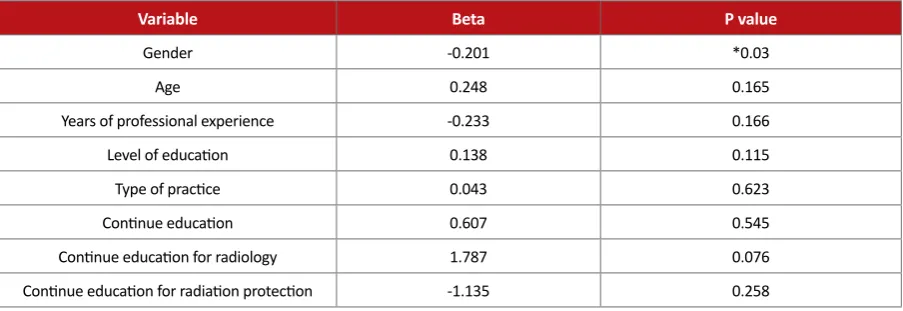
ANOVA test showed that there were statistically signifi-cant differences between dentists workplace and the mean score for CBCT indications (P=0.02) (Tables 5, 6).
Also, regression analysis showed that there were statisti-cally significant differences between the period of protec-tion from radiaprotec-tion and the mean score for CBCT indica-tions, (P=0.03) (Tables 5, 6).
4. Discussion
Cone beam computed tomography is a medical imag-ing technique consistimag-ing of X-ray computed tomography where the X-rays are divergent, forming a cone [18]. CBCT has become increasingly important in treatment planning and diagnosis in implant dentistry, interventional radiology [IR], among other things. Perhaps because of the increased access to such technology, CBCT scanners have
found many of its uses in dentistry, such as in the fields of oral surgery, endodontics and orthodontics [18, 19].
This study was done for assessment of dentists’ aware-ness for indications of cone beam computed tomography. The mean score for CBCT indications was 75.76±3.41 out of 100 score. Kamburog et al. shows that the level of knowledge in dental student about CBCT indications was poor and that these results are similar to findings by many other investigations [20, 21, 22].
Shetty et al. [17] shows that all of dentists were aware of CBCT and considered it to be a useful diagnostic tool in dentistry. Balabaskaran and Srinivasan [23] shows that 18% (n=9) of dentists are not aware of cone beam com-puted tomography used for dentomaxillofacial region.
Recent advances in cone beam computed tomography in dentistry have identified the importance of providing out-comes related to the appropriate use of this innovative tech-nology to practitioners, educators, and investigators [24].
P value Beta
Variable
0.716
-0.035 Gender
0.784 0.05
Age
0.195 0.213
Years of professional experience
0.201
-0.115 Level of education
0.929
-0.008 Type of practice
0.690 0.037
Continue education
0.114
-0.169 Continue education for radiology
0.067 0.184
Continue education for Radiation protection
Table 5. Relationship between demographic characteristics with mean score of administration CBCT.
P value Beta
Variable
*0.03
-0.201 Gender
0.165 0.248
Age
0.166
-0.233 Years of professional experience
0.115 0.138
Level of education
0.623 0.043
Type of practice
0.545 0.607
Continue education
0.076 1.787
Continue education for radiology
0.258
-1.135 Continue education for radiation protection
*P<0.05 is significant
Most of the dentists (89%) prefer CBCT due to the im-plant that is higher from research by Shetty et al. [17].
Sudhakara Reddy et al. [16] Shows that most of the den-tists preferred CBCT referrals for dental implant planning (23.6%), tumors and cyst (8.1%), endodontic treatment (4.3%), orthodontic assessment (3.1%) and impacted tooth (0.6%). Chau et al. compared typical patient radiation dos-es delivered in implant imaging with spiral CT, conven-tional spiral tomography and CBCT in their study. They reported that CBCT delivers the lowest radiation dose to the organs, whereas spiral multi slice CT delivers the high-est dose [25].
Current research indicated good knowledge of dentists about new imaging techniques [16]. Published researches show that CBCT is important in detection process and plays an important role in the management and outcome assessment [24].
Researches show that CBT is used in the treatment of dental implants, especially in linear measurement, three-dimensional topography of alveolar ridge and proximity to vital anatomical structures and surgical guide design [26[. Also, CBCT is used in implantology in a wide range of as-sessments prior to treatment, such as anatomical variation, and assessments related to complications after surgery with a focus on the harm to neurovascular structure [27].
In the present study, root fracture was the most common indications of CBCT. Vertical root fractures can be seen in 3.69 to 20% of tooth root canal therapy. Two-dimensional radiographs proved fracture only if it is on the radiation path [28]. Hassan and his colleagues demonstrated a higher accuracy of CBCT than periapical on vertical root fractures [29].
In this study, 69.8% of the dentists know that radiographs CBC have better the diagnostic value about root fracture than periapical. According to the results of this study in-formation of participating in this investigation is high. However, it should be noted that if the diagnosis cannot be reached with conventional radiography, CBCT is an effec-tive aid [30].
The most common reason for prescribing CBCT was re-lationship between impacted teeth with normal landmarks (sinus/mandibular canal). In many cases, two-dimensional radiographs aren’t able to demonstrate real connection im-pacted teeth with adjacent anatomical (sinus/mandibular canal) that this is important particularly in making deci-sions about orthodontic treatment of impacted canines.
The results of this study is compatible with Suomalainen et al. Cone-beam CT revealed the number of roots of teeth more reliably than panoramic radiographs. Also CBCT examination was highly reliable in locating the inferior alveolar canal. Suomalainen et al. recommend CBCT ex-amination for preoperative radiographic evaluation of com-plicated impacted lower third molars [31].
Also, Pecker et al. and Ishak and colleagues shows that CBCT is more useful than panoramic radiography for de-tecting multiple roots of impacted mandibular third molars [32, 33]. Pertl et al. show that OPG using steel balls as a calibration reference seems reliable in a standard situation [31]. Evidently better assessment of impacted teeth started with panoramic radiography.
45.1% of dentists believe that the diagnostic value of in-traoral radiographs is better in the diagnosis of inflammato-ry lesions than CBCT and panoramic. Although periapical are preferred to panoramic radiographs, but research has shown that the limitations of periapical radiographs may hinder the detection of periapical lesions and more roots need to be assessed, and secondly, more periapical lesions need to be detected with CBCT [34]. Also, Patel et al. shows a 14 times increase in failure rate when teeth with no pre-operative periapical radiolucencies were assessed with CBCT compared with periapical radiographs at 1year [35].
In this study, only 3.2 percent of dentists corrected their answer about CBCT indications in systemic patients. In patients with systemic disease, conventional radiographs are enough except as in cases disputed (impacted teeth near anatomic landmarks, the failure to detect suspicious canal).
In this study, 3.3% dentists were corrected their about suspected cases of salivary stones. Occlusal radiographs, panoramic and sialographic are methods appropriate in the case of salivary gland stones. CBCT is not administrated in assessing the growth and development of teeth before and after 6 years that there is few correct answers about the administration of CBCT in the evaluation before and after orthodontic treatment. This could be due to a very small percentage of dentists in orthodontic field.
The diagnostic value mean of CBCT was 15.2±17.16 of 20, which indicated a good knowledge in this study and there were significant differences between specialists and general dentists (experience, experts and visited patients who have a need to be CBCT). Also, women have been informed about the diagnostic value CBCT.
showed that dentists in Kerman city had an average level of knowledge regarding CBCT. It is recommended that qualification programs must be held for dentists to increase their awareness toward cone beam computed tomography.
References
[1] Arai Y, Tammisalo E, Iwai K, Hashimoto K, Shinoda K. Development of a compact computed tomographic appa-ratus for dental use. Dentomaxillofacial Radiology. 1999; 28(4):245-8.
[2] Mozzo P, Procacci C, Tacconi A, Martini PT, Andreis IA. A new volumetric CT machine for dental imaging based on the conebeam technique: preliminary results. European Radiology. 1998; 8(9):1558-64.
[3] Dölekoğlu S, Fişekçioğlu E, İlgüy M, İlgüy D. The usage of digital radiography and cone beam computed tomography among Turkish dentists. Dentomaxillofacial Radiology. 2011; 40(6):379-84.
[4] Guo YC, Wei L, Zhu F, Yan CX, Chen T. Development of CBCT technique and its application on dental age assess-ment. Fa Yi Xue Za Zhi. 2014; 30(4):279-81.
[5] Miles DA, Razzano MR. The future of digital imaging in dentistry. Dental Clinics North America. 2000; 44(2):427-38.
[6] Davies C, Grange S, Trevor MM. Radiation protection prac-tices and related continuing professional education in den-tal radiography: a survey of practitioners in the North-east of England. Radiography. 2005; 11(Issue 4):255-261.
[7] Pauwels R, Beinsberger J, Collaert B, Theodorakou C, Rog-ers J, Walker A, et al. Effective dose range for dental cone beam computed tomography scanners. European Journal of Radiology. 2012; 81(2):267-71
[8] American Dental Association Council on Scientific Affairs. The use of cone-beam computed tomography in dentistry: an advisory statement from the American Dental Associa-tion Council on Scientific Affairs. Journal of American Den-tal Association. 2012; 143(8):899-902.
[9] Scarfe WC, Farman AG. What is cone-beam CT and how does it work? Dental Clinics North America. 2008; 52(4):707-30.
[10] White SC. Cone-beam imaging in dentistry. Health Physics Journal. 2008; 95:628-37.
[11] Miles DA, Razzano MR. The future of digital imaging in dentistry. Dental Clinics North America. 2000; 44(2):427-38.
[12] Wenzel A, Møystad A. Decision criteria and characteris-tics of Norwegian general dental practitioners selecting digital radiography. Dentomaxillofacial Radiology. 2001; 30(4):197-202.
[13] Wenzel A, Møystad A. Experience of Norwegian general dental practitioners with solid state and storage phosphor detectors. Dentomaxillofacial Radiology. 2001; 30(4):203-8.
[14] Berkhout WE, Sanderink GC, Van der Stelt PF. A compari-son of digital and film radiography in Dutch dental prac-tices assessed by questionnaire. Dentomaxillofacial Radiol-ogy. 2002; 31(2):93-9.
[15] Tofangchiha M, Faraz Arianfar F, Bakhshi M, Khorasani M. The assessment of dentists’ knowledge regarding indi-cations of cone beam computed tomography in Qazvin, Iran. Biotechnology and Health Sciences. 2015; 2(1):e25815.
[16] Reddy RS, Kiran CS, Ramesh T, Kumar BN, Naik RM, Ramya K . Knowledge and attitude of dental fraternity to-wards cone beam computed tomography in south India: A questionnaire study. Indian Journal of Dentistry. 2013; 4(2):88-94.
[17] Shetty SR, Castelino RL, Babu SG, Laxmana AR, Roopashri K. Knowledge and attitude of dentists towards cone beam computed tomography in Mangalore – A questionnaire survey. Austin Journal of Radiology. 2015; 2(2):1-5.
[18] Scarfe WC, Farman AG, Sukovic P. Clinical applications of cone-beam computed tomography in dental practice. Jour-nal of the Canadian Dental Association. 2006; 72(1):75–80.
[19] Hatcher DC. Operational principles for cone-beam com-puted tomography. Journal of American Dental Associa-tion. 2010; 141(Suppl 3):3S-6S.
[20] Ito K, Gomi Y, Sato S, Arai Y, Shinoda K. Clinical appli-cation of a new compact CT system to assess 3-D images for the preoperative treatment planning of implants in the posterior mandible A case report. Clinical Oral Implants Research. 2001; 12(5):539-42.
[21] Mahdi-Zadeh M, Fazaeli-Pour M, Namdari A. [Evaluation of den¬tists’ awareness of how to prescribe correct radio-graphs in Isfahan in 2010-2011 (Persian)]. Journal of Isfahan Dental School. 2012; 7:637-42.
[22] Ezoddini-Ardakani F, Sarayesh V. Knowledge of correct prescription of radiographs among dentists in Yazd, Iran. Journal of Dental Research, Dental Clinics, Dental Pros-pects. 2008; 2(3):95-8.
[23] Balabaskaran K, Arathy Srinivasan L. Awareness and at-titude among dental professional towards CBCT. IOSR Journal of Dental and Medical Sciences. 2013; 10(5):55-59.
[24] Adibi S, Zhang W, Servos T, O’Neill PN. Cone beam com-puted tomography in dentistry: what dental educators and learners should know. Journal of Dental Education. 2012; 76(11):1437-42.
[25] Chau ACM, Fung K. Comparison of radiation dose for implant imaging using conventional spiral tomography, computed tomography, and cone-beam computed tomog-raphy. Oral Surgery, Oral Medicine, Oral Pathology, Oral Radiology, and Endodontology. 2009; 107(4):559-65.
[26] Benavides E1, Rios HF, Ganz SD, An CH, Resnik R, Rear-don GT, et al. Use of cone beam computed tomography in implant dentistry: the International Congress of Oral Im-plantologists consensus report. Implant Dentistry. 2012; 21(20):78-86.
[28] Ezzodini-Ardakani F, Razavi SH, Tabrizi-Zadeh M. Di-agnostic value of cone-beam computed tomography and periapical radiography in detection of vertical root fracture. Iranian Endodontic Journal. 2015; 10(2):122-6.
[29] Hassan B, Metska ME, Ozok AR, van der Stelt P, Wesselink PR. Detection of vertical root fractures in endodontically treated teeth by a cone beam computed tomography scan. Journal of Endodontics. 2009; 35(5):719-22.
[30] Wang P, He W, Sun H, Lu Q, Ni L. Evaluation of horizon-tal/oblique root fractures in the palatal roots of maxillary first molars using cone-beam computed tomography: a re-port of three cases. Dental Traumatology. 2011; 27(6):464-7.
[31] Suomalainen A, Ventä I, Mattila M, Turtola L, Vehmas T, Peltola JS. Reliability of CBCT and other radiographic methods in preoperative evaluation of lower third molars. Oral Surgery, Oral Medicine, Oral Pathology, Oral Radiol-ogy, and Endodontology. 2010; 109(2):276-84.
[32] Peker I, Sarikir C, Alkurt MT, Zor ZF. Panoramic radiog-raphy and cone-beam computed tomogradiog-raphy findings in preoperative examination of impacted mandibular third molars. BMC Oral Health. 2014; 14:71.
[33] Ishak MH, Zhun OC, Shaari R, Rahman SA, Hasan MN, Alam MK. Panoramic radiography in evaluating the rela-tionship of mandibular canal and impacted third molars in comparison with cone-beam computed tomography. My-mensingh Medical Journal. 2014; 23(4):781-6.
[34] Patel S, Wilson R, Dawood A, Mannocci F. The detection of periapical pathos is using periapical radiography and cone beam computed tomography – part 1: pre-operative status. International Endodontic Journal. 2012; 45(8):702-10.