INTRODUCTION
We are living in an Electromagnetic world. Modern lifestyle is so technology driven that for every other need we are dependent on electrical appliances such as televisions, computers, microwave ovens, mobile telephony and many other devices. These devices run with the help of supply frequency of 50/60 Hz, which emits electromagnetic fields of few orders of millitesla. There have been reports in the literature that this ELF EMF affects the various biochemical processes. Various surveys1-3 and epidemiological studies4-5, have been carried out to find the effects of these low frequency electromagnetic fields. Several studies have been carried out to investigate the effects on DNA6-7 enzyme activity8-13and cells14-15. Enzymes play a vital role in the biological processes; also cell communication is facilitated by these biocatalysts. Any alteration in the activity of the enzyme may affect these biological processes. The α-amylases
Material Science Research India Vol. 5(1), 113-118 (2008)
Effect of 50 Hz electromagnetic fields
on alpha amylase activity
K.S. PRASHANTH¹, T.R.S. CHOUHAN² and SNEHALATHA NADIGER³
¹Lecturer, Department of Physics New Horizon college of Engineering, Outer Ring Road, Panathur post Bangalore - 560 087 (India)
²Deccan institute of advanced studies, #17 first Cross, First main Domlur Bangalore - 560 072 (India)
³HOD, Department of Biotechnology, New Horizon College of Engineering, Outer Ring Road, Panathur post Bangalore - 560 087 (India)
(Received: April 06, 2008; Accepted: June 10, 2008) ABSTRACT
The effect of extremely low frequency (ELF) electromagnetic field (EMF) (50 Hz 0.5 mT) on the activity of alpha amylase (EC 3.2.1.1) was studied. In addition factors effecting the enzyme activity, temperature, pH, substrate concentration were also investigated. Our results show there is an increase in the enzyme activity when it is exposed to ELF EMF and also the velocity of the reaction increases. But there is no significant effect on optimal temperature and pH under the presence of the electromagnetic field.
Key words: ELF EMF, alpha amylase, enzyme activity.
are calcium metalloenzymes, completely unable to function in the absence of calcium. By acting at random locations along the starch chain, α-amylase breaks down long-chain carbohydrates, ultimately yielding maltotriose and maltose from amylose, or maltose, glucose and “limit dextrin” from amylopectin. Because it can act anywhere on the substrate, α-amylase tends to be faster acting than
β-amylase. In animals, it is a major digestive enzyme.In this paper we have chosen to study the effect of ELF EMF on the activity of alpha amylase enzyme.
MATERIAL AND METHODS
10 cm. The system arrangement generated a unifor m magnetic field of 0.53 mT ac r ms. The distance between the two coils was 7 cm. At the centre of the arrangement a shelf was placed to hold the samples to be exposed. The signal was provided using step down ac transformer 6 V, 50 Hz duty cycle and the field intensity 0.53 mT rms measured by a gauss meter GMO5 Figure 1(Hirst
magnetic instr uments UK, range 0 mT-3 T, frequency 15 Hz to 10 KHz and with an accuracy of ± 1% ) the gauss meter was connected to a computer (laptop) with a RS 232 interface and using Microsoft Visual Basic software programming tool the real time data was captured and stored in the system.
Fig. 1a: Laptop Fig. 1b: Gauss meter
The magnetic field of 0.53 mT was provided within Helmholtz coils supplied by electric generator able to deliver 50 Hz electric current. A PVC test tube stand to hold the samples was positioned in the middle of the Helmholtz coils. This arrangement allowed an uniform magnetic field of ac rms 0.53 mT. The test tubes containing the reaction mixture were exposed for 20 min. Constant temperature of 370C was maintained. Controls were run with no EMF.
Measurement of enzyme activity
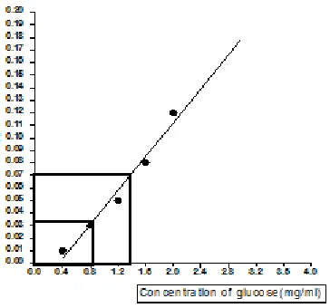
α amylase EC (3.2.1.1) {(Loba Chemie pvt Ltd Diastase (fungal) www.lobachemie.com)} catalysed endohydrolysis of 1,4-alpha-D-glucosidic linkages in oligosaccharides and polysaccharides, acts on starch and hydrolyzes it into glucose, which reacts with 3,5 dinitroaminosalicylic acid to produce a red coloured complex of nitroaminosalicylic acid. The intensity of the colour is proportional to the activity of the enzyme, which is measured spectrophotometrically at 520 nm, 1.5 ml of 0.1M phosphate buffer (pH 6.8), 0.5 ml of starch was added in test tubes labeled as test, control and blank. 1.0 ml of the enzyme was added to the test tube labeled as test and incubated at room temperature for 5 minutes. 1.0 ml of distilled water was added to all tubes; mixed thoroughly and incubated at 37°C for 20 min. 1.0 ml of enzyme was added to the test tube labeled control after the incubation period. The reaction was stopped by adding 1 ml of DNS (dinitrosalicylic acid) reagent to all the tubes. The test tubes were then mixed and allowed to stand for 5 minutes and filtered. Test tubes are heated in a boiling water bath for 15 min. The contents were cooled and absorbance was read at 520 nm. Standard graph was plotted using different concentrations of glucose solution versus OD (optical density at 520 nm) [16]
Effect of substrate concentration
Different concentrations of Starch (400 mg/ 100 ml) were pipetted out into test tubes and volume was made upto 2 ml with distilled water. 2.0 ml of phosphate buffer (pH 6.8) was added to each of the test tubes and placed in an incubator at 37°C for 20 minutes. 0.6 ml of enzyme solution was added to all the test tubes .The reaction was stopped by adding 1.0 ml of DNS reagent and absorbance was
read at 520 nm. The reaction mixture was exposed to EMF and controls were run with no EMF. Effect of pH
To study the effect of pH on enzyme alpha amylase under EMF the following reaction was set up. 0.5 ml of the substrate and 0.5 ml of enzyme solution was added to phosphate buffer of different pH. The test tubes were incubated at 37°C for 20 minutes and reaction was stopped by adding 0.5 ml of DNS to all the tubes .The absorbance was measured at 520 nm. Controls were run with no EMF. Effect of temperature
The reaction mixture containing 0.5 ml of phosphate buffer (pH 6.8), 0.25 ml of substrate and 0.25 ml of enzyme alpha amylase was subjected to different temperature and incubated for 20 minutes. The reaction was stopped by adding 1.0 ml of DNS reagent and absorbance was read at 520 nm. The reaction mixture was exposed to EMF and controls were run with no EMF.
Statistical analysis
Statistical analysis was done using Graph pad prism version 5.0.Two tailed paired t-tests were applied to compare the enzyme activities which were exposed to electromagnetic fields and the control samples which were not exposed to electromagnetic fields.
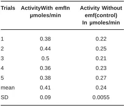
Table 1: Amylase activity after 20 minutes P value=0.0051 (paired t-test)
Trials ActivityWith emfIn Activity Without µmoles/min emf(control)
In µmoles/min
1 0.38 0.22
2 0.44 0.25
3 0.5 0.21
4 0.36 0.23
5 0.38 0.27
mean 0.41 0.24
SD 0.09 0.0055
RESULTS
Phosphate buffer solutions of 0.1 M, alpha amylase with the concentration of 1 g/100ml and the Starch solution of 100 mg/ml were exposed to the field (50 Hz, 0.5 mT). After exposure, the activity of the enzyme solutions was measured according to the methods explained above; parallel control experiments were performed with samples of enzyme solutions not exposed to the electromagnetic field. The result obtained for the measurement of enzyme activity is represented in Table 1. In the experiment, the reaction mixture
containing the enzyme and the substrate was kept in the field for 20 minutes, and enzymatic activity was measured for 20 minutes in the presence of the field with the help of the standard graph (Fig. 2). Control samples were run in the same experimental conditions as above but in the absence of the field. The enzymatic activity exposed to the field showed a two fold increase with SD 10%. The activity of the enzyme was measured in function of the concentration of their substrates and the resulting Michaelis-Menten kinetics showed both Vmax and Km was affected by the field exposure. Fig. 3 and 4 represents a Lineweavear Berk plot
Fig. 2: Standard graph of optical density as a function of concentration of glucose
Fig. 3: Lineweavear-Berk plot with EMF (p=0.0001)
Fig. 4: Lineweavear -Berk plot without EMF (p=0.0006)
with control and exposed samples, which indicates there is an increase in the velocity of the reactionVmax and Km values when exposed to the field. Fig. 5 and 6 represents the effect of pH on enzyme activity
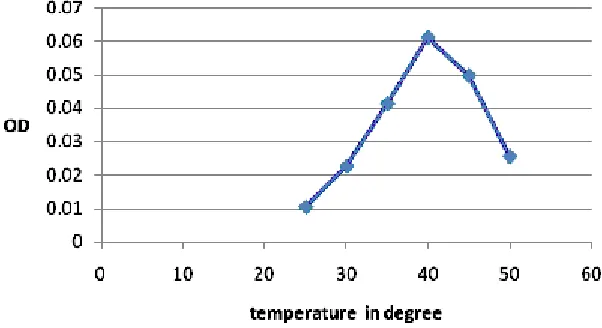
and Figures 7and 8 represents the effect of temperature on enzyme activity. It was evident from the graph that the optimal pH is 6.8 and optimal temperature is 40°C.
Fig. 6: Effect of pH without EMF, OD as function of pH
Fig. 7: Effect of temperature without EMF, OD as a function of temperature
Fig. 8: Effect of temperature with EMF, OD as function of temperature
DISCUSSION
The data reported in this paper show that ELF EMFs of 50 Hz, 0.5 mT produces an increase of 50 % of the enzymatic activity. The enzymes were exposed to the EMF for 20 minutes at 37 0C at pH 6.8 placed at the center of the Helmholtz coils. The action of this field seems to switch the enzyme to a state of an increase in activity. Optimal pH and temperature are very essential for activity of enzymes. Changes in pH and temperature may not only affect the shape of an enzyme but may also change the shape or charge properties of the substrate so that either the substrate cannot bind to the active site or it cannot undergo catalysis. We inspected whether elf emf substantially altered the optimal pH and optimal temperature. However there was a change in OD values when the samples were exposed to EMF at different pH and temperature which indicates there was alteration in the enzyme activity. But there was no significant effect of ELF EMF on optimal pH and temperature.
The ELF EMF had no effect on the activities of either integral membrane enzymes such as Ca ATPase, Na/K ATPase and Succinic dehydrogenase or per ipheral membranes8. A significant increase in lactate dehydrogenase enzyme activity was demonstrated in serum and liver17, as well as a significant elevation of gamma-glutamyltransferase enzyme activity in the liver. The glutathione S-transferase enzyme activity and lipid peroxidation level in the liver were significantly increased while a significant decrease in hepatic glutathione content was recorded. The findings indicate that there is a association between the exposure to extremely low frequency electromagnetic fields and the oxidative stress through distressing redox balance leading to physiological disturbances. Therefore we speculate that field of 50 Hz which alters the enzyme activity may have cascade effect in the biochemical processes.
REFERENCES
1. J. Brix, H. Wettemann, O. Scheel, F. Feiner, and R. Matthes. J. Bioelectromagnetics., 22: 323-332 (2001).
2. Michael A. Kelsh, T. Dan Bracken, Jack D. Sahl, Mona Shum, and Kristie L. Ebi. J. Bioelectromagnetics., 24: 316-326 (2003).
3. S.I. Henderson and M.J.Bangay. J. Bioelectromagnetics., 27: 73-76 (2006). 4. J. Szabo, K.Mezei, G.Thuro Czy, and G.
Mezei. J. Bioelectromagnetics., 27: 451-457 (2006).
5. W.T. Kaune, T.D.Bracken, R.S. Senior, R.F. Rankin, J.C. Niple, and R. Kavet. J. Bioelectromagnetics., 21: 197-213 (2000). 6. Nikolai. K. Chemeris et al. J. Mutation
Research., 558: 27-34 (2004).
7. Sabine Ivancsits, Elisabeth Diem, Alexander Pilger, Hugo W. Rüdiger, Oswald Jahn. J. Mutation Research., 519: 1-13 (2002). 8. A. Morelli et al.Arch. of Biochemistry and
Biophysics., 441,191-198 (2005).
9. A. Manoliu et al. J. Magnetism and Magnetic
Materials., 300: e323–e326 (2006). 10. J.M. Farrell et al. J. Bioelectrochemistry and
Bioenergetics., 43: 91-96 (1997).
11. M. Blank, L. Soo, V. Papstein. J. Bioelectrochemistry and Bioenergetics., 38: 267-273 (1995).
12. M. Blank, L. Soo. J.Bioelectrochemistry and Bioenergetics., 45: 253-259 (1998). 13. Martin Blank. J. Bioelectromagnetics., 26:
677-683 (2005).
14. Kyle Chang, Walter Hong-Shong Chang, Sherry Huang, Smile Huang, Chung Shih. J. Orthopaedic Research., 23: 1308-1314 (2005).
15. Ming Jin , Hana Lin , Li Han , Mark Opler , Stuar t Maurer ,Mar tin Blank , Reba Goodman. J. Bioelectrochemistry and Bioenergetics., 44: 111 -120(1997). 16. Peter Bernfield In., Methods of Enzymology,
Academic Press, New York, (1955). 17. Torres-Duran PV, Ferreira-Hermosillo A,