R E S E A R C H
Open Access
Oxidative stress and redox state-regulating
enzymes have prognostic relevance in diffuse
large B-cell lymphoma
Pekka Peroja
1, Anna Kaisa Pasanen
1, Kirsi-Maria Haapasaari
2, Esa Jantunen
3, Ylermi Soini
4,
Taina Turpeenniemi-Hujanen
1, Risto Bloigu
5, Laura Lilja
1, Outi Kuittinen
1†and Peeter Karihtala
1*†Abstract
Background:Oxidative stress and redox-regulating enzymes may have roles both in lymphomagenesis and
resistance to lymphoma therapy. Previous studies from the pre-rituximab era suggest that antioxidant enzyme expression is related to prognosis in diffuse large B-cell lymphoma (DLBCL), although these results cannot be extrapolated to patient populations undergoing modern treatment modalities. In this study we assessed expression of the oxidative stress markers 8-hydroxydeoxyguanosine (8-OHdG) and nitrotyrosine and the antioxidant enzymes thioredoxin (Trx), manganese superoxide dismutase (MnSOD) and glutamate-cysteine ligase (GCL) via
immunohistochemistry in 106 patients with DLBCL. All patients were treated with CHOP-like therapy combined with rituximab. Immunostaining results were correlated with progression-free survival, disease-specific survival and traditional prognostic factors of DLBCL.
Results:Strong 8-OHdG immunostaining intensity was associated with extranodal involvement (p = 0.00002), a high International Prognostic Index (p = 0.002) and strong Trx (p = 0.011) and GCL (p = 0.0003) expression. Strong Trx staining intensity was associated with poor progression-free survival (p = 0.046) and poor disease-specific survival (p = 0.015). Strong GCL immunostaining intensity predicted poor progression-free survival (p = 0.049). Patients with either strong Trx or strong nitrotyrosine expression showed significantly poorer progression-free survival (p = 0.003) and disease-specific survival (p = 0.031) compared with the other patients.
Conclusions:The redox state-regulating enzymes GCL and Trx are promising markers in the evaluation of DLBCL prognosis in the era of modern immunochemotherapy.
Keywords:Antioxidant enzyme, Nitrotyrosine, Prognosis, Reactive oxygen species, Thioredoxin
Background
Of lymphomas, diffuse large B-cell lymphoma (DLBCL) accounts for 30% to 40%. DLBCL comprises three differ-ent subtypes as determined by gene expression profiling: the germinal center (GC) type, the activated B-cell type and a third heterogeneous type that cannot be included in either of the two other categories [1]. In routine prac-tice the subtypes are classified by means of by immuno-histochemical grouping (Hans’ algorithm) into GC and non-GC subtypes [2]. Standard treatment for DLBCL
consists of CHOP (cyclophosphamide, doxorubicin, vin-cristine and prednisone) or CHOP-type regimens and rituximab. Rituximab has revolutionized the treatment of DLBCL by increasing survival in all International Prognostic Index (IPI) classes [3]. New molecular mar-kers predicting the outcome of DLBCL are still impor-tant in order to optimize treatment.
Production of reactive oxygen species (ROS) is com-mon to all aerobic cells, but excessive oxidative stress may lead to variable pathological conditions. The most important factor in the redox environment is the creation and elimination of ROS. These are important cellular mediators and their formation is strictly controlled under physiological circumstances. They are mostly formed in * Correspondence: [email protected]
†Contributed equally 1
Department of Oncology and Radiotherapy, University of Oulu and Oulu University Hospital, Oulu, Finland
Full list of author information is available at the end of the article
the mitochondria as a by-product of oxygen metabolism. The ROS generated in mitochondria plus, for example, hydrogen peroxide and superoxide, can affect cellular sig-naling pathways by activating sigsig-naling cascades or redox-sensitive transcriptional factors. In many patholo-gical conditions, e.g. atherosclerosis, diabetes and cancer, ROS creation and elimination is out of control, leading to excessive amounts [4-8]. Antioxidants are important in the elimination of ROS, thus maintaining the normal physiological state [9].
Redox proteins can be used in the evaluation of oxida-tive stress. Manganese superoxide dismutase (MnSOD) is a mitochondrial enzyme and it is the most important superoxide dismutase in physiological conditions. Nor-mally, MnSOD makes ROS less harmful by reducing superoxide anions to hydrogen peroxide, which is then neutralized to water and oxygen [10]. Thioredoxin (Trx) functions by reducing oxidized proteins by way of cysteine thiol-disulfide exchange and therefore it limits the damage caused by oxidative stress [11]. Gamma cysteine ligase (GCL) is a rate-limiting enzyme in the creation of glutathione (GSH). Glutathione reduces disul-fide bonds and thus repairs oxidative damage by serving as an electron donor [12]. 8-Hydroxydeoxyguanosine (8-OHdG) and nitrotyrosine are markers of oxidative damage. 8-OHdG is an end-product of oxidative damage to DNA [13]. Nitrotyrosine in its free form and in pro-teins marks oxidative damage to protein structures [14]. Nitrotyrosine can also by itself cause DNA damage [15] and bring about apoptosis [16].
An oxidatively balanced redox status may affect carcino-genesis by modulating DNA in particular, but also other cellular structures [10]. On the other hand, oxidative stress mediates the effects of many cytostatic drugs by causing sublethal DNA damage and thus activating apoptosis. Oxi-dative stress markers are prognostically important, e.g. in breast cancer [17,18], ovarian carcinoma [19], head and neck squamous cell carcinoma [20] and acute myeloid leu-kemia [21]. The prognostic role of oxidative stress in DLBCL has been evaluated in two previous studies in the pre-rituximab era, with conflicting results. The present study was undertaken to evaluate the role of oxidative stress and counteracting enzymes in more detail in DLBCL patients treated with modern immunochemotherapy.
Results
Strong cytoplasmic 8-OHdG staining intensity was asso-ciated with extranodal involvement (p = 0.00002) and a high IPI score (divided into two groups, 0-2 and 3-5) (p = 0.002). There were no other significant associations between clinical prognostic parameters and staining intensities of the studied markers. The staining intensities of Trx, GCL and nitrotyrosine correlated with the per-centage of malignant positive cells. Intensities were
discovered to be better suited to assess survival estimates. Staining patterns were discovered and analyzed in our previous study [22]. The staining intensity of cytoplasmic 8-OHdG correlated with high Trx (p = 0.011) and GCL (p = 0.0003) intensities. There were no other associations between the studied oxidative stress markers and antioxi-dant enzymes.
Overall 5-year progression-free survival was 81.4% and overall 5-year DLBCL-specific survival was 87.6%. Strong Trx staining intensity was associated with poor progres-sion-free survival (p = 0.046) and poor disease-specific sur-vival (p = 0.015) (Figure 1). The rate of 5-year progression-free survival was 85.1% in patients with negative to moder-ate Trx intensity and 57.4% in those with strong Trx intensity. Patients with strong cytoplasmic nitrotyrosine immunostaining had poor progression-free survival (p = 0.006; 5-year survival 85.8% in patients with negative to moderate staining and 55.7% in those with strong positiv-ity). Moderate to strong GCL staining intensity was asso-ciated with worse prognosis, 5-year progression-free survival being 87.0% versus 70.8% (p = 0.049). Likewise, 5-year disease-specific survival was 94.6% (negative to light GCL immunostaining) versus 77.5% (moderate to strong GCL immunostaining) (p = 0.062). MnSOD and 8-OHdG staining results were not associated with progression-free or disease-specific survival.
Patients with either strong Trx or strong nitrotyrosine immunostaining intensities had significantly poorer pro-gression-free survival (p = 0.003) and disease-specific sur-vival (p = 0.031) compared with the other patients. In patients with a non-GC phenotype, strong Trx expression was associated with progression-free survival (p = 0.010); this was not observed in patients with a GC-phenotype. The rate of 5-year progression-free survival was 78.4% in patients with negative to moderate Trx immunostaining and it was 31.6% in those with strong Trx intensity in the non-GC phenotype group.
Discussion
This is the first study in which the prognostic value of oxi-dative stress markers and antioxidant enzymes in DLBCL patients treated by means of modern immunochemother-apy has been assessed. We discovered that low immunoex-pression of GCL, Trx and nitrotyrosine was associated with favorable progression-free survival and Trx was also significant as regards disease-specific survival. Intensive Trx immunostaining also revealed a high-risk group when only non-GC phenotype patients were included. Staining of GCL was almost significant as regards prediction of poor disease-specific survival. Our results suggest that excessive antioxidant and high nitrotyrosine expression are associated with adverse prognosis.
study by Tomeet al. (2005) [23] concerned analysis of the levels of antioxidant enzymes and redox-associated proteins. Their results suggested that the group with the worst prognosis were those with decreased expression of antioxidant enzymes, including catalase, glutathione per-oxidase and MnSOD, and increased expression of Trx. In a study by Andreadis et al. (2007) [24], the expres-sion of genes in the glutathione family was examined. Their data suggested that overexpression of these genes correlated with worse prognosis. They used the redox signature score created by Tome et al. [23] and found that their results were exactly the opposite, i.e. overex-pression of antioxidants and redox-state proteins corre-lated with poor prognosis. However, in neither of these studies had the patients received modern immunochem-otherapy. Because prognostic markers are highly asso-ciated with the therapeutic modality used, the results cannot be extrapolated to patient populations under-going modern treatments. We have previously carried out thorough evaluation of the expression of oxidative stress markers in different forms of lymphoma. In a recent study [22] we found that high levels of GCL and nitrotyrosine, and also low levels of MnSOD correlated with poor prognosis, but the number of patients with DLBCL in that study was only 18. The aim of the pre-sent study was to assess the relationships between anti-oxidant enzymes, oxidative stress markers and prognosis in cases of diffuse large B-cell lymphoma treated in the rituximab era.
At first glance the results reported by Andreadiset al. and Tomeet al.seem to be contradictory, but when eval-uating the data according to the expression of individual enzymes, the differences are not so striking. Comparing the results reported by Tomeet al.and those in the cur-rent study, most are contradictory, as their data indicated that antioxidant function would be a cellular protector. Both our study and theirs showed that strong intensity of Trx was associated with worse prognosis. In the study by Andreadiset al.the authors came to the same conclusion as ourselves, i.e. that the GSH system is related to poor prognosis and excessive antioxidant expression worsens the prognosis. Because the biological functions are com-plex and antioxidant enzymes may have both positive and negative effects on malignant cell growth, the expres-sion of these markers should probably be analyzed indivi-dually and also possibly disease-specifically. Redox state-regulating enzymes may have prognostic value regardless of treatment, since our results are largely in line with
those in two previous studies where different treatments were applied. The expression of 8-OHdG, the most com-monly used marker of oxidative DNA damage, is a signif-icant prognostic factor in various solid malignancies, but in DLBCL it does not seem to have a prognostic role. Nevertheless, high cytoplasmic 8-OHdG expression, which reflects ROS-derived mitochondrial DNA adduct formation, was associated with features of aggressive dis-ease such as high IPI class and extranodal involvement. Expression of 8-OHdG was associated with high Trx and GCL expression and this suggests therefore that these enzymes might be induced under heavy oxidative stress in DLBCL cells.
Rituximab has revolutionized DLBCL treatment and patient prognosis. However, some patients still succumb to their lymphomas. IPI scoring is at the moment the only clinically relevant prognostic scoring system. Results regarding the prognostic value of gene expression profil-ing-based grouping are contradictory [25,26]. Unfortu-nately, IPI scoring can no longer identify a patient population with a 5-year prognosis of less than 50%. Therefore, reliable biological prognostic factors and espe-cially factors describing basic biological phenomena which might serve as predictors of treatment results are needed. Our results indicate that oxidative stress markers have correlations with prognosis in cases of DLBCL. Pre-clinical data indicate that GSH induces resistance against key lymphoma chemotherapeutic agents.In vitro, drugs such as indomethacin can counteract this adverse effect by reducing GSH levels [27]. This implies that drugs that suppress GSH might be therapeutically beneficial in lym-phomas with high GSH expression. This may also have relevance to the Trx system [28]. Trx is an activator of nuclear factorB (NF-B), which inhibits apoptosis and increases proliferation [29]. Non-GC-type DLBCL is characterized by constitutional activation of NF-B. It has been suggested that high levels of NF-B may be a sign of adverse prognosis [30,31]. Our observation of strong Trx intensity and poor prognosis in non-GC phe-notype patients might be explained by activation of
B and its anti-apoptotic properties through Trx overexpression.
Resistance to doxorubicin [27] and vincristine [32] has been linked to increased amounts of intracellular GSH. Cyclophosphamide treatment depletes GSH and increases the amount of oxidative stress in human ovarian granulosa cells [33]. Therefore, it is plausible that an increased
amount of GSH limits the effectiveness of
Figure 1Progression-free survival (A) and DLBCL-specific survival (B) of all patients. Strong Trx intensity was associated with both poor progression-free (C) and disease-specific (D) survival. Nitrotyrosine as a marker of nitrosative stress was a marker of poor prognosis (E). GCL-positive patients had worse disease-specific survival (F) and a trend towards shorter disease-specific survival (G). Patients with strong
cyclophosphamide and might be connected to lymphoma chemoresistance. Our results may partly be explained by activation of GCL, which increases the amount of GSH and may therefore bring about resistance to chemother-apy. Doxorubicin sensitivity has also been linked to an excessive amount of Trx [34].
Conclusions
In conclusion, oxidative stress and cellular redox state-reg-ulating enzymes seem to play an important role in DLBCL also in the era of modern therapies. They are of prognostic relevance and may participate in chemoresistance. These results should be authenticated in a prospective study and the role of chemoresistance should be studied in lym-phoma cell culture models. If the results can be confirmed
in vitrothey probably should be taken into account in phase I-II clinical trials dealing with patients with antioxi-dant enzyme-overexpressing chemoresistant DLBCL.
Methods
Patient material
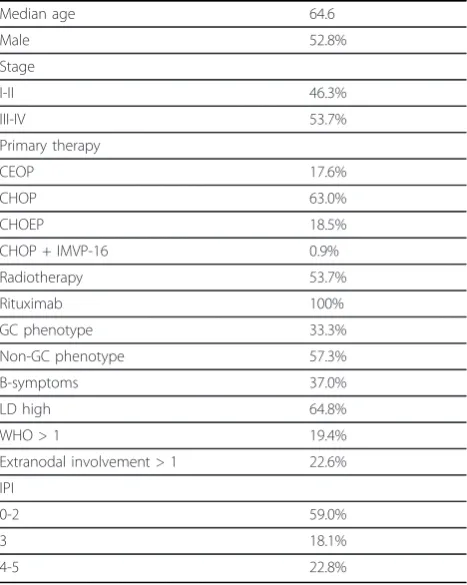
Table 1 summarizes the characteristics of the patients included in the study. There were 106 patients with
histologically confirmed DLBCL from whom paraffin-embedded tissue sections from diagnostic lymph nodes, extralymphatic tumor site samples or coarse needle biopsy samples were available. Detailed patient informa-tion was collected in each case. The lymphomas were diagnosed and treated at Oulu University Hospital and Kuopio University Hospital between the years 2003-2009. Diagnoses were reviewed by an experienced hema-topathologist. The diagnostic work-up included history and physical examination, blood chemistry, bone mar-row biopsy and aspiration and whole body computer tomography. The median age of the patients was 64.6 years (21-90 yr). CHOP-like therapy combined with rituximab antibody treatment was given to all patients. Involved field radiotherapy was applied after immuno-chemotherapy at the sites of bulk disease with residual masses according to individual discretion. DLBCL phe-notypes were divided into three groups according to Hans’algorithm: (i) a GCB phenotype with positive CD-10 and/or bcl-6 and negative MUM-1 immunostaining; (ii) a non-GCB phenotype with positive MUM-1 and positive or negative bcl-6 and negative CD-10 immunos-taining; (iii) a third type consisting of cases with charac-teristics of both previous types [2]. The ethical committee of the Northern Ostrobothnia Hospital Dis-trict has approved the study design (reference number 42/2010).
Immunohistochemical staining and sample evaluation
We used immunohistochemistry to study the expression of 8-OHdG, nitrotyrosine, Trx, MnSOD and GCL, using identical methods as in our recent work with different lymphoma entities. We have previously characterized the expression of the used antibodies in several malignant and pre-malignant tissues [18,19,22,35,36]. Sample evaluation in lymphomas has also been described earlier [22]. The cytoplasmic expression was diffuse in 8-OHdG, nitrotyro-sine, Trx and GCL. MnSOD expression was granulated, in line with its previously described mitochondrial location. The nuclear staining was throughout negative in nitrotyro-sine, MnSOD and GCL. 8-OHdG and Trx showed positive and diffuse staining in some of the malignant cells. The nuclear membrane was pronounced in some of the sam-ples stained with 8-OHdG. 8-OHdG and nitrotyrosine expression in malignant cells was uniform. Trx, MnSOD and GCL showed variable intensity in malignant cell stain-ing in few cases. These samples were coded by usstain-ing the more dominant intensity. The staining results were evalu-ated only in malignant lymphoma cells, since in our pre-vious work stromal staining extent or intensity did not show any clinical significance. The staining intensity of 8-OHdG, nitrotyrosine, Trx and MnSOD was divided into two groups: negative to moderate, and strong. The stain-ing intensity of cytoplasmic GCL was divided into two Table 1 Patient characteristics.
Median age 64.6
Male 52.8%
Stage
I-II 46.3%
III-IV 53.7%
Primary therapy
CEOP 17.6%
CHOP 63.0%
CHOEP 18.5%
CHOP + IMVP-16 0.9%
Radiotherapy 53.7%
Rituximab 100%
GC phenotype 33.3%
Non-GC phenotype 57.3%
B-symptoms 37.0%
LD high 64.8%
WHO > 1 19.4%
Extranodal involvement > 1 22.6%
IPI
0-2 59.0%
3 18.1%
4-5 22.8%
groups: negative to light, and moderate to strong. We also created a combination group from Trx and nitrotyrosine immunostaining results: (i) either nitrotyrosine or Trx immunostaining intensity strong; (ii) all other immunos-taining results.
Statistical analyses
Progression-free survival was calculated from the date of diagnosis to the date of relapse or date of disease-associated death. Disease-specific survival was calculated from the date of diagnosis to the date of death caused by lymphoma. To assess the relationship between clinical parameters and oxidative stress marker expression levels we used Pearson’s chi-square test. Survival estimates were calculated by using Kaplan-Meier analyses and statistical significance was determined by the log-rank test. Values of p under 0.05 were considered statistically significant. All statistical ana-lyses were performed using the Statistical Package for the Social Sciences, v. 18.0 (Chicago, IL, USA).
Acknowledgements
The authors would like to thank Anne Bisi for her skillful technical assistance. This work was supported by the Cancer Society of Northern Finland (AKP), the Northern Finnish Medical Society Duodecim (OK), the Finnish Medical Foundation (PK) and the Orion-Farmos foundation (PK).
Author details
1Department of Oncology and Radiotherapy, University of Oulu and Oulu University Hospital, Oulu, Finland.2Department of Pathology, University of Oulu, Oulu, Finland.3Institute of Clinical Medicine and Department of Medicine, University of Eastern Finland and Kuopio University Hospital, Kuopio, Finland.4Department of Clinical Pathology and Forensic Medicine, School of Medicine, University of Eastern Finland, Cancer Center of Eastern Finland and Kuopio University Hospital, Kuopio, Finland.5Medical Informatics Group, University of Oulu, Oulu, Finland.
Authors’contributions
PP, LL and AKP participated in the design of the study, contributed to the evaluation of immunostainings and drafted the manuscript. KMH, EJ and YS helped in study design, evaluated immunostaining results, and and critically revised the manuscript. TTH contributed to study design and was also responsible for manuscript preparation. RB undertook statistical analyses. PK and OK participated in the design of the study, helped in statistical analyses and wrote the different stages of the manuscript. All authors read and approved the final manuscript.
Competing interests
The authors declare that they have no competing interests.
Received: 20 February 2012 Accepted: 26 March 2012 Published: 26 March 2012
References
1. Alizadeh AA, Eisen MB, Davis RE, Ma C, Lossos IS, Rosenwald A,et al: Distinct types of diffuse large B-cell lymphoma identified by gene expression profiling.Nature2000,403:503-511.
2. Hans CP, Weisenburger DD, Greiner TC, Gascoyne RD, Delabie J, Ott G,et al: Confirmation of the molecular classification of diffuse large B-cell lymphoma by immunohistochemistry using a tissue microarray.Blood 2004,103:275-282.
3. Ziepert M, Hasenclever D, Kuhnt E, Glass B, Schmitz N, Pfreundschuh M, et al:Standard International prognostic index remains a valid predictor of outcome for patients with aggressive CD20+ B-cell lymphoma in the rituximab era.J Clin Oncol2010,28:2373-2380.
4. Haidari M, Ali M, Gangehei L, Chen M, Zhang W, Cybulsky MI:Increased oxidative stress in atherosclerosis-predisposed regions of the mouse aorta.Life Sci2010,87:100-110.
5. Doran DE, Weiss D, Zhang Y, Griendling KK, Taylor WR:Differential effects of AT1 receptor and Ca2+ channel blockade on atherosclerosis, inflammatory gene expression, and production of reactive oxygen species.Atherosclerosis2007,195:39-47.
6. Baynes JW:Role of oxidative stress in development of complications in diabetes.Diabetes1991,40:405-412.
7. Kinnula VL, Crapo JD:Superoxide dismutases in malignant cells and human tumors.Free Radic Biol Med2004,36:718-744.
8. Karihtala P, Winqvist R, Syvaoja JE, Kinnula VL, Soini Y:Increasing oxidative damage and loss of mismatch repair enzymes during breast
carcinogenesis.Eur J Cancer2006,42:2653-2659.
9. Mates JM, Segura JA, Alonso FJ, Marquez J:Intracellular redox status and oxidative stress: implications for cell proliferation, apoptosis, and carcinogenesis.Arch Toxicol2008,82:273-299.
10. Karihtala P, Soini Y:Reactive oxygen species and antioxidant mechanisms in human tissues and their relation to malignancies.APMIS2007, 115:81-103.
11. Holmgren A, Lu J:Thioredoxin and thioredoxin reductase: current research with special reference to human disease.Biochem Biophys Res Commun2010,396:120-124.
12. Franklin CC, Backos DS, Mohar I, White CC, Forman HJ, Kavanagh TJ: Structure, function, and post-translational regulation of the catalytic and modifier subunits of glutamate cysteine ligase.Mol Aspects Med2009, 30:86-98.
13. Kasai H:Analysis of a form of oxidative DNA damage, 8-hydroxy-2’ -deoxyguanosine, as a marker of cellular oxidative stress during carcinogenesis.Mutat Res1997,387:147-163.
14. Pfeiffer S, Schmidt K, Mayer B:Dityrosine formation outcompetes tyrosine nitration at low steady-state concentrations of peroxynitrite. Implications for tyrosine modification by nitric oxide/superoxide in vivo.J Biol Chem 2000,275:6346-6352.
15. Murata M, Kawanishi S:Oxidative DNA damage induced by nitrotyrosine, a biomarker of inflammation.Biochem Biophys Res Commun2004, 316:123-128.
16. Moulian N, Truffault F, Gaudry-Talarmain YM, Serraf A, Berrih-Aknin S:In vivo and in vitro apoptosis of human thymocytes are associated with nitrotyrosine formation.Blood2001,97:3521-3530.
17. Sova H, Jukkola-Vuorinen A, Puistola U, Kauppila S, Karihtala P: 8-Hydroxydeoxyguanosine: a new potential independent prognostic factor in breast cancer.Br J Cancer2010,102:1018-1023.
18. Karihtala P, Kauppila S, Soini Y, Jukkola-Vuorinen A:Oxidative stress and counteracting mechanisms in hormone receptor positive, triple-negative and basal-like breast carcinomas.BMC Cancer2011,11:262.
19. Karihtala P, Soini Y, Vaskivuo L, Bloigu R, Puistola U:DNA adduct 8-hydroxydeoxyguanosine, a novel putative marker of prognostic significance in ovarian carcinoma.Int J Gynecol Cancer2009,19:1047-1051. 20. Salzman R, Pacal L, Kankova K, Tomandl J, Horakova Z, Tothova E,et al:
High perioperative level of oxidative stress as a prognostic tool for identifying patients with a high risk of recurrence of head and neck squamous cell carcinoma.Int J Clin Oncol2010,15:565-570.
21. Zhou FL, Zhang WG, Wei YC, Meng S, Bai GG, Wang BY,et al:Involvement of oxidative stress in the relapse of acute myeloid leukemia.J Biol Chem 2010,285:15010-15015.
22. Pasanen AK, Kuitunen H, Haapasaari KM, Karihtala P, Kyllonen H, Soini Y, et al:Expression and Prognostic Evaluation of Oxidative Stress Markers in an Immunohistochemical Study of B-cell Derived Lymphomas.Leuk Lymphoma.
23. Tome ME, Johnson DB, Rimsza LM, Roberts RA, Grogan TM, Miller TP,et al: A redox signature score identifies diffuse large B-cell lymphoma patients with a poor prognosis.Blood2005,106:3594-3601.
24. Andreadis C, Gimotty PA, Wahl P, Hammond R, Houldsworth J, Schuster SJ, et al:Members of the glutathione and ABC-transporter families are associated with clinical outcome in patients with diffuse large B-cell lymphoma.Blood2007,109:3409-3416.
l’Estudi dels Limfomes de Catalunya I Balears (GELCAB):Gene-expression profiling and not immunophenotypic algorithms predicts prognosis in patients with diffuse large B-cell lymphoma treated with
immunochemotherapy.Blood2011,117:4836-4843.
26. Ott G, Ziepert M, Klapper W, Horn H, Szczepanowski M, Bernd HW,et al: Immunoblastic morphology but not the immunohistochemical GCB/ nonGCB classifier predicts outcome in diffuse large B-cell lymphoma in the RICOVER-60 trial of the DSHNHL.Blood2010,116:4916-4925. 27. Asano T, Tsutsuda-Asano A, Fukunaga Y:Indomethacin overcomes
doxorubicin resistance by decreasing intracellular content of glutathione and its conjugates with decreasing expression of
gamma-glutamylcysteine synthetase via promoter activity in doxorubicin-resistant leukemia cells.Cancer Chemother Pharmacol2009,64:715-721. 28. Zhang H, Go YM, Jones DP:Mitochondrial thioredoxin-2/peroxiredoxin-3
system functions in parallel with mitochondrial GSH system in protection against oxidative stress.Arch Biochem Biophys2007, 465:119-126.
29. Hirota K, Murata M, Sachi Y, Nakamura H, Takeuchi J, Mori K,et al:Distinct roles of thioredoxin in the cytoplasm and in the nucleus. A two-step mechanism of redox regulation of transcription factor NF-kappaB.J Biol Chem1999,274:27891-27897.
30. Djavaheri-Mergny M, Javelaud D, Wietzerbin J, Besancon F:NF-kappaB activation prevents apoptotic oxidative stress via an increase of both thioredoxin and MnSOD levels in TNFalpha-treated Ewing sarcoma cells.
FEBS Lett2004,578:111-115.
31. Pavan A, Spina M, Canzonieri V, Sansonno S, Toffoli G, De Re V:Recent prognostic factors in diffuse large B-cell lymphoma indicate NF-kappaB pathway as a target for new therapeutic strategies.Leuk Lymphoma2008, 49:2048-2058.
32. Tsai SY, Sun NK, Lu HP, Cheng ML, Chao CC:Involvement of reactive oxygen species in multidrug resistance of a vincristine-selected lymphoblastoma.Cancer Sci2007,98:1206-1214.
33. Tsai-Turton M, Luong BT, Tan Y, Luderer U:Cyclophosphamide-induced apoptosis in COV434 human granulosa cells involves oxidative stress and glutathione depletion.Toxicol Sci2007,98:216-230.
34. Yokomizo A, Ono M, Nanri H, Makino Y, Ohga T, Wada M,et al:Cellular levels of thioredoxin associated with drug sensitivity to cisplatin, mitomycin C, doxorubicin, and etoposide.Cancer Res1995,55:4293-4296. 35. Soini Y, Haapasaari KM, Vaarala MH, Turpeenniemi-Hujanen T, Kärjä V,
Karihtala P:8-hydroxydeguanosine and nitrotyrosine are prognostic factors in urinary bladder carcinoma.Int J Clin Exp Pathol2011,3:267-275. 36. Pylväs M, Puistola U, Kauppila S, Soini Y, Karihtala P:Oxidative
stress-induced antioxidant enzyme expression is an early phenomenon in ovarian carcinogenesis.Eur J Cancer2010,46:1661-1667.
doi:10.1186/2162-3619-1-2
Cite this article as:Perojaet al.:Oxidative stress and redox state-regulating enzymes have prognostic relevance in diffuse large B-cell lymphoma.Experimental Hematology & Oncology20121:2.
Submit your next manuscript to BioMed Central and take full advantage of:
• Convenient online submission
• Thorough peer review
• No space constraints or color figure charges
• Immediate publication on acceptance
• Inclusion in PubMed, CAS, Scopus and Google Scholar
• Research which is freely available for redistribution