RESEARCH PAPER
Efficient Synthesis of CeVO
4Nanoparticles Using Combustion
Route and Their Antibacterial Activity
Damayanti Ramchandra Kamble 1, Sachin Vasant Bangale 2 *, Suresh Kushinath Ghotekar 3, Sambhaji Rau Bamane 4
1 Department of Chemistry, Shankarrao Mohite Mahavidyala, Akluj 416 101, University of Solapur, Maharashtra, India.
2 Department of Chemistry, G. M. Vedak College of Science, Tala 402 111, University of Mumbai, Maharashtra, India
3 Department of Applied Science & Humanities, G. M. Vedak Institute of Technology, Tala 402 111, University of Mumbai, Maharashtra, India.
4 Department of Chemistry, Raja Shripatrao Bhagwantrao College, Aundh 415 510, Shivaji University, Maharashtra, India.
*Corresponding Author Email: [email protected] ARTICLE INFO
Article History:
Received 29 February 2018 Accepted 18 March 2018 Published 01 April 2018
Keywords:
Antibacterial activity CeVO4
Nanotechnology
ABSTRACT
How to cite this article
Kamble D. R, Bangale S. V, Ghotekar S. K, Bamane S. R. Efficient Synthesis of CeVO4 Nanoparticles Using Combustion Route and Their Antibacterial Activity. J Nanostruct, 2018; 8(2):144-151. DOI: 10.22052/JNS.2018.02.004
CeVO4 (Cerium orthovanadate) nanoparticles were synthesized by urea-assisted simple and efficient combustion method. Phase formations of synthesized nanoparticles were characterized by thermogravimetric-differential thermal analyzer (TG-DTA). X-ray diffraction (XRD) pattern revealed the crystal planes and size of synthesized CeVO4 nanoparticles. The average size, morphological shape and the crystalline nature of the nanoparticles were determined by scanning electron microscopy (SEM), transmission electron microscopic with selected area electron diffraction (TEM-SAED). Energy-dispersive X-ray spectroscopy (EDX) confirmed the presence of elemental composition and purity of the synthesized nanoparticles. Fourier transform Infrared spectroscopy (FT-IR) confirmed the possible stretching frequency on the surface of CeVO4 nanoparticles. Surface area and porosity studies of synthesized nanoparticles were analyzed by Brunauer-Emmett-Teller (BET) and Barrett-Joyner-Halenda (BJH) curve. Moreover, CeVO4 nanoparticles evinced excellent antibacterial activity against Escherichia coli, Klebsiella pneumoniae, Pseudomonas aeruginosa, Proteus vulgaris, Salmonella typhi, Staphylococcus aureus, Streptococcus pyogenus, Bacillus subtilis, Streptococcus pneumoniae and Staphylococcus epidermidis. The studies describing the synthesis of CeVO4 nanoparticles by efficient combustion method followed by the investigation of antibacterial activities may be useful for research opening a new arena in the field of nanobiotechnology.
INTRODUCTION
Nowadays, the synthesis of nanosized rare earth orthovanadates (RVO4)/metal oxides has
attracted much curiosity due to their miraculous and extensive applications in the field of catalysis,
bioengineering and material science [1-5]. Amongst
145
J Nanostruct 8(2): 144-151, Spring 2018
[12] and batteries fields [13]. Considering CeVO4
nanoparticles have excellent physical and chemical properties, it was particularly important to seek a facile, efficient and energy saving preparation method. The scrutiny of the literature revealed some notable methods were used for synthesis
of CeVO4. For example, sol-gel method [14], microwave assisted synthesis [8], ultrasound method [15], hydrothermal method [16], solid-state reaction method [17] and sonochemical
method [18] have been reported.Therewith, metal
oxide nanoparticles can be used as an anti-biotic, antioxidant, pesticide formulation, antimicrobial and antifungal agent when incorporated in textiles, coatings and plastics [19-22].
In this work, we rapidly synthesized CeVO4
nanoparticles by simple and efficient combustion method using urea as a fuel. These synthesized nanoparticles were evaluated for antimicrobial activities by employing against some selected human pathogens. It was found that efficiently
synthesized CeVO4 nanoparticles evinced good biomedical application in nanobiotechnology.
MATERIALS AND METHODS
Synthesis of CeVO4 nanoparticles
In this study, CeVO4 powder was successfully
synthesized by solution combustion route
using the starting regents as Ce(NO3)3.6H2O,
V(NO3)3.6H2O and Urea as a fuel. Urea possesses
a high heat of combustion. It is an organic fuel providing a platform for redox reactions during the course of combustion. Initially cerium nitrate, vanadium nitrate and urea are taken in the 1:1:4 stoichiometric amount and homogenous paste was made. The paste formed was evaporated on
hot plate at about 70 to 80 0C to get thick gel.
This kept on a hot plate for auto combustion and
heated at 170 to 180 0C. To obtain nanocrystalline
CeVO4 powder, this was sintered at 800 0C for 4 hrs.
A fine dark brown colored material was obtained and this was carefully collected and packed for further characterization purposes.
Characterization techniques
The synthesized CeVO4 nanoparticles
were characterized using thermogravimetric- Differential thermal analyzer (TG-DTA, PERKIN ELMER, USA, Diamond TG/DTA). The crystallinity and crystal phases were characterized by X-ray diffraction (XRD, Brukar, D8-Advanced Diffractometer) pattern measured with Cu-Kα
Radiation (λ= 1.5406 Å) in the range of 20–90o. The
morphology and composition of the synthesized
CeVO4 nanoparticles were examined by scanning
electron microscopy (SEM, JEOL, JSM-6360), SEM coupled energy-dispersive X-ray spectroscopy (EDX, Bruker, XFlash 6I30). Find the exact morphological
structures and size of the CeVO4 nanoparticles
using transmission electron microscopic (TEM) with selected area electron diffraction (SAED) analysis is done by using a PHILIPS, CM200 with an accelerating voltage of 200 kV. The Fourier transform Infrared (FTIR) spectrum was recorded
by JASCO 4100 in the range of 4000–400 cm-1. The
specific surface area, pore size and volume were characterized by Brunauer-Emmett-Teller (BET) and Barrett-Joyner-Halenda (BJH) analysis method at 77.40 deg. K (NOVA-100 Ver. 3.70).
Antibacterial activity of synthesized CeVO4 nanoparticles
146 J Nanostruct 8(2): 144-151, Spring 2018 active in primary screening was similarly diluted
to obtain 100, 50, 25, 12.5, 6.250, 3.125 and
1.5625 µg/ml concentrations. The highest dilution showing at least 99 % inhibition is taken as MIC.
RESULTS AND DISCUSSION
TG-DTA
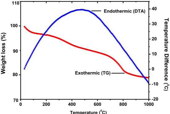
TG-DTA curve recorded for thermal
decomposition of CeVO4 is shown in Fig. 1. The
curve indicates that the slight weight loss in CeVO4
powder due to little loss of moisture, nitrogen
and carbon dioxide gas. The DTA curve of CeVO4
recorded in static air and is shown in Fig. 1. The
curve exhibited that CeVO4 did not decompose,
but weight loss was due to dehydrogenation, decarboxylation and denitration and yield final
product at 8000C. This weight change was in the
synthesized powder was almost remain stable
from the beginning as shown in reaction.
Ce(NO3)3 + V(NO3)3 + 4CON2H4 CeVO4 + 4CO2 + 8H2O + 5N2
Structural & crystallographic analysis
The synthesized CeVO4 formation was confirmed by the characteristic peaks observed in the XRD patterns, as shown in Fig. 2. Such a powder XRD was carried out using monochromatic CuKα1 radiation (wavelength 1.5406Å), operating at a voltage of 40 KV and a current of 40 mA, in
20 30 40 50 60 70 80 90
0 50 100 150 200 250 300 350 (42 4) (51 2) (22 4) (33 2) (40 0) (31 2) (30 1) (22 0) (11 2) (20 0)
2Degree)
Inte ns ity (a rbita ry unit)
Fig. 2. XRD pattern of synthesized CeVO4 nanoparticles sintered at 800 ºC for 4 hrs.
0 200 400 600 800 1000
80 100
Temperature (0
C) Weigh t los s ( %) 70 110 -20 -10 0 10 20 30 40 Exothermic (TG) Te mp era tur e Dif fer en ce ( 0 C ) 90 Endothermic (DTA)
147
J Nanostruct 8(2): 144-151, Spring 2018
the angular range 2θ of 20-90 deg. XRD analysis showed a series of diffraction peaks at 24.0º, 32.40º, 34.30º, 39.10º, 47.90º, 49.20º, 60.10º, 67.80º, 71.10º and 83.30º, corresponding to (200), (112), (220), (301), (312), (400), (332), (224), (512) and (424) crystal planes of tetragonal CeVO4 nanostructures (JCPDS No. 12-0757). The sharp XRD peaks exposed that synthesized CeVO4 nanoparticles are good crystalline in nature.
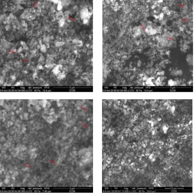
SEM microphotographs
Morphologies and sizes of synthesized CeVO4
nanoparticles were examined by SEM techniques. It can be seen that the average crystal grain size
of the CeVO4 nanoparticles was mainly 51-93 nm except slightly agglomeration (Fig. 3). This result exceed to the literature result which tetragonal
structure of CeVO4 nanoparticles was prepared by
ultrasound method [13].
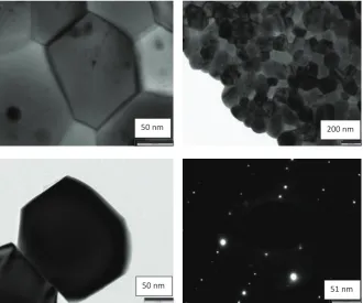
TEM images
TEM images and SAED pattern of the synthesized
CeVO4 nanoparticles were characterized. Fig.
4 indicates the hexagonal shape of CeVO4
nanoparticles with size 45-95 nm. The SAED pattern shows a number of bright spots, which confirmed
that, the as-synthesized CeVO4 nanoparticles are
single phase nature of the material.
EDX studies
The composition of synthesized CeVO4
nanoparticles has been analyzed by investigating the energy-dispersive X-ray spectroscopy (EDX), as shown in Fig.5. This was carried out to understand the composition of the cerium,
148 J Nanostruct 8(2): 144-151, Spring 2018 vanadium and oxygen in the synthesized
material. There was no unidenti fi ed observed in EDX. This quanti tati ve data confi rms the purity, compositi on and formati on of CeVO4
nanoparti cles.
Vibrati onal properti es
Fig. 6 represents the FTIR spectrum of CeVO4
nanoparti cles synthesized from combusti on
method. The IR band at 810 cm-1 is att ributed to
the Re-O-V vibrati ons of ReVO4. Residual water
200 nm
50 nm
50 nm 51 nm
Fig. 4. TEM images with SAED pattern of synthesized CeVO4 nanoparticles.
149
J Nanostruct 8(2): 144-151, Spring 2018
and -OH group are detected around 3449 cm-1,
corresponding to the O-H stretching frequency due to the bending vibration of associated water.
It suggested that the CeVO4 nanoparticle was
successfully fabricated by facile and efficient combustion method.
Surface area and porosity studies
The specific surface area of the CeVO4
nanoparticles calculated using multipoint BET
equation is 16.52 m2/g and according that the
CeVO4 nanoparticles have solid and smooth
surface. Furthermore, the corresponding BJH analyses indicate that the cumulative pore
volume was 0.005 cm3/g and the pore size of
which estimated from the peak position was 3.32 nm possesses a relatively narrow pore size distribution. Most of the pores ranged from 1.36 nm to 34.79 nm, as shown in the inset of Fig. 7. Therefore, these particles are polycrystalline in
4000 3500 3000 2500 2000 1500 1000 500
0 20 40 60 80 100
Tran
smi
ttan
ce
(%)
Wavenumber (cm-1)
810 cm-1
3449 cm-1
Fig. 6. FTIR spectra of synthesized CeVO4 nanoparticles.
35 0
2 4 6 8 10 12 14
30 25
20 15
10 0
Pore diameter (nm)
Pore vol
ume (
cc
/g)
5
150 J Nanostruct 8(2): 144-151, Spring 2018 nature.
Antimicrobial activity of CeVO4 nanoparticles
The results of antibacterial activity of the synthesized CeVO4 nanoparticles are presented in Table 1. Moderate to good antibacterial activity is observed against some bacteria. Synthesized CeVO4 nanoparticles exhibited potent and good antibacterial activity against E. coli, P. aeruginosa, S. aureus, B. subtilis, S. pneumonia and moderate activity against other bacteria with ampicillin was used as the reference drug.
CONCLUSION
The convenient utilization of urea as a fuel for
the efficient synthesis CeVO4 nanoparticles through
a combustion method to obtain significantly
biologically active material. The synthesized CeVO4 nanoparticles were hexagonal in shape as
observed in TEM analysis. The synthesized CeVO4
nanoparticles have shown potential upshot against selected pathogenic bacteria. This encouraging result provides useful information for designing a much better medicinal compound using a synthesis
of CeVO4 nanoparticles with minimal side effects and play significant role in biomedicine.
ACKNOWLEDGEMENT
Authors are thankful to SAIF IIT Powai, Shivaji University Kolhapur and Microcare Laboratory Gujrat for providing the technical, instrumental and biological activities supports.
CONFLICT OF INTEREST
The authors declare that there are no conflicts of interest regarding the publication of this manuscript.
REFERENCES
1. Phuruangrat A, Kuntalue B, Thongtem S. Thongtem T. Effect of PEG on phase, morphology and photocatalytic activity of
CeVO4 nanostructures. Mate. Lett. 2016; 174: 138–141.
2. Aher YB, Jain GH, Patil GE, Savale AR, Ghotekar SK, Pore DM, Pansambal SS, Deshmukh KK. Biosynthesis of copper oxide nanoparticles using leaves extract of Leucaena leucocephala L. and their promising upshot against the selected human pathogen. Int. Jou. Mol. Clin. Micro. 2017; 7: 776-786. 3. Pande SN, Bharati KT, Wakchure SK, Ghotekar SK, Gujrathi
DB, Phatangare ND. Green synthesis of silver nanoparticles by caralluma fimbriata L. and its characterization. Ind. Jou. App. Res. 2015; 5: 749-750.
4. Nguyen T, Dinh C, Do T. Monodisperse samarium and cerium orthovanadate nanocrystals and metal oxidation states on the nanocrystal surface. Langmuir. 2009; 25: 11142–11148. 5. Bangale SV, Bamane SR. Preparation and electrical properties
of nanostructured spinel ZnCr2O4 by combustion route. J
Mater. Sci: Mater. Elect. 2013; 24: 277-281.
6. Wang H, Meng Y, Yan H. Rapid synthesis of nanocrystalline
CeVO4 by microwave irradiation. Inorg. Chem. Comm. 2004;
7: 553–555.
7. Bangale SV, Patil DR, Bamane SR. Nanostructured spinel ZnFe2O4 for the detection of chlorine gas. Sensors &
Transducers. 2011; 134: 107-119.
8. Bangale SV, Dhapte VV, Patil DR, Bamane SR. A novel combustion route for the preparation of nanocrystalline LaAlO3 oxide based electronic nose sensitive to NH3 at room temperature. Sensors & Transducers. 2015; 146: 145-155. 9. Chen L. Hydrothermal synthesis and ethanol sensing
properties of CeVO4 and CeVO4-CeO2 powders. Mater. Lett. 2006; 60: 1859–1862.
10. Fan C, Liu Q, Ma T, Shen J, Yang Y, Tang H, Wang Y, Yang J. Fabrication of 3D CeVO4/grapheme aerogels with efficient
visible-light photocatalytic activity. Ceram. Inter. 2016; 42:
Test pathogens MIC (µg/ ml) of CeVO4 MIC (µg/ ml) of Reference drug
E. coli (MTCC-443) 50 100
K. pneumoniae (MTCC-109) 125 100
P. aeruginosa (MTCC-1688) 62.5 100
P. vulgaris (MTCC-8427) 100 100
S. typhi (MTCC-98) 100 100
S. aureus (MTCC-96) 125 250
S. pyogenus (MTCC-442) 200 100
B. subtilis (MTCC-441) 62.5 250
S. pneumoniae (MTCC-6305) 100 250
S. epidermidis (MTCC-12228) 100 100
151
J Nanostruct 8(2): 144-151, Spring 2018
10487-10492.
11. Fengzhen L, Xin S, Yibin Y, Limin Z, Zhuwei S, Xuehua L, Xianhua M. Shape controlled synthesis and tribological properties of CeVO4 nanoparticles as lubricating additive. J
Rare Ear. 2011; 29: 688-691.
12. Zhu L, Li Q, Li J, Liu X, Meng J, Cao X. Selective synthesis of mesoporous and nanorod CeVO4 without template. J Nano.
Res. 2007; 9: 261–268.
13. Picardia G, Varsanoa F, Deckera F, Opara-Krasovecb U, Surcab A, Orel B. Electrochemical characterization of optically passive CeVO4 counter electrodes. Elect. Acta.
1999; 44: 3157–3164.
14. Opara-Krasovecb U, Orel B, Surca A, Bukovec N, Reisfeld R. Structural and spectro electrochemical investigations of
tetragonal CeVO4 and Ce/V-oxide sol-gel derived ion-storage
films. Solid State Ionics. 1999; 118: 195–214.
15. Zheng Y, Qian Y, Jiqi J, Piesong T. Synthesis and characterization of nanoparticulate CeVO4 by ultrasound
method and its photocatalytic activity. Asia-Paci. Ene. Equip. Eng. Res. Conf. 2015: 229-232.
16. Guang L, Xuejun Z, Fei W, Hui W, Wei L. Facile fabrication of CeVO4 microspheres with efficient visible-light photocatalytic activity. Mater. Lett. 2017; 195: 168–171. 17. Akiteru W. Highly conductive oxides, CeVO4, Ce1−
xMxVO4−0.5x(M=Ca, Sr, Pb) and Ce1−yBiyVO4, with zircon- type
structure prepared by solid-state seaction in air. J Sol. Sta. Chem. 2000; 153: 174-179.
18. Mosleh M, Mahinpour A. Sonochemical synthesis and characterization of cerium vanadate nanoparticles and investigation of its photocatalyst activity. J Mate Sci: Mater in Elec. 2016; 27: 8930-8934.
19. Borkow G, Zatcoff RC, Gabbay J. Reducing the risk of skin pathologies in diabetics by using copper impregnated socks. Med. Hypotheses. 2009: 1–4.
20. Savale A, Ghotekar S, Pansambal S, Pardeshi O. Green synthesis of fluorescent CdO nanoparticles using Leucaena leucocephala L. extract and their biological activities. J Bacteriol Mycol open acess. 2017; 5: 00148.
21. Ghotekar SK, Vaidya PS, Pande SN, Pawar SP. Synthesis of silver nanoparticles by using 3-methyl pyrazol 5-one (chemical reduction method) and its characterizations. Int. J. Multidis. Res and Deve. 2015; 2(5): 419-422.
22. Pansambal S, Deshmukh K, Savale A, Ghotekar S, Pardeshi O, Jain G, Aher Y, Pore D. Phytosynthesis and biological activities of fluorescent CuO nanoparticles using Acanthospermum hispidum L. extract. J Nanostruct. 2017; 7: 165-174. 23. Rattan A. Antimicrobials in laboratory medicine. Churchill B