Combined biomarker testing
for the prediction of left ventricular
remodelling in ST-elevation
myocardial infarction
Sebastian Johannes Reinstadler,1Hans-Josef Feistritzer,1Martin Reindl,1 Gert Klug,1Agnes Mayr,2Johannes Mair,1Werner Jaschke,2Bernhard Metzler1
To cite:Reinstadler SJ, Feistritzer H-J, Reindl M,
et al. Combined biomarker
testing for the prediction of left ventricular remodelling in ST-elevation
myocardial infarction.Open
Heart2016;3:e000485.
doi:10.1136/openhrt-2016-000485
Received 9 June 2016 Revised 10 August 2016 Accepted 22 August 2016
1Department of Cardiology and Angiology, University Clinic of Internal Medicine III, Medical University of Innsbruck, Innsbruck, Austria 2Department of Radiology, Medical University of Innsbruck, Innsbruck, Austria
Correspondence to Dr Bernhard Metzler; [email protected]
ABSTRACT
Objective:The utility of different biomarkers for the prediction of left ventricular remodelling (LVR) following ST-elevation myocardial infarction (STEMI) has been evaluated in several studies. However, very few data exist on the prognostic value of combined biomarkers. The aim of this study was to
comprehensively investigate the prognostic value for LVR of routinely available biomarkers measured after reperfused STEMI.
Methods:Serial measurements of N-terminal pro-B-type natriuretic peptide (NT-proBNP), high-sensitivity cardiac troponin T (hs-cTnT), aspartate
aminotransferase (AST), alanine aminotransferase (ALT), lactate dehydrogenase (LDH) and high-sensitivity C reactive protein (hs-CRP) were performed in 123 patients with STEMI treated with primary percutaneous coronary intervention in this prospective observational study. Patients underwent cardiac MRI at 2 (1–4) and 125 (121–146) days after infarction. An increase in end-diastolic volume of≥20% was defined as LVR.
Results:LVR occurred in 16 (13%) patients. Peak concentrations of the following biomarkers showed significant areas under the curves (AUCs) for the prediction of LVR—NT-proBNP: 0.68 (95% CI 0.59 to 0.76, p=0.03), hs-cTnT: 0.75 (95% CI 0.66 to 0.82, p<0.01), AST: 0.72 (95% CI 0.63 to 0.79, p<0.01), ALT: 0.66 (95% CI 0.57 to 0.75, p=0.03), LDH: 0.78 (95% CI 0.70 to 0.85, p<0.01) and hs-CRP: 0.63 (95% CI 0.54 to 0.72, p=0.05). The combination of all biomarkers yielded a significant increase in AUC to 0.85 (95% CI 0.77 to 0.91) (all vs NT-proBNP: p=0.02, all vs hs-cTnT: p=0.02, all vs AST: p<0.01, all vs ALT: p<0.01, all vs hs-CRP: p<0.01 and all vs LDH: p=0.04). Conclusions:In patients with reperfused STEMI, the combined assessment of peak NT-proBNP, hs-cTnT, AST, ALT, hs-CRP and LDH provide incremental prognostic information for the prediction of LVR when compared with single-biomarker measurement.
INTRODUCTION
Outcome of patients suffering an
ST-elevation myocardial infarction (STEMI)
has markedly improved over the past
decades.1Nonetheless, survivors of the acute event are still at increased risk for further cardiovascular events.2
Adverse left ventricular remodelling (LVR) is a dynamic process at tissue, cellular and molecular level, characterised by changes in ventricular geometry, stiffness and function,
KEY QUESTIONS
What is already known about this subject?
▸ The development of adverse left ventricular remodelling (LVR) is associated with heart failure and mortality in patients surviving acute ST-elevation myocardial infarction (STEMI). Determination of routine biomarkers during the acute phase after reperfusion therapy might be a simple method to estimate the risk of LVR for the individual patient. It is, however, unclear whether the combination of routine biomarkers improves risk stratification for LVR after STEMI.
What does this study add?
▸ This study demonstrates that peak concentra-tions of N-terminal pro-B-type natriuretic peptide, high-sensitivity cardiac troponin T, aspartate aminotransferase, alanine transamin-ase, high-sensitivity C reactive protein and lactate dehydrogenase have significant ability to distinguish patients who develop LVR from those who do not. Importantly, the combination of all biomarkers provided incremental prognos-tic information for LVR when compared with single-biomarker testing.
How might this impact on clinical practice?
▸ Combined measurement of routine biomarkers improves early risk assessment for the develop-ment of LVR in patients with STEMI treated with primary percutaneous coronary intervention. Randomised studies are necessary to evaluate whether a biomarker-guided treatment strategy targeting LVR might improve patients outcome.
on September 12, 2020 by guest. Protected by copyright.
which affects the infarcted and non-infarcted myocar-dium.3 4It is well established that the degree of LVR is a major determinate of adverse clinical events after infarc-tion.3–5Therefore, early identification of patients at risk for LVR is regarded as an important step in post-STEMI risk stratification.
Biomarker measurement during the (sub-)acute phase after STEMI might be a simple and cost-effective tool to estimate the risk for LVR in clinical practice. To date, >50 biomarkers were described as potentially useful for the prediction of LVR.6 However, most of them were tested in relatively small number of patients (<100 parti-cipants) and only very few of them are available in daily clinical routine. Moreover, the potential incremental prognostic value for the combination of these biomar-kers is largely unknown.7 Natriuretic peptides, cardiac troponins, inflammatory markers, transaminases and lactate dehydrogenase (LDH) are often determined in the subacute phase after STEMI and associated with ejection fraction and myocardial damage,8–12 which are major determinates of LVR. Moreover, they have been suggested for prognosis assessment in patients with STEMI.8 10 11 13
Cardiac MRI (CMRI) provides high spatial and tem-poral resolution as well as high blood–tissue contrast and is therefore considered as the most accurate imaging technique to longitudinally assess changes in myocardial morphology and function after STEMI.14
Therefore, we aimed to investigate the prognostic value of routinely available biomarkers (N-terminal
pro-B-type natriuretic peptide (NT-proBNP),
high-sensitivity cardiac troponin T (hs-cTnT), aspartate aminotransferase (AST), alanine aminotransferase (ALT), LDH and high-sensitivity C reactive protein (hs-CRP)) measured during the (sub-)acute phase after STEMI, to predict CMRI-determined LVR. Moreover, we sought to evaluate a potential multimarker approach.
METHODS Study population
One hundred and thirty-three consecutive patients pre-senting at our University Hospital with acute STEMI were enrolled in this prospective single-centre, observa-tional study. Only patients presenting with first STEMI, as defined by the redefined ESC/ACC committee cri-teria,15 and treated with primary percutaneous coronary intervention (PPCI) were recruited. Exclusion criteria included age <18 years, history of a previous myocardial infarction or angiographically proven coronary artery disease, Killip-class III or IV, severe renal failure (defined as an estimated glomerular filtration rate <30 mL/min/1.73 m2) or contraindications for CMRI. Patient demographics and medications were prospect-ively obtained during hospitalisation for the index event and at 4 months follow-up using a standardised question-naire. Furthermore, all patients underwent physical examination. The study was approved by the local ethic
committee of Medical University of Innsbruck and written informed consent was obtained from each par-ticipant prior to inclusion in the study.
Cardiac MRI
Patients underwent CMRI on a 1.5 T scanner
(Magnetom AVANTO-scanner, Siemens, Erlangen,
Germany) within the first week and 4 months post-PPCI for STEMI. The CMRI protocol and postprocessing was published in detail previously.14 Briefly, cine CMRI images in short axis (10–12 slices, covering the heart from the base to the apex) were obtained using breath-hold, retrospective ECG-triggered trueFISP bright-blood sequences. Evaluation of cine images was performed using commercially available software (ARGUS,
Siemens). Late enhancement CMRI images were
obtained 10 min after a bolus injection of gadolinium (0.1 mmol/kg) by applying an ECG-triggered phase-sensitive inversion recovery single-shot trueFISP sequence with consecutive short-axis slices. Infarct characteristics were evaluated as reported previously.16 An increase in end-diastolic volume of 20% or more between baseline and follow-up CMRI investigation was defined as adverse remodelling.16
Biomarker testing
Venous blood samples were obtained on admission and subsequently once daily from day 1 to day 4 after PPCI. hs-cTnT was additionally measured 6 and 12 hours post-PCI. All samples were immediately analysed at the central laboratory of the University Hospital of Innsbruck by personnel blinded to study data. NT-proBNP concentrations were calculated using a com-mercially available assay with an E170 instrument ( proBNP II assay using monoclonal antibodies on a Modular, Roche Diagnostics, Vienna, Austria). The ana-lytical limit of detection of NT-proBNP is 5 ng/L and the limit of quantification is 50 ng/L. The intra-assay coefficient of variations (CV) are 1.9% at a concentra-tion of 64 ng/L and 1.2% at a concentraconcentra-tion of 2105 ng/L, and the interassay CV are 3.1% at a concen-tration of 46 ng/L and 2.7% at a concenconcen-tration of 2170 ng/L according to the package insert. hs-cTnT con-centrations were also measured using an assay with an E170 instrument (Roche Diagnostics).17 The analytical limit of detection and the 99th centile upper reference limit are 5 and 14 ng/L, respectively. The CV value is <10% at 13 ng/L. LDH activities, AST and ALT concen-trations as well as hs-CRP concenconcen-trations were measured by routine assays as described previously.9 Peak levels of each biomarker were calculated.
Statistical analysis
Distribution of continuous variables was tested with the Shapiro-Wilk test. Results of continuous variables are expressed as mean±SD or median with IQR. Results of categorical variables are shown as number and corre-sponding percentages. The Pearson or Spearman rank
on September 12, 2020 by guest. Protected by copyright.
correlation was calculated as indicated. Differences in location between groups were tested accordingly by
means of the t-test (Welch) and the
Wilcoxon-Mann-Whitney U test. Proportions were com-pared byχ2test and Fisher’s exact test as indicated. The utility of biomarkers for the prediction of LVR was tested with the use of receiver operating characteristic (ROC) analysis. The area under the curve (AUC), sensitivity and specificity were calculated. C-statistics according to DeLong et al18 were applied to evaluate the combined prognostic value in predicting LVR. A two-sided α=5% was determined overall. We used SPSS Statistics V.22.0.0.1 (IBM Corp., Armonk, New York, USA) and MedCalc V.15.8 (MedCalc Software, Ostend, Belgium) for statistical analysis.
RESULTS
Baseline characteristics and CMRI findings
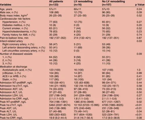
Of 133 enrolled patients with STEMI, 10 (92.5%) patients had no complete CMRI evaluation and/or bio-marker sampling and were therefore excluded. Thus, data of 123 (92.5%) patients were available for the ana-lysis (figure 1).Table 1shows the baseline characteristics (including biomarker concentrations) of the entire study cohort as well as for patients with and without LVR. There was no significant correlation between pain-to-balloon time and the investigated biomarkers (all p>0.05). All patients underwent CMRI 2 (1–4) and 125 (121–146) days after STEMI. LVR occurred in 16 patients (13%) at 4 months follow-up. CMRI results at baseline and follow-up are summarised in table 2. Of note, only patients without LVR showed a significant increase in LVEF ( p<0.01), whereas patients with LVR had no significant improvement in LVEF ( p=0.20). Baseline infarct size was larger in the LVR group ( p<0.01), but significantly decreased in both groups from baseline to follow-up (all p<0.05). The presence of microvascular obstruction was also significantly more likely in the LVR group ( p=0.02).
Biomarkers and LV remodelling
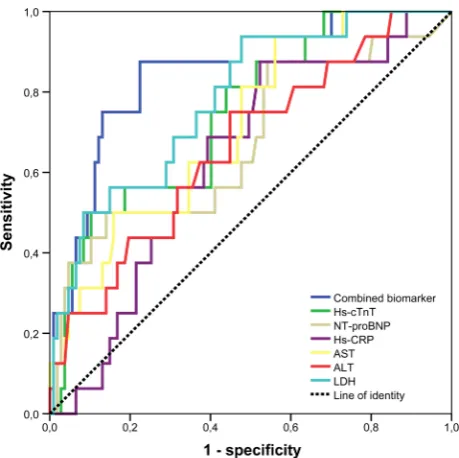
Patients with LVR had higher peak concentrations of NT-proBNP (1385 (616–3949) ng/L vs 677 (151–1267), p=0.02), hs-cTnT (10 153 (5722–15 995) ng/L vs 4709 (1985–8025), p<0.01), AST (375 (240–646) U/L vs 231 (126–349), p<0.01), ALT (73 (49–141) U/L vs 52 (38– 81), p=0.04) and LDH (917 (654–1530) U/L vs 523 (324–791), p<0.01) when compared with patients without LVR. The predictive value of biomarkers for LVR was assessed by ROC analysis (figure 2). The AUCs of significant predictors were 0.68 (95% CI 0.52 to 0.83, p=0.03) for NT-proBNP, 0.75 (95% CI 0.62 to 0.87, p<0.01) for hs-cTnT, 0.72 (95% CI 0.59 to 0.84, p<0.01) for AST, 0.66 (95% CI 0.57 to 0.75, p=0.03) for ALT, 0.63 (95% CI 0.54 to 0.72, p=0.05) for hs-CRP and 0.78 (95% CI 0.67 to 0.90, p<0.01) for LDH. Cut-off values with highest sensitivity and specificity of biomarkers are shown intable 3. The combination of all significant bio-markers in addition to NT-proBNP resulted in a signifi -cant increase in the c-statistics from 0.68 (95% CI 0.59 to 0.76) to 0.85 (95% CI 0.77 to 0.91, p=0.02), thus showing an incremental predictive value of combined
biomarker assessment over NT-proBNP alone.
Furthermore, the combination of biomarkers in addition to hs-cTnT ( p=0.02), AST ( p<0.01), ALT ( p<0.01), hs-CRP ( p<0.01) and LDH ( p=0.04) also resulted in a significant increase in the c-statistics.
In contrast to peak concentrations, admission concentra-tions of all investigated biomarkers were not significantly different between patients who developed LVR compared with patients who did not develop LVR (table 1). In line with this, none of these biomarkers showed a significant discriminatory ability for the prediction of LVR on admis-sion (AUC of NT-proBNP=0.57, AUC of hs-cTnT=0.54, AUC of AST=0.55, AUC of ALT=0.55, AUC of LDH=0.59 and AUC of hs-CRP=0.44, all p>0.05).
DISCUSSION
This study sought to comprehensively investigate the value of routine biomarkers for the prediction of LVR after reperfused STEMI. Peak concentrations of NT-proBNP, hs-cTnT, AST, ALT, hs-CRP and LDH, mea-sured during the (sub-)acute phase after PPCI, showed significant ability to distinguish patients who developed LVR from those who did not. Importantly, the combin-ation of all six biomarkers provided incremental prog-nostic information on LVR when compared with single-biomarker testing. In contrast, admission values of the investigated biomarkers were not useful for the pre-diction of LVR. Together, our data highlight the use of a multimarker approach for the prediction of LVR which should be evaluated in further larger trials.
STEMI is one of the main causes of heart failure and LVR plays a central role in this process. LVR rapidly occurs following STEMI, and it is well established that progressive postinfarction remodelling is an important predictor of poor clinical outcome, including mortality.3 4 Figure 1 Flow diagram of study patients. CMRI, cardiac
MRI, LVR, left ventricular remodelling.
on September 12, 2020 by guest. Protected by copyright.
In the current study, LVR occurred in∼13% of patients. This is in line with other recent reports that found a similar rate of LVR in comparable patient’s cohorts.7 19 20 Patients were largely treated according to recommended therapy, including a high use of ACE inhibitors and
β-blockers. Our results therefore underline that even in the modern era of PPCI and upstream heart failure therapy, LVR still occurs in a significant proportion of patients. Consequently, LVR remains a major clinical problem in the current era of STEMI management and Table 1 Baseline patient characteristics
Variables
All patients (n=123)
LV remodelling (n=16)
No LV remodelling
(n=107) p Value
Age, years 57±11 60±11 57±10 0.24
Male sex, n (%) 108 (88) 12 (75) 96 (90) 0.09
Body mass index, kg/m2 26 (25–28) 27 (25–30) 26 (25–28) 0.52
Cardiovascular risk factors
Hypertension, n (%) 77 (63) 12 (75) 65 (61) 0.27
Diabetes mellitus, n (%) 10 (8) 0 (0) 10 (9) 0.20
Current smoker, n (%) 63 (51) 7 (44) 56 (52) 0.52
Hypercholesterolaemia, n (%) 78 (63) 8 (50) 70 (65) 0.23
Family history for AMI, n (%) 35 (29) 4 (25) 31 (29) 0.74
Pain-to-balloon time, min 192 (137–352) 214 (132–421) 192 (137–351) 0.87
Infarct-related artery 0.03
Right coronary artery, n (%) 58 (47) 5 (31) 53 (49)
Left anterior descending artery, n (%) 50 (41) 11 (69) 39 (36)
Left circumflex coronary artery, n (%) 15 (12) 0 (0) 15 (14)
Number of diseased vessels 0.09
1, n (%) 64 (52) 9 (56) 55 (51)
2, n (%) 44 (36) 3 (18) 41 (38)
3, n (%) 15 (12) 4 (25) 11 (10)
Medication at discharge
Acetylsalicylic acid, n (%) 123 (100) 16 (100) 107 (100) 1.0
β-Blocker, n (%) 104 (85) 14 (87) 90 (84) 0.99
ACE-I or ARB, n (%) 105 (86) 14 (87) 91 (85) 0.41
Statin, n (%) 117 (95) 16 (100) 101 (94) 0.57
Admission NT-proBNP, ng/L 100 (58–401) 125 (63–838) 99 (54–371) 0.44
Admission hs-cTnT, ng/L 144 (21–1835) 132 (18–10 263) 153 (25–1617) 0.58
Admission AST, U/L 74 (33–223) 97 (36–410) 73 (33–213) 0.55
Admission ALT, U/L 37 (27–65) 37 (28–90) 36 (27–65) 0.50
Admission LDH, U/L 237 (196–343) 241 (204–586) 234 (196–324) 0.27
Admission hs-CRP, mg/L 2.1 (1.1–5.0) 1.8 (1.1–3.6) 2.1 (1.1–5.6) 0.41
Peak NT-proBNP, ng/L 704 (196–1391) 1385 (616–3949) 677 (151–1267) 0.02
Peak hs-cTnT, ng/L 5464 (2337–8574) 10 153 (5722–15 995) 4709 (1985–8025) <0.01
Peak AST, U/L 245 (138–369) 375 (240–646) 231 (126–349) <0.01
Peak ALT, U/L 57 (40–88) 73 (49–141) 52 (38–81) 0.04
Peak LDH, U/L 593 (353–832) 917 (654–1530) 523 (324–791) <0.01
Peak hs-CRP, mg/L 19.9 (9.2–44.4) 31.8 (16.7–56.4) 17.0 (9.0–38.8) 0.08
Continuous variables are shown as mean±SD or as median plus IQR.
ACE-I, ACE inhibitors; ALT, alanine transaminase; AMI, acute myocardial infarction; ARB, angiotensin receptor blocker; AST, aspartate aminotransferase; hs-CRP, high-sensitivity C reactive protein; hs-cTnT, high-sensitivity cardiac troponin T; LDH, lactate dehydrogenase; LV, left ventricular; NT-proBNP, N-terminal pro B-type natriuretic peptide.
Table 2 Cardiovascular MRI results
Characteristics Baseline Follow-up p Value
LVEF (%) 55 (49–61) 60 (54–66) <0.01
Infarct size (%LVMM) 16 (9–25) 11 (6–16) <0.01
LV stroke volume (mL) 79 (68–92) 88 (80–99) <0.01
LV end-diastolic volume (mL) 150 (128–166) 152 (128–170) 0.05
LV end-systolic volume (mL) 68 (50–81) 58 (47–75) <0.01
Microvascular obstruction: n (%) 65 (52) NA NA
Data are presented as median plus IQR.
LV, left ventricular; LVEF, left ventricular ejection fraction; LVMM, left ventricular myocardial mass; NA, not applicable.
on September 12, 2020 by guest. Protected by copyright.
better identification of patients at risk for LVR is essential to guide treatment and follow-up. Biomarkers may allow such an early prediction of LVR that could easily be used in a broad range of patients with STEMI. Several biomar-kers, including natriuretic peptides,13 cardiac tropo-nins,10 inflammatory markers,21 transaminases11 and LDH,22 are readily available, are routinely measured in patients hospitalised for STEMI and their measurement is relatively cheap. These biomarkers have been described to be associated with myocardial function, infarct size and microvascular injury,9–12 23 which are known determinants of LVR after STEMI. Most of them, but not all (transaminases and LDH), have also been investigated for their usefulness in predicting the occur-rence of LVR after myocardial infarction.6However, these studies were hampered by several limitations, including small numbers of included patients (most enrolled <100
patients),6the inclusion of highly selected patient groups (eg, patients with anterior Q-wave MI only)24and the use of echocardiography rather than CMRI,6 7 24which is the reference standard for the assessment of LVR.14 Finally, studies comparing the predictive value of multiple bio-markers within the same study are missing. Thus, to the best of our knowledge, this is thefirst study to investigate
a multimarker approach for the prediction of
CMRI-determined LVR after PPCI for STEMI. We found that peak concentrations of NT-proBNP, hs-cTnT, AST, ALT hs-CRP and LDH were significant predictors of LVR. Therefore, we not only confirm the significant relation of NT-proBNP, hs-cTnT and hs-CRP with LVR in a large cohort of patients with STEMI, but also show that AST, ALT and LDH are useful for the prediction of LVR in this setting. Importantly, we also observed that the combined measurement of these biomarkers might further increase the discriminative capacity. Our results therefore suggest that combining these biomarkers could constitute a strong tool for refining risk stratification after STEMI. This could have therapeutic implications, since patients at high risk for LVR could be treated more aggressively and followed more closely. In the light of the lack of trials, however, designed to prospectively investigate the value of a biomarker-guided treatment strategy, their use for risk stratification of the postinfarction patient awaits further validation and research.
Conclusions on the pathophysiological associations between investigated biomarkers and LVR as well as spe-cific processes leading to their elevation are beyond the scope of this study, but the following is appreciable to mention. In patients with acute MI, AST, ALT and LDH are both related with adverse clinical outcome, even in the contemporary era of mechanical reperfusion.25 26 There is recent evidence showing that circulating levels of AST, ALT and LDH are not only an indicator of infarct size, but may also reflect more extensive microvascular injury in reperfused STEMI.9 11 Infarct size and micro-vascular injury are strong predictors of LVR27 and subse-quently adverse clinical outcome.28 29 Although most evidence indicate that AST and ALT release is primarily of myocardial origin, circulatory failure with subsequent hypoxic live injury might in part also explain these eleva-tions.8In particular, AST was superior to cardiac troponin I in predicting outcome in STEMI in this study. Figure 2 ROC curve analysis for the prediction of LVR by
investigated routine biomarkers and their combination. NT-proBNP, N-terminal pro B-type natriuretic peptide, hs-cTnT, high-sensitivity cardiac troponin T, AST, aspartate aminotransferase, ALT, alanine transaminase, hs-CRP, high-sensitivity C reactive protein, LDH, lactate
dehydrogenase, LVR, left ventricular remodelling; ROC, receiver operating characteristic.
Table 3 Predictive accuracy of routine biomarkers for left ventricular remodelling
Biomarker AUC Cut-off concentration Sensitivity (%) Specificity (%)
NT-proBNP 0.68 (0.59–0.76) 1931 ng/L 50 86
hs-cTnT 0.75 (0.66–0.82) 5919 ng/L 75 60
AST 0.72 (0.63–0.79) 300 U/L 63 65
ALT 0.66 (0.57–0.75) 85 U/L 75 55
hs-CRP 0.63 (0.54–0.72) 22.5 mg/L 88 48
LDH 0.78 (0.70–0.85) 757 U/L 70 71
ALT, alanine transaminase; AST, aspartate aminotransferase; AUC, area under the curve; hs-CRP, high-sensitivity C reactive protein; hs-cTnT, high-sensitivity cardiac troponin T; LDH, lactate dehydrogenase; LVR, left ventricular remodelling; NT-proBNP, N-terminal pro B-type natriuretic peptide.
on September 12, 2020 by guest. Protected by copyright.
Although, it is well established that cardiac troponins are useful for the prediction of LVR,6 30there are no studies that used high-sensitivity troponin assays. The introduc-tion of hs-cTnT assays not only resulted in a significant increase in the sensitivity of cardiac troponins at an early diagnostic stage, but might also improve risk stratifi ca-tion.31 Nonetheless, we found no association between admission hs-cTnT and LVR in the current analysis. On the other hand, peak hs-cTnT was confirmed to be signifi -cantly related with LVR. NT-proBNP, which is released fol-lowing increased myocardial wall stress, is mainly used as an indicator of heart failure progression.32 Our study underscores that NT-proBNP is also useful for the predic-tion of LVR, a finding which is consistent with several other investigations,6 such as the study by Orn et al.33 Finally, the degree of inflammation, as reflected by circu-lating hs-CRP, is associated with LVR.7Our finding of a prognostic significance of circulating hs-CRP for the pre-diction of LVR is in line with these previous results.
Our study has several limitations. Although this study represents the largest CMRI trial on the prognostic value of combined routine biomarkers for LVR after STEMI so far, further confirmation in larger studies is very import-ant. Moreover, the potential impact of several important determinants of LVR (eg, microvascular obstruction or time to treatment) on the predictive value of biomarkers could not be investigated in the current trial. These spe-cific issues also need clarification in larger investigations. As most other studies, we preferred peak concentrations over AUC values since the first approach is a much more feasible measurement in daily clinical routine. Inclusion and exclusion criteria might limit generalisa-tion of study results, as, for example, patients presenting with Killip class ≥3 were not eligible for this study. Nevertheless, patient and CMRI characteristics are com-parable with other larger multicentre CMRI STEMI trials.28 Finally, there is a plethora of other potential useful biomarkers for the prediction of LVR,6which are, however, not available in daily routine and were there-fore not incorporated in the current analysis.
In conclusion, we found that peak concentrations of NT-proBNP, hs-cTnT, AST, ALT, hs-CRP and LDH after PPCI for STEMI provide prognostic information on the development of LVR as visualised by CMRI. Combined sampling of these routinely available biomarkers could facilitate the identification of patients at increased risk of LVR that might benefit from more intensive postin-farction care. However, considering the lack of rando-mised trials evaluating a biomarker-guided treatment strategy, this issue awaits further validation and research.
AcknowledgementsThis study was supported by grants from the‘Austrian Society of Cardiology’as well as by the intramural funding programme of Medical University Innsbruck for young scientists MUI-START,‘Project 2015-06-013’.
Contributors SJR designed the study; analysis and interpretation of data; drafting of the manuscript. H-JF contributed to interpretation of the data; drafting of the manuscript and revising critically for important intellectual content. MR contributed to analysis and interpretation of the data; revising critically for important intellectual content. GK contributed to analysis and
interpretation of the data; revising critically for important intellectual content. AM contributed to image analysis; revising critically for important intellectual content. JM contributed to interpretation of the data; revising critically for important intellectual content. WJ contributed to interpretation of the data; revising critically for important intellectual content. BM (corresponding author) designed the study; analysis and interpretation of data; drafting of the manuscript; revising critically for important intellectual content.
Funding Austrian Society of Cardiology, Medical University Innsbruck “MUI-START”(Project 2015-06-013).
Competing interests None declared.
Provenance and peer review Not commissioned; externally peer reviewed.
Data sharing statement No additional data are available.
Open Access This is an Open Access article distributed in accordance with the Creative Commons Attribution Non Commercial (CC BY-NC 4.0) license, which permits others to distribute, remix, adapt, build upon this work non-commercially, and license their derivative works on different terms, provided the original work is properly cited and the use is non-commercial. See: http:// creativecommons.org/licenses/by-nc/4.0/
REFERENCES
1. McManus DD, Gore J, Yarzebski J,et al. Recent trends in the incidence, treatment, and outcomes of patients with STEMI and NSTEMI.Am J Med2011;124:40–7.
2. Jernberg T, Johanson P, Held C,et al. Association between adoption of evidence-based treatment and survival for patients with ST-elevation myocardial infarction.JAMA2011;305:1677–84. 3. Cohn JN, Ferrari R, Sharpe N. Cardiac remodeling—concepts and
clinical implications: a consensus paper from an international forum on cardiac remodeling. Behalf of an International Forum on Cardiac Remodeling.J Am Coll Cardiol2000;35:569–82.
4. Altara R, Manca M, Sabra R,et al. Temporal cardiac remodeling post-myocardial infarction: dynamics and prognostic implications in personalized medicine.Heart Fail Rev2015:21;25–47.
5. Kramer DG, Trikalinos TA, Kent DM,et al. Quantitative evaluation of drug or device effects on ventricular remodeling as predictors of therapeutic effects on mortality in patients with heart failure and reduced ejection fraction: a meta-analytic approach.J Am Coll Cardiol2010;56:392–406.
6. Fertin M, Dubois E, Belliard A,et al. Usefulness of circulating biomarkers for the prediction of left ventricular remodeling after myocardial infarction.Am J Cardiol2012;110:277–83.
7. Urbano-Moral JA, Lopez-Haldon JE, Fernandez M,et al. Prognostic value of different serum biomarkers for left ventricular remodelling after ST-elevation myocardial infarction treated with primary percutaneous coronary intervention.Heart2012;98:1153–9. 8. Moon J, Kang W, Oh PC,et al. Serum transaminase determined in
the emergency room predicts outcomes in patients with acute ST-segment elevation myocardial infarction who undergo primary percutaneous coronary intervention.Int J Cardiol2014;177:442–7. 9. Mayr A, Klug G, Schocke M,et al. Late microvascular obstruction
after acute myocardial infarction: relation with cardiac and inflammatory markers.Int J Cardiol2012;157:391–6.
10. Hall TS, Hallén J, Krucoff MW,et al. Cardiac troponin I for prediction of clinical outcomes and cardiac function through 3-month follow-up after primary percutaneous coronary intervention for ST-segment elevation myocardial infarction.Am Heart J2015;169:257–65.e1. 11. Reinstadler SJ, Reindl M, Feistritzer HJ,et al. Prognostic
significance of transaminases after acute ST-elevation myocardial infarction: insights from a cardiac magnetic resonance study.Wien Klin Wochenschr2015;127:843–50.
12. Reindl M, Reinstadler SJ, Feistritzer HJ,et al. Relation of inflammatory markers with myocardial and microvascular injury in patients with reperfused ST-elevation myocardial infarction.Eur Heart J Acute Cardiovasc Care2016. Published Online First: 20 Jul 2016. doi:2048872616661691
13. Helanova K, Littnerova S, Kubena P,et al. Prognostic impact of neutrophil gelatinase-associated lipocalin and B-type natriuretic in patients with ST-elevation myocardial infarction treated by primary PCI: a prospective observational cohort study.BMJ Open2015;5: e006872.
14. Klug G, Metzler B. Assessing myocardial recovery following ST-segment elevation myocardial infarction: short- and long-term perspectives using cardiovascular magnetic resonance.Expert Rev Cardiovasc Ther2013;11:203–19.
on September 12, 2020 by guest. Protected by copyright.
15. Thygesen K, Alpert JS, Jaffe AS,et al. Third universal definition of myocardial infarction.Eur Heart J2012;33:2551–67.
16. Reinstadler SJ, Klug G, Feistritzer HJ,et al. Association of copeptin with myocardial infarct size and myocardial function after ST segment elevation myocardial infarction.Heart2013;99:1525–9. 17. Feistritzer HJ, Klug G, Reinstadler SJ,et al. Aortic stiffness is
associated with elevated high-sensitivity cardiac troponin T concentrations at a chronic stage after ST-segment elevation myocardial infarction.J Hypertens2015;33:1970–6.
18. DeLong ER, DeLong DM, Clarke-Pearson DL. Comparing the areas under two or more correlated receiver operating characteristic curves: a nonparametric approach.Biometrics1988;44:837–45. 19. Feistritzer HJ, Klug G, Reinstadler SJ,et al. Fetuin-A is related to
infarct size, left ventricular function and remodelling after acute STEMI.Open Heart2015;2:e000244.
20. Dominguez-Rodriguez A, Abreu-Gonzalez P, Avanzas P,et al. Neopterin predicts left ventricular remodeling in patients with ST-segment elevation myocardial infarction undergoing primary percutaneous coronary intervention.Atherosclerosis2010;211:574–8. 21. Seropian IM, Sonnino C, Van Tassell BW,et al. Inflammatory
markers in ST-elevation acute myocardial infarction.Eur Heart J Acute Cardiovasc Care2016;5:382–95.
22. Sjauw KD, van der Horst IC, Nijsten MW,et al. Value of routine admission laboratory tests to predict thirty-day mortality in patients with acute myocardial infarction.Am J Cardiol2006;97:1435–40. 23. Reinstadler SJ, Feistritzer HJ, Klug G,et al. High-sensitivity troponin
T for prediction of left ventricular function and infarct size one year following ST-elevation myocardial infarction.Int J Cardiol
2016;202:188–93.
24. Fertin M, Hennache B, Hamon M,et al. Usefulness of serial assessment of B-type natriuretic peptide, troponin I, and C-reactive
protein to predict left ventricular remodeling after acute myocardial infarction (from the REVE-2 study).Am J Cardiol
2010;106:1410–16.
25. Lofthus DM, Stevens SR, Armstrong PW,et al. Pattern of liver enzyme elevations in acute ST-elevation myocardial infarction.
Coron Art Dis2012;23:22–30.
26. Wei S, Mao L, Liu B,et al. Serum biomarkers and the prognosis of AMI patients.Herz2014;39:384–9.
27. Mather AN, Fairbairn TA, Ball SG,et al. Reperfusion haemorrhage as determined by cardiovascular MRI is a predictor of adverse left ventricular remodelling and markers of late arrhythmic risk.Heart
2011;97:453–9.
28. Eitel I, de Waha S, Wöhrle J,et al. Comprehensive prognosis assessment by CMR imaging after ST-segment elevation myocardial infarction.J Am Coll Cardiol2014;64:1217–26.
29. Reinstadler SJ, Baum A, Rommel KP,et al. ST-segment depression resolution predicts infarct size and reperfusion injury in ST-elevation myocardial infarction.Heart2015;101:1819–25.
30. Hallén J, Jensen JK, Fagerland MW,et al. Cardiac troponin I for the prediction of functional recovery and left ventricular remodelling following primary percutaneous coronary intervention for ST-elevation myocardial infarction.Heart2010;96:1892–7. 31. Mair J. High-sensitivity cardiac troponins in everyday clinical
practice.World J Cardiol2014;6:175–82.
32. van der Velde AR, Meijers WC, de Boer RA. Biomarkers for risk prediction in acute decompensated heart failure.Curr Heart Fail Rep
2014;11:246–59.
33. Orn S, Manhenke C, Squire IB,et al. Plasma MMP-2, MMP-9 and N-BNP in long-term survivors following complicated myocardial infarction: relation to cardiac magnetic resonance imaging measures of left ventricular structure and function.J Card Fail2007;13:843–9.
on September 12, 2020 by guest. Protected by copyright.