Copyright © 2012 IJECCE, All right reserved
Classification of MRI Brain Images Using Neuro Fuzzy
Model
Mr. Lalit P. Bhaiya, Ms. Suchita Goswami
Abstract
—
It is difficult to identify the abnormalities in brain specially in case of Magnetic Resonance Image brain image processing. This paper presents a hybrid technique for the classification of MRI human brain images. The proposed hybrid technique consists of three stages namely feature extraction, feature reduction and classification. The feature extraction and reduction is done by Principal Component Analysis and the classification is done by a hybrid Neuro-fuzzy classifier (ANFIS). ANFIS classifier combines the merits of both the neuro classifier and the fuzzy classifier and overcomes the demerits of both the classifiers. Artificial neural networks employed for brain image classification are being computationally heavy and also do not guarantee high accuracy. The major drawback of ANN is that it requires a large training set to achieve high accuracy. On the other hand fuzzy logic technique is more accurate but it fully depends on expert knowledge, which may not always available. Fuzzy logic technique needs less convergence time but it depends on trial and error method in selecting either the fuzzy membership functions or the fuzzy rules. These problems are overcome by the hybrid model namely, neuro-fuzzy model. This system removes essential requirements since it includes the advantages of both the ANN and the fuzzy logic systems. In this paper the classification of different brain images using Adaptive neuro-fuzzy inference systems (ANFIS technology) is done. Experimental results illustrate promising results in terms of classification accuracy and convergence rate.Keywords — ANFIS, Convergence rate, Classification
Accuracy, Fuzzy logic, Neural network
I.
I
NTRODUCTIONWith the growing age, there is advancement in each and every field. As far as the medical field is concerned, it also has everyday progress. The medical imaging field in particular, has grown substantially in recent years, and has generated additional interest in methods and tools for the management, analysis, communication of medical image data. Medical imaging technology facilitates the doctors to see the interior portions of the body for easy diagnosis. It also helped doctors to make keyhole surgeries for reaching the interior parts without really opening too much of the body. CT scanners, ultra sound and magnetic resonance imaging took over X-ray imaging by making the doctors to look at the body’s elusive third dimension.MRI picks up signals from the body’s magnetic particles spilling to its magnetic tune and with the help of its powerful computer, convert scanner data into revealing pictures of internal organs. MRI differs from CT scan as it does not use radiations.
MRI is a noninvasive medical test that helps physicians diagnose and treat medical conditions. It is a technique based on the measurement of magnetic field vectors generated after an appropriate excitation with strong
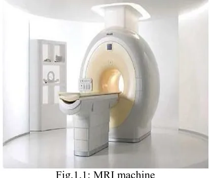
magnetic fields and radio frequency pulses in the nuclei of hydrogen atoms present in water molecules of a patient’s tissues. We know that the content of water differ for each tissue, by using this fact one can quantify the differences of radiated magnetic energy and have elements to identify each tissue. When we measure the specific magnetic vector components under controlled conditions, different images can be taken and we can obtain the information related to tissue contrast which reveals the details that can be missed in other measurements[11]. Detailed MRI image allows the physicians to better evaluate various parts of the body and determine the presence of certain abnormalities that may not be accessed adequately with other imaging methods such as X-ray, CT scan, and ultra sound. Currently, MRI is the most sensitive imaging test of the head in routine clinical practice. MRI can detect a variety of conditions of the brain such as cysts, tumors, bleeding, swelling, developmental and structural abnormalities, infections, inflammatory conditions or problems with the blood vessels.MRI can provide clear images of parts of the brain that can not be seen as well with an X-ray, CAT scan, or ultrasound, making it particularly valuable for diagnosing problems with the pituitary gland and brain stem. Figure 1.1 shows the typical MRI machine used to scan the human brain image.
Fig.1.1: MRI machine
Copyright © 2012 IJECCE, All right reserved Fig.1.2: Functional diagram of MRI machine
Applications of MRI segmentation include the diagnosis of brain trauma where a signature of brain injury, white matter lesions may be identified in moderate and mild cases. MRI segmentation methods are also useful in diagnosing multiple sclerosis, including the detection of lesions and the quantization of lesion volume using multispectral methods.[5]
In MRI, water molecules give off radio signals which are converted into high resolution images that look like a picture shown in figure 1.3.
Fig.1.3: Brain MRI
II.
M
ETHODOLOGYThe work involves the feature extraction of MRI images of brain, dimensionality reduction and finally developing a suitable neuro fuzzy classifier to classify the normal and abnormal brain images. Images of brain are obtained from MRI and the textural features are extracted using principal
component analysis (PCA) technique. These features are used to train the neuro fuzzy classifier. The neuro fuzzy classifier is used for classification is the Adaptive Network based Fuzzy Inference system(ANFIS).The developed neuro fuzzy classifier is tested for classification of different brain MRI samples. Thus, the proposed work emphasizes on development of Neural Network and Fuzzy logic based method for the classification of MRI brain images. The block schematic diagram shown in figure 1 is the proposed architecture for classification of MRI brain images.
Fig.2.1: Proposed Methodology for Classification of MRI brain images
A. MRI image data set
For the classification of normal and abnormal brain images a data set is collected from different sources one of the source is the Harvard medical school website. [http://www.med.harvard.edu/aanlib/home.html] The various types of brain images includes Axial, T2-weighted, 256-256 pixels MR brain images. Figure shows one of the database considered for the classification. The images are classified as normal and abnormal brain images.
Fig.2.2: A typical example of the used MRI
B. Feature Extraction
Copyright © 2012 IJECCE, All right reserved for training the database. The obtained trained feature is
compared with the test sample feature obtained and classified as one of the extracted character. [2] The feature extraction is done using principal component analysis (PCA).This technique is mostly used for the image recognition and reduction. It reduces the large dimensionality of the data. The feature extraction of the test image is done. The memory of an MR image recognizer is generally simulated by a training set. The training database is a set of MR images. The task of MR image recognizer is to find the most similar feature vector among the training set image and test image. In the training phase, feature vectors are extracted for each image in the training set. Let_1 be a training image of image 1 which has a pixel resolution of MxN (M rows, N columns). In order to extract PCA features of_1, first convert the image into a pixel vector Φ1 by concatenating each of M rows into a single vctor. The length of the vector Φ1 will be MxN.
Fig.2.3: Schematic diagram of a MR image recognizer
C. Neuro-Fuzzy Classifier
A neuro-fuzzy classifier is used to detect the abnormalities in the MRI brain images. Generally the input layer consist of seven neurons corresponding to the seven features. The output layer consist of one neuron indicating whether the MRI is of a normal brain or abnormal and the hidden layer changes according to the number of rules that give best recognition rate for each group of features.[3]
Here the neuro-fuzzy classifier used is based on the ANFIS technique. An ANFIS system is a combination of neural network and fuzzy systems in which that neural network is used to determine the parameters of fuzzy system. ANFIS largely removes the requirement for manual optimization of parameters of fuzzy system. The neuro-fuzzy system with the learning capabilities of neural
network and with the advantages of the rule-base fuzzy system can improve the performance significantly and neuro-fuzzy system can also provide a mechanism to incorporate past observations into the classification process. In neural network the training essentially builds the system. However, using a neuro-fuzzy technique, the system is built by fuzzy logic definitions and it is then refined with the help of neural network training algorithms.
Some advantages of ANFIS systems are:
It refines if-then rules to describe the behavior of a complex system.
It does not require prior human expertise
It uses membership functions plus desired dataset to approximate.
It provides greater choice of membership functions to use.
Very fast convergence time.[7]
Fig.2.4 : ANFIS architecture
ANFIS GUI- The ANFIS Editor GUI menu bar can be
Copyright © 2012 IJECCE, All right reserved Fig.2.5: ANFIS Editor GUI
III. C
OMPARATIVE PERFORMANCEA
NALYSIS OFN
EURAL&
F
UZZYM
ODELThe parameters for comparing the performance of the above three mentioned classification techniques are classification accuracy, convergence rate and mean square error. The classification accuracy is the ratio of the number of correctly classified images to the number of incorrect classified images. The classification accuracy of fuzzy classifier is superior to neural classifier and inferior to Neuro-fuzzy classifier. The neural classifier has least classification accuracy because it requires large training data set which is not practically feasible. The fuzzy classifier yields inferior classification accuracy to Neuro-fuzzy classifier because the initial seed points are randomly selected. The Convergence rate is the average time (in CPU secs) required for testing the images. The convergence time period of Neuro-fuzzy classifier is 10 times better than the neural and fuzzy classifier. The mean squared error of a classifier is one of many ways to quantify the difference between values implied by a classifier and the true values of the quantity being estimated. The error rate of fuzzy classifier and the neural classifier are high as they suffer from the drawbacks of random initial cluster center selection and the requirement of large training data set respectively. The Neuro-fuzzy classifier provides a minimum error rate. The neuro-fuzzy classifier combines the merits of both the neural classifier and fuzzy classifier therefore it yields superior results than the other classifiers.[7]
The neuro-fuzzy classifier is consistent on its outputs, provided sufficient training is given. Furthermore, it has the capability of making smart decisions on the inputs that may be lightly different from ones it is trained on.[3]
IV. C
ONCLUSIONNeural fuzzy models are performing successfully where other methods do not. The classification accuracy of combined neuro fuzzy classifier is comparatively higher than the individual fuzzy and neural classifiers. The convergence time period of neuro fuzzy classifier is 10 times better than the neural and fuzzy classifier. The future scope of this work is to enhance the ANFIS architecture to achieve high classification accuracy at a lower convergence rate. ANFIS is adopted for classification because it has fast speed on training and simple structure. The traditional connectionist systems do not provide explicit fuzzy interface, the proposed hybrid systems have wider scope/acceptability and presents dual advantages of a fuzzy logic based decision support and an ANN technique. The proposed system provides the following advantages: it is rapid, easy to operate, non invasive & inexpensive.
V. R
EFERENCE[1] H. Selvaraj, S. Thamarai Selvi, D. Selvathi, L. Gewali “Brain
MRI Slices Classification Using Least Squares Support Vector Machine” Vol. 1, No. 1, Issue 1, Page 21 of 33 ICMED 2007
[2] Youness Aliyari Ghassabeh, Nosratallah Forghani, Mohamad
Forouzanfar, Mohammad Teshnehlab “ MRI Fuzzy
Segmentation of Brain Tissue Using IFCM Algorithm with
Genetic Algorithm Optimization” 2007 IEEE
[3] Laxmi Ghodke, Apeksha Naik, Rahul Konale, Suchit Mehta
“Brain Cancer Detection using Neuro Fuzzy Logic” IJESS 2011
[4] Xinyu Du, Yongjie Li, Dezhong Yao”A Support Vector Machine
Based Algorithm for Magnetic Resonance Image Segmentation”
2008 IEEE
[5] G Vijay Kumar, Dr GV Raju “Biological early brain cancer
detection using artificial neural network”, 2010 International journal of computer applications Vol. 02, No. 08, 2010.
[6] J.Jaya, K.Thanushkodi, M.Karnan “Tracking algorithm for
De-noising of MR brain images”IJCSNS,Vol.9 no. 11, Nov. 2009
[7] D. Jude Hemanth, C.Kezi Selva Vijila and J.Anitha “Application
of Neuro-Fuzzy Model for MR Brain Tumor Image Classification”, International Journal of Biomedical Soft
Computing and Human Sciences, Vol.16,No.1,2010
[8] T.Logeswari and M.Karnan “An Enhanced Implementation of
Brain Tumor Detection Using Segmentation Based on Soft Computing” International Journal of Computer Theory and
Engineering, Vol. 2, No. 4, August, 2010
[9] M.Taherdangkoo, M.Yazdi, and M.H. Rezvani Segmentation of
MR Brain Images Using FCM improved by Artificial Bee
Colony (ABC) Algorithm. IEEE 2010
[10] P.Vasuda, S.Satheesh “Improved Fuzzy C-Means Algorithm for
MR Brain Image Segmentation (IJCSE)” Vol. 02, No. 05, 2010
[11] D. B. Kadam “Neural Network Based Brain Tumor Detection
Using Mr Images” IJCSC Vol. 2, No. 2, July-December 2011,
pp. 325-331
[12] El-Sayed A. El-Dahshan, Abdel-Badeeh M. Salem, And Tamer
H. Younis “A Hybrid Technique For automatic MRI brain Images Classification” studia univ. babes_{bolyai, informatica,
Volume LIV, 2009
[13] Rajeev Ratan, Sanjay Sharma, S. K. Sharma ―Brain tumor
detection sbased on multi-parameter MRI image analysis” ICGST-GVIP journal,ISSN 1687-398x, vol. [9],issue [III],june 2009
[14] Changjing Shang and Qiang Shen” Aiding Neural Network
Based Image Classification with Fuzzy-Rough Feature
Copyright © 2012 IJECCE, All right reserved
A
UTHOR’
SP
ROFILEMr. Lalit Kumar P. Bhaiya
is an Associate Professor & Head of Electronics & Telecommunication Department at Rungta College of Engineering & Technology, Bhilai. He did his B.E. & M.E in Electronics engineering from Amravati university (MH) in 1988 and 2004 respectively. He is pursuing his Ph.D. in Electronics Engineering from Chhattisgarh Swami Vivekanand Technical University, Bhilai. His research interest includes biomedical imaging, digital signal processing, image processing and soft computing. He has published around 42 papers in national and international journals and conferences. He is also the member of various professional societies like IETE, ISTE, IE(I). He has a vast experience of about 23 years in teaching profession.
Ms. Suchita Goswami
is an Assistant Professor in Chhattisgarh Institute of Technology, Rajnandgaon. She has passed BE (Hons) from CSIT, Durg in 2009 and
currently pursuing M.Tech. in Digital