PRIMARY RESEARCH
Prognostic value of Kindlin-2 expression
in patients with solid tumors: a meta-analysis
Sheng Liu
†, Sheng Chen
†, Kaige Ma and Zengwu Shao
*Abstract
Background: Kindlin-2 is one of the Kindlin family members which are evolutionarily conserved focal adhesion
proteins with integrin β-binding affinity. Recently, accumulative studies have suggested that Kindlin-2 plays important roles in tumor biology. However, the prognostic significance of Kindlin-2 in patients with solid tumors remains contro-versial. Therefore, this study aimed to clarify the prognostic value of Kindlin-2 in solid tumors via meta-analysis.
Methods: A comprehensive search was performed in PubMed, Embase, Web of Science and EBSCO for all relevant
studies reporting the prognostic significance of Kindlin-2 expression in solid cancer patients. The summary hazard ratio (HR) and corresponding 95% confidence interval (CI) were calculated to estimate the association between Kind-lin-2 expression with survival of solid cancer patients.
Results: We included 14 eligible studies containing 1869 patients in our meta-analysis. The pooled results indicated
that high Kindlin-2 expression was significantly associated with poor overall survival (OS) (pooled HR 1.66, 95% CI 1.44–1.92, P < 0.0001), disease-free survival (DFS)/recurrence-free survival (RFS)/progression-free survival (PFS) (pooled HR 1.73, 95% CI 1.16–2.57, P = 0.0067). For certain tumor types, high Kindlin-2 expression was significantly correlated with a poor outcome in patients with solid tumors, including pancreatic ductal adenocarcinoma (DFS/RFS/PFS), esophageal squamous cell carcinoma (OS, DFS/RFS/PFS), hepatocellular carcinoma (OS), clear cell renal cell carcinoma (OS), bladder cancer (OS, DFS/RFS/PFS), chondrosarcoma (OS), osteosarcoma (OS), gastric cancer (DFS/RFS/PFS), and glioma (OS).
Conclusions: Our meta-analysis demonstrated that high Kindlin-2 expression might indicate poor outcome in
patients with solid tumors and could serve as a prognostic biomarker for solid cancer patients.
Keywords: Kindlin-2, Solid tumor, Cancer, Prognosis, Meta-analysis
© The Author(s) 2018. This article is distributed under the terms of the Creative Commons Attribution 4.0 International License
(http://creat iveco mmons .org/licen ses/by/4.0/), which permits unrestricted use, distribution, and reproduction in any medium,
provided you give appropriate credit to the original author(s) and the source, provide a link to the Creative Commons license, and indicate if changes were made. The Creative Commons Public Domain Dedication waiver (http://creat iveco mmons .org/
publi cdoma in/zero/1.0/) applies to the data made available in this article, unless otherwise stated.
Background
Cancer is one of the leading contributors to heavy health care burden and disease-related mortality world-wide, with approximately 1,735,350 new cancer cases and 609,640 cancer-related deaths in the United States in 2018 [1, 2]. Although great advances in early detec-tion and treatments have been made in recent years, the prognosis of cancer patients is still poor [3, 4]. Therefore, novel prognostic biomarkers are urgently needed for
precisely predicting the outcome and providing thera-peutic targets for cancer patients.
The Kindlin family is composed of three members of evolutionarily conserved focal adhesion proteins (Kind-lin-1, -2 and -3) in mammal, which share the same 4.1-ezrin-radixin-moesin (FERM) domain, but have dif-ferent expression distribution [5]. Kindlins can exert extensive biological functions in cell proliferation, migra-tion, differentiation and cell death through binding with integrin β cytoplasmic tails and activating integrins, which have been linked to many hereditary disease and acquired disease of human [6]. Kindlin-1 (also known as FERMT1) is highly expressed in the skin and other tis-sues, whose deficiency and mutation can cause Kindler Syndrome [7, 8]. Kindlin-3 (also known as FERMT3) is
Open Access
*Correspondence: [email protected]
†Sheng Liu and Sheng Chen contributed equally to this work
generally expressed in the notochord, central nervous system, cement gland, and etc., mutations in which can contribute to leukocyte adhesion deficiency type III [8, 9].
Kindlin-2 (also known as FERMT2) was detected in various cell types, including fibroblast cells, smooth mus-cle cells and endothelial cells [10]. As a broadly distrib-uted focal adhesion protein, Kindlin-2 has binding sites for various interaction partners, such as integrin, actin, the filamin-binding protein migfilin, integrin-linked kinase (ILK) [11, 12]. Previous studies demonstrated that Kindlin-2 could interact with integrin and these partners to activate Wnt signaling, transforming growth factor β (TGF-β) signaling,epidermal growth factor receptor (EGFR) signaling, Hedgehog and extracellular regulated protein kinases (ERK) signaling pathways, which play vital roles in tumor progression [13]. Recently, increas-ing evidences indicated the correlation between Kind-lin-2 expression and prognosis in various types of solid tumors [14–28]. However, several studies demonstrated negative role or no significant association [14, 24, 28, 29]. Therefore, we performed this meta-analysis to explore the prognostic value of Kindlin-2 expression in patients with solid tumors.
Materials and methods Study strategy
This meta-analysis study was based on the Preferred Reporting Items for Systematic Reviews and Meta-Anal-yses (PRISMA) guidelines [30]. Two authors (Sheng Liu and Sheng Chen) independently carried out the search. PubMed, Embase, Web of Science and EBSCO were searched for articles reporting the prognostic role of Kindlin-2 expression in patients with solid tumors. The search strategy based on MeSH words was “Kindlin-2 OR FEMRT2 OR pleckstrin homology domain-containing family C member 1 (PLEKHC1) OR uncoordinated pro-tein 112 (UNC112) OR mitogen-inducible gene-2 (MIG-2) OR UNC112 related protein 2 short form (URP2SF)” AND “tumor OR neoplasm OR cancer OR carcinoma OR malignancy” AND “prognosis OR prognostic OR sur-vival”. The retrieval ended on 10 July, 2018. The references lists in identified articles were screened carefully lest rel-evant studies should be omitted.
Inclusion and exclusion criteria
We included all articles meeting the criteria as fol-lows: (1) cohort study; (2) Kindlin-2 expression in can-cer tissue or relevant tissue; (3) the prognostic outcome of Kindlin-2 different expression group; (4) available data such as Kaplan–Meier (KM) plot, the hazard ratio (HR) and 95% confidence intervals (CI). Studies of non-human research, reviews, letters, case reports, laboratory
articles, non-English articles and conference abstracts were excluded. Two authors (Sheng Liu and Sheng Chen) independently screened the titles and abstracts of iden-tified articles, and excluded those considered irrelevant. Further evaluation was conducted by viewing the full text carefully. Disagreements were resolved by consulting with a third author (Zengwu Shao).
Data extraction
Two researchers (Sheng Liu and Sheng Chen) indepen-dently extracted the relevant data from all eligible arti-cles. The following data of each study was extracted: first author, publication year, original country, number of enrolled patients, tumor type, detected methods, cut-off value, high expression presentations, follow-up time, and HR and 95% CI of the high Kindlin-2 expression group versus the low one for various outcomes. The HR and 95% CI were extracted preferentially from multivariable analyses such as Cox proportional-hazards model. When the HRs were not provided, we extracted the survival information from the original study data (KM plot or the required data) using the software Engauge Digitizer 10.5 [31] and estimated the survival data by Tierney’s method [32].
Quality assessment
The quality of each study was assessed by two investiga-tors (Sheng Liu and Sheng Chen) independently using the Newcastle–Ottawa Quality Assessment Scale (NOS). Any disagreement was resolved by discussing with another investigator (Kaige Ma). The scales allocate the total score for each study ranged from 0 to 9 for the qual-ity of selection, comparabilqual-ity, exposure and outcomes of included studies. The studies with scores ≥ 6 were con-sidered as high-quality studies.
Statistical analysis
The statistical analysis was performed using the software R 3.4.4 [33], meta package [34] and meta for package [35]. Pooled HRs and their corresponding 95% CIs were used to describe the prognostic value of Kindlin-2 expression. The heterogeneity was assessed using the Cochran Q-test and I-squared test. If I2 < 50% or P > 0.05, it was indi-cated that no heterogeneity existed among studies, and a fixed-effects model was performed. Otherwise, it was considered as significant heterogeneity and the random-effects model was applied. Meta-regression and subgroup analysis were performed with the studies sorted into sub-groups according to similar variables. Sensitivity analysis was applied to evaluate the stability of the results. Funnel plot and Egger’s test were applied to assess the potential publication bias. Statistical significance was defined as P
Results
Eligible studies and their characteristics
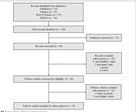
According to the searching strategy above mentioned, 120 records were retrieved from the databases. After 72 duplicated records were removed, the remaining arti-cles were screened. Then, 22 of 48 records were excluded because of several reasons: nine articles did not report Kindlin-2 expression as a prognostic variable; three did not involve a tumor; the remaining articles were six meeting articles, two patent articles and two review articles. When the further full-text review was finished, eleven basic research articles and one in non-English were excluded. Finally, the meta-analysis was performed for the remaining 14 articles (Fig. 1).
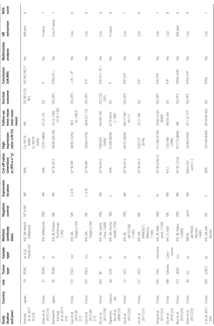
The included articles all had cohort study and pub-lished in the recent decade (2008–2017). In total, 1869 patients in the 16 cohorts were enrolled from China,
Japan and Greece. They were diagnosed with pancreatic ductal adenocarcinoma (PDAC), esophageal squamous cell carcinoma (ESCC), bladder cancer (BC), chondro-sarcoma (CHS), hepatocellular carcinoma (HCC), osteo-sarcoma (OSS), glioma, serous epithelial ovarian cancers (sEOC), gastric cancer (GC), or clear cell renal cell carci-noma (ccRCC). The expression of Kindlin-2 was detected by immunohistochemistry (IHC) or Western Blot (WB) in these studies, although the cut-off value varied in dif-ferent studies. At least overall survival (OS) was used as the prognostic outcome in every study. HRs with their 95% CIs based on Cox proportional-hazards model (Cox) were reported in 11 studies directly. In the remaining three studies, the data were calculated from the KM plots or the P-value of log-rank test. Every study’s NOS score was more than 6 points, which meant favorable method-ology. The main characteristics of the eligible studies were summarized in Table 1. And the main clinicopathologic
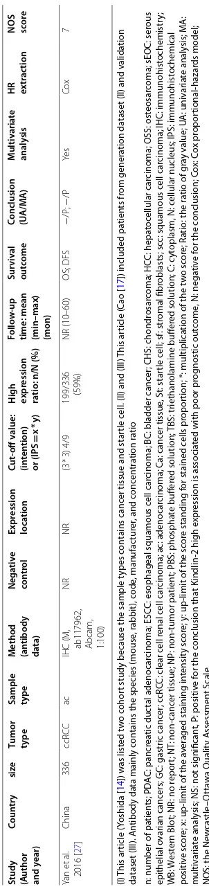
features and their distribution of patients in these stud-ies were shown on Table 2. Kindlin-2 expression was reported to have a significant association with several variables, including age, tumor size, stage, tumor cat-egory, lymphatic and vascular invasion, metastasis and response to chemotherapy (P < 0.05) (Table 2).
Correlation between Kindlin‑2 expression and survival outcomes of solid tumors
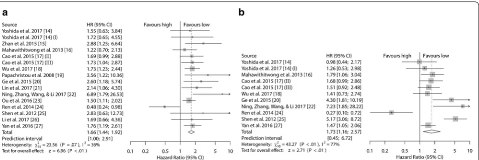
According to the protocol described above, the meta-analysis was performed and its main results were listed in Table 3. There were four survival outcomes evaluated in the included studies, including OS, disease-free survival (DFS), recurrence-free survival (RFS), progression-free survival (PFS). Given that they are similar in definition and number of studies evaluating RFS and PFS was lim-ited (Table 1), we combined the latter three ones together as DFS/RFS/PFS. Thus, this meta-analysis was conducted with two groups: OS and DFS/RFS/PFS.
For the first group, there was no significant statisti-cal heterogeneity (I2= 36.3%, P = 0.0729). Then, we pooled the HRs and 95% CIs by the fixed-effects model. It was indicated that high Kindlin-2 expression in cancer patients was significantly associated with a poor outcome (for OS, HR 1.66, 95% CI 1.44–1.92, P < 0.0001) (Fig. 2 and Table 3).
For the second group, there was obvious heterogeneity (I2= 76.9%, P < 0.0001). Hence, the random-effects model was performed, and the correlation between high Kind-lin-2 expression and poor outcomes was still statistically significant (for DFS/RFS/PFS, HR 1.73, 95% CI 1.16–2.57,
P = 0.0067) (Fig. 2 and Table 3).
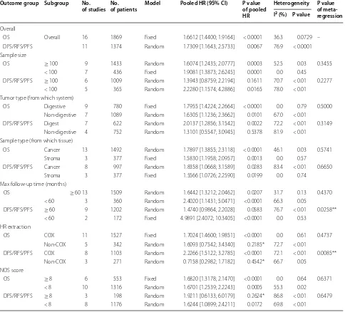
Subgroup analysis and meta‑regression analysis
In order to identify factors that could explain the het-erogeneity of the two above groups, subgroup analysis was performed focusing on six features able to analyze: number of patients in single study (less than 100 or not), tumor type (from digestive system or not), sample type (from cancer tissue or stroma tissue), maximum follow-up time (less than 60 months or not), HR extraction (from COX model or not), NOS score (less than 8 or not) (Fig. 3 and Table 3). However, other features were not analyzed due to the deficient report or inconsistent cut-off value. Through the subgroup analysis, we found that the correlation between high expression of Kindlin-2 and OS or DFS/RFS/PFS of solid tumor patients remained significant in all features above except for the subgroup of studies with the following features: patient quantity more than 100 (for OS, HR 1.39, 95% CI 0.88–2.22, P = 0.1611); tumor type not from digestive system (for OS, HR 1.31, 95% CI 0.55–3.09, P = 0.5378); HR not extracted from COX model (for OS, HR 1.60, 95% CI 0.75–3.43,
P = 0.2185; for DFS/RFS/PFS, HR 0.72, 95% CI 0.30–1.72,
P = 0.4542); NOS score no less than 8 (for OS, HR 1.92, 95% CI 0.61–6.02, P = 0.2624) (Table 3). To explore the potential sources of heterogeneity, meta-regression anal-ysis was performed according to the covariates including above features. The result illustrated that the above fea-tures might be not the source of heterogeneity as mod-erators except for maximum follow-up time (for DFS/ RFS/PFS, P = 0.0258) and HR extraction (for DFS/RFS/ PFS, P = 0.0085) (Table 3). Importantly, the pooled data from 11 cohorts and 1527 patients showed that Kind-lin-2 could be an independent factor for prognosis of solid tumor patients (for OS, HR 1.70, 95% CI 1.46–1.98,
P < 0.0001; for DFS/RFS/PFS, HR 2.23, 95% CI 1.51–3.28,
P < 0.0001) (Table 3).
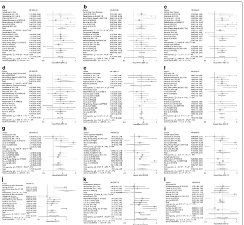
Correlation between Kindlin‑2 expression and survival outcomes of specific tumor types
The prognostic value of Kindlin-2 expression in dif-ferent tumors was further investigated. We found that high expression of Kindlin-2 in PDAC patients showed an obvious correlation with poor OS (HR 1.60, 95% CI 1.10–2.34, P= 0.015) (Fig. 4), but showed no statistically significant association with poor DFS/RFS/PFS (HR 1.44, 95% CI 0.972–2.13, P= 0.069) (Fig. 4). Through meta-analysis, we also observed that high Kindlin-2 expression significantly correlated with poor OS in patients with ESCC (HR 1.71, 95% CI 1.19–2.47, P= 0.004), HCC (HR 2.33, 95% CI 1.38–3.93, P = 0.002), ccRCC (HR 1.75, 95% CI 1.22–2.52, P = 0.003) (Fig. 4). The pooled data also showed statistically association between high Kindlin-2 expression with poor RFS/DFS/PFS in ESCC (HR 1.59, 95% CI 1.10–2.28, P = 0.0129), HCC (HR 4.30, 95% CI 1.81–10.19), ccRCC (HR 1.47, 95% CI 1.05–2.06) (Fig. 4).
Consistent with their original article, the remaining HRs and their 95% CI showed that high Kindlin-2 expres-sion had a significant relation with a worse prognosis in BC (for OS, HR 1.73, 95% CI 1.23–2.44; for DFS/RFS/ PFS, HR 1.41, 95% CI 0.73–2.74), CHS (for OS, HR 3.56, 95% CI 1.22–10.36), GC (for OS, HR 2.83, 95% CI 0.63– 12.73; for DFS/RFS/PFS, HR 5.17, 95% CI 3.06–8.72), glioma (for OS, HR 1.50, 95% CI 1.11–2.02), OS (for OS, HR 6.89, 95% CI 1.79–26.53; for DFS/RFS/PFS, HR 7.23, 95% CI 1.85–28.22), while it had a significant association with the better prognostic outcome of SEOC (for OS, HR 0.48, 95% CI 0.24–0.98; for DFS/RFS/PFS, HR 0.27, 95% CI 0.10–0.72) (Fig. 4).
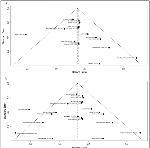
Publication bias assessment and sensitivity analysis
Table
1
T
he main char
ac
teristics of
the
eligible studies
Study (Author and
y ear) C oun tr y siz e
Tumor type Sample type
M ethod (an tibody da ta) Nega tiv e con tr ol Expr ession loca tion C ut ‑off v alue: (in ten tion) or (IPS = x * y) H igh expr ession ra
tio: n/N (%)
Follo
w
‑up
time: mean (min–max) (mon)
Sur viv al out come C
onclusion (UA/M
A ) Multiv aria te analy sis
HR extr
ac
tion
NOS sco
re
Yoshida et
al . 2017 (I) [ 14 ] Japan 79 PDA C ac ( Ca) Star tle (St)
IHC (M, M
er ck M illipor e) NT + NP NR 50%
Ca: 54/79 (68%) St: 49/79 (62%)
NR
OS; RFS OS; RFS
NS; NS; NS; P
No KM plot 8 Zhan et al . 2015 [ 15 ] China 31 PDA C ac IHC (M illipor e) PBS NR 50% 15/31 (48%) 47 (3–73) OS P No P-value 7 M aha with -itw ong et al . 2013 [ 16 ] Japan 95 PDA C sf
IHC (R, P
rot ein TechGr oup , 1:100) NR NR (4 * 3) 4/12 34/95 (64.2%)
24 (3–136); 14 (0–136)
OS; DFS P/NS; P/ − Ye s Co x, P -value 7 Cao et al .
2015 (II) [17
] China 110 ESC C scc
IHC (M, Or
igen,1:50) NR C + N (3 * 4) NR 34/65 (52%)
36.5 (0–148.7)
OS; DFS − /P ; − /P Ye s Cox 6 Cao et al .
2015 (III) [17
] China 147 ESC C scc
IHC (M, Or
igen,1:50) NR C + N (3 * 4) NR 20/64 (31%) 28.8 (27–72) OS; DFS P; P Ye s Cox 6 W u et al . 2017 [ 18 ] China 203 BC sf
IHC (M, Santa Cruz, 1:500)
PBS NR (3 * 4) 6/12
109/203 (54%)
64 (49–78)
OS; CSS; DFS
P/P ; P/ − ; P/ − Ye s Cox 6 Papachr is -tou et al . 2008 [ 19 ] Gr
eece etc.
60
CHS
sf
IHC (M, home
-made , 1/50) TBS C 33% 51/60 (85%)
67.9 (40.9, 2–180)
OS P No P-value 8 G e et al . 2015 [ 20 ] China 72 H CC ac
IHC (R, ab152106, 1:100)
PBS NR (4 * 3) 4/12 43/72 (60%)
NR (17.96– 43.11)
OS; DFS P/P ; P/P Ye s Cox 7 Lin et al . 2017 [ 21 ] China 127 H CC ac
IHC (M, M
AB2617, Biller ica, 1:100) NR C (3 * 4) 4/12
103/127 (81%)
22 (1–94) OS P/P Ye s Cox 8 N ing et al . 2017 [ 22 ] China 100 OSS Sar coma
IHC (R, M
illi -por e, 1:150) PBS N (3 * 4) 4.56/12 51/100 (51%)
29.82 (5.26– 38.89)
OS; DFS P/P ; P/P Ye s Cox 7 Ou et al . 2016 [ 23 ] China 188 Glioma Car ci -noma IHC (1:100) NR NR 4/12
132/188 (70%)
NR (0–39) OS P/P Ye s Cox 8 Ren et al . 2014 [ 24 ] China 113 sEOC scc
IHC (R, Dak
o, 1:2000) PBS NR (4 * 4) 12/16 91/113 (80%) NR OS; PFS N/NS; N/N Ye s KM -plot 6 Shen et al . 2012 [ 25 ] China 40 GC ac
WB (R, ab74030, Abcam, 1:600)
ac
tin
NR
Ratio: K2/ ac
tin > 2 22/40 (55%) 37.1 (5–77) OS; PFS P/NS; P/P Ye s Cox 8 Li et al . 2017 [ 26 ] China 109 ccR CC ac
IHC (M, M
(I) This ar ticle ( Yoshida [ 14 ]) w as list ed t w o c ohor
t study because the sample t
ypes c
on
tains canc
er tissue and star
tle c
ell
. (II) and (III)
This ar
ticle (
C
ao [
17
]) included pa
tien
ts fr
om gener
ation da
taset (II) and v
alida
tion
da
taset (III). An
tibody da
ta mainly c
on
tains the species (mouse
, r abbit), c ode , manufac tur er
, and c
onc
en
tr
ation r
atio
n: number of pa
tien ts; PD A C: pancr ea tic duc tal adenocar cinoma; ESC
C: esophageal squamous c
ell car
cinoma; BC: bladder canc
er
; CHS: chondr
osar
coma; HC
C: hepa
toc
ellular car
cinoma; OSS: ost
eosar
coma; sEOC: ser
ous
epithelial o
var
ian canc
ers; GC: gastr
ic canc
er
; c
cRC
C: clear c
ell r
enal c
ell car
cinoma; ac: adenocar
cinoma; C
a: canc
er tissue
, St: star
tle c
ell; sf
: str
omal fibr
oblasts; sc
c: squamous c
ell car
cinoma; IHC: immunohist
ochemistr y; WB: W est er
n Blot; NR: no r
epor
t; NT
:
non-canc
er tissue; NP
: non-tumor pa
tien
t; PBS: phospha
te buff er ed solution; TBS: tr iethanolamine buff er
ed solution; C: c
yt
oplasm, N: c
ellular nucleus; IPS: immunohist
ochemical
positiv
e sc
or
e; x: up
-limit of the a
ver
aged staining in
tensit
y sc
or
e; y
: up
-limit of the sc
or
e standing f
or stained c
ells pr
opor
tion; *: multiplica
tion of the t
w
o sc
or
e; R
atio: the r
atio of g
ra y v alue; U A: univ ar ia te analy sis; M A: multiv ar ia te analy
sis; NS: not sig
nifican
t, P
: positiv
e f
or the c
onclusion tha
t K
indlin-2 high expr
ession is associa
ted with poor pr
og
nostic out
come
, N: nega
tiv
e f
or the c
onclusion; C ox: C ox pr opor tional-hazar ds model;
NOS: the New
castle –O tta w a Qualit y A ssessmen t S cale Table 1 (c on tinued)
Study (Author and
y ear) C oun tr y siz e
Tumor type Sample type
M ethod (an tibody da ta) Nega tiv e con tr ol Expr ession loca tion C ut ‑off v alue: (in ten tion) or (IPS = x * y) H igh expr ession ra
tio: n/N (%)
Follo
w
‑up
time: mean (min–max) (mon)
Sur viv al out come C
onclusion (UA/M
A ) Multiv aria te analy sis
HR extr
ac
tion
NOS sco
re Yan et al . 2016 [ 27 ] China 336 ccR CC ac
IHC (M, ab117962, Abcam, 1:100)
NR NR (3 * 3) 4/9
199/336 (59%)
Table
2
T
he main clinic
opa thologic f ea tur es of pa tien ts and
their distribution in
the eligible studies (I) This ar ticle ( Yoshida [ 14 ]) w as list ed t w o c ohor
t study because the sample t
ypes c
on
tains canc
er tissue and star
tle c
ell
. (II) and (III)
This ar
ticle (
C
ao [
17
]) included pa
tien
ts fr
om gener
ation da
taset (II) and v
alida
tion
da
taset (III)
n: number of pa
tien
ts; NR: no r
epor t; C ap: capillar y in vasion; M ic: micr ov ascular in
vasion; C: chemother
ap
y; R: r
adiother
ap
y; RC: r
esponse f or chemother ap y *M eans tha t K indlin-2 expr ession w as r epor ted t o ha
ve a sig
nifican
t r
ela
tion with the v
ar
iable in the study
Study ( A uthor and y ear) n A ge (y ears or numbers):
[mean or median (range)] (cut
‑ off : lo w/high) Se x (M/F) H ist olog ical diff er en tia tion (I/II/III) Tumor siz e (cm) (cut ‑off ) (lo w/high)
Tumor categor
y (g rade) Lympha tic in vasion ( ∓ )
Vascular invasion (lo
w/ high) M etastasis ( ∓ ) Stag ing method Stage (cut ‑ off ) O ther ther ap y (no/y es) Yoshida et al .
2017 (I) [
14
]
79
65 (mean) (41–85) (65):39/40
51/28 9/63/7 NR NR 19/60 32/47 NR NR NR
C: 9/70 R: 68/11
Zhan et al . 2015 [ 15 ] 31 NR NR NR NR NR NR NR NR NR NR NR M aha withit -w ong et al . 2013 [ 16 ] 95
65 (mean) (36–86) (65): 52/43
58/37
10/33/52
NR
(T1/2/3/4) 9/3/82/1
34/61* 38/57 NR UIC C NR
C: 10/85 R: 78/17
Cao et al . 2015 (II) [ 17 ] 110 (58): 55/55 80/30 33/67/10
(3, 5) 32/45/11
(T1, 2/3, 4) 7/103
57/53 NR NR TNM (IIB/IIIA) 59/51 99/12 Cao et al . 2015 (III)[ 17 ] 147 (58): 79/68 113/34 23/109/15
(3, 5) 38/71/36
(T1, 2/3, 4) 20/127
64/83 NR NR TNM (IIB/IIIA) 70/77 104/43 W u et al . 2017 [ 18 ] 203 (65): 109/94 165/38 (L
ow/high) 96/107*
(3) 140/63 NR NR NR NR TNM (I/II) 8/115* NR Papachr ist ou et al
. 2008 [
19
]
60
54 (mean) (21–85)
34/26 20/29/11* (8) 23/37 NR NR NR NR NR NR NR G e et al . 2015 [ 20 ] 72 (53): 35/37 60/12 NR (5) 29/43* NR NR
Cap: 44/28* Mic: 49/23*
NR TNM (II/III) 41/31 NR Lin et al . 2017 [ 21 ] 127 (60): 111/16 17/110 NR (3) 10/117 NR NR
Cap: 40/87 Mic: 66/61*
9/115* NR (II/III) 11/116 NR N ing et al . 2017 [ 22 ] 100 (18): 40/60 68/32 (L
ow/high) 15/85*
NR NR NR NR 60/40* NR NR RC: 50/50* Ou et al . 2016 [ 23 ] 188
39 (mean) (39): 98/90*
103/85 NR NR NR NR NR NR NR (II/III) 85/103* NR Ren et al . 2014 [ 24 ] 113 (50): 28/85* − /113 (L ow/high) 26/87* NR NR NR NR 49/34 FIGO (I/II/III/IV ) 9/13/73/10 RC: 21/68 Shen et al . 2012 [ 25 ] 40
67 (mean) (47–93) (60): 14/26
30/10
4/8/28
NR
(T1, 2/3, 4) 8/32* N1/2/3 21/10/9*
NR 37/3 Patholog ic (II/III) 8/32* NR Li et al . 2017 [ 26 ] 109 (60):62/47 67/42 36/41/32* NR (T
x/1/2/34) 4/68/20/17
N x/0/1 2/99/8* NR NR A JCC (II/III) 70/39 NR Yan et al . 2016 [ 27 ] 336 (65):177/159 240/96 NR (4) 176/160
(T1, 2/3, 4) 167/169
significant (Begger’s P = 0.105, Egger’s P = 0.207). Then, we introduced trim-and-filled model to neutralize the potential bias (Fig. 5), and statistical significance of the correlation still existed (for OS, HR 1.55, 95% CI 1.35– 1.77, P < 0.0001). Hence, no significant publication bias existed and exerted a strong impact on the pooled results in this meta-analysis.
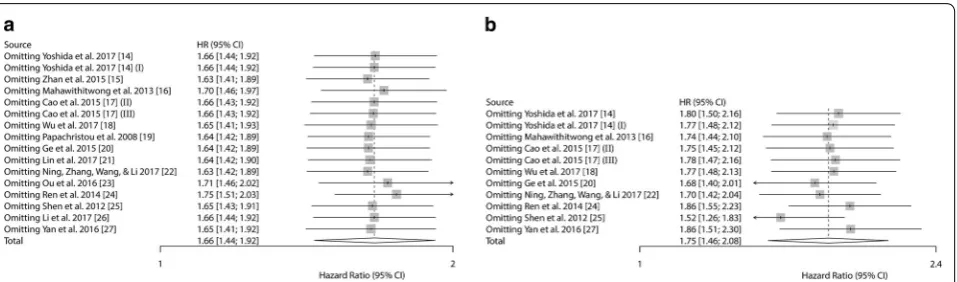
To evaluate the effect of each study on the pooled results, we performed sensitivity analysis by omitting each single study sequentially. No study displayed an apparent influence on the overall results of OS and DFS/ RFS/PFS (Fig. 6).
Table 3 The pooled HR and 95% CI for the prognostic value of Kindlin-2 expression
*Means that the P value of pooled HR is more than 0.05
**Means the P value from the test of moderators in the meta-regression is lower than 0.05
Outcome group Subgroup No.
of studies No. of patients Model Pooled HR (95% CI) P value of pooled HR
Heterogeneity P value
of meta‑ regression I2 (%) P value
Overall
OS Overall 16 1869 Fixed 1.6612 [1.4400; 1.9164] < 0.0001 36.3 0.0729 – DFS/RFS/PFS 11 1374 Random 1.7309 [1.1643; 2.5733] 0.0067 76.9 < 0.0001 Sample size
OS ≥ 100 9 1433 Random 1.6074 [1.2435; 2.0777] 0.0003 52.5 0.03 0.3455
< 100 7 436 Fixed 1.9081 [1.3873; 2.6245] 0.0001 0.0 0.45
DFS/RFS/PFS ≥ 100 6 1009 Random 1.3943 [0.8759; 2.2194] 0.1611 70.7 < 0.01 0.2277 < 100 5 365 Random 2.2280 [1.1574; 4.2886] 0.0165 78.0 < 0.01
Tumor type (from which system)
OS Digestive 9 780 Fixed 1.7955 [1.4224; 2.2664] < 0.0001 0.0 0.79 0.5000 Non-digestive 7 1089 Random 1.6305 [1.1236; 2.3662] 0.0101 67.0 < 0.01
DFS/RFS/PFS Digest 7 622 Random 2.0137 [1.2856; 3.1542] 0.0022 72.2 < 0.01 0.3149 Non-digestive 4 752 Random 1.3101 [0.5547; 3.0945] 0.5378 81.9 < 0.01
Sample type (from which tissue)
OS Cancer 13 1492 Random 1.7897 [1.3855; 2.3118] < 0.0001 46.1 0.03 0.5741
Stroma 3 377 Fixed 1.5830 [1.1958; 2.0957] 0.0013 0.0 0.57
DFS/RFS/PFS Cancer 8 997 Random 1.8358 [1.0668; 3.1589] 0.0283 83.4 < 0.01 0.6650
Stroma 3 377 Fixed 1.5566 [1.0726; 2.2590] 0.0199 0.0 0.74
Max follow-up time (months)
OS ≥ 60 13 1509 Random 1.6442 [1.3212; 2.0462] 0.0207 31.7 0.13 0.4370
< 60 3 360 Random 2.4020 [1.1431; 5.0471] < 0.0001 66.3 0.05
DFS/RFS/PFS ≥ 60 9 1202 Random 1.4740 [0.9864; 2.2028] 0.0583 76.7 < 0.01 0.0258** < 60 2 172 Fixed 4.9891 [2.4072; 10.3405] < 0.0001 0.0 0.53
HR extraction
OS COX 11 1527 Fixed 1.7024 [1.4600; 1.9851] < 0.0001 0.0 0.61 0.4737
Non-COX 5 342 Random 1.6093 [0.7542; 3.4340] 0.2185* 72.7 < 0.01
DFS/RFS/PFS COX 8 1103 Random 2.2266 [1.5122; 3.2785] < 0.0001 72.1 < 0.01 0.0085** Non-COX 3 271 Random 0.7158 [0.2982; 1.7182] 0.4542* 66.7 0.05
NOS score
OS ≥ 8 6 553 Fixed 1.6820 [1.3178; 2.1470] < 0.0001 0.0 0.64 0.6371
< 8 10 1316 Random 1.6701 [1.2539; 2.2243] 0.0005 55.3 0.02
Discussion
The human Kindlin-2 gene, also known as mitogen inducible gene-2 (MIG-2), was originally identified in the human diploid fibroblast cell line WI-38 by differential cDNA library screening and is located on chromosome 14q22.1 [20, 36]. Recently, increasing evidences have sug-gested that Kindlin-2 expression levels significantly cor-relate with tumor invasion, lymph node metastasis and worse survival in different cancers, such as breast can-cer, bladder cancer [5]. However, Ren et al. reported that Kindlin-2 inhibited the growth and migration of colorec-tal cancer cells [29], and Shi et al. found that Kindlin-2 could act as a suppressor of mesenchymal cancer cell invasion [37]. Owing to limited numbers of patients and conflicting conclusion in existing studies, the associa-tion between Kindlin-2 and prognosis of cancer patients remains controversial.
To our knowledge, there is no systemic review focus-ing on the correlation between Kindlin-2 expression and prognosis of cancer patients. Therefore, we performed this meta-analysis for critically assessing the prognostic significance of Kindlin-2 expression and to determine whether high Kindlin-2 expression is associated with poor prognosis of cancer patients or not. Our results showed that high Kindlin-2 expression was significantly associated with poor OS of patients with various solid tumors. Meanwhile, the correlation between high Kind-lin-2 expression and poor DFS/RFS/PFS was not homog-enous, but still significant. Then, we performed the
subgroup analysis for potential heterogeneity according to number of patients in single study, tumor type, sam-ple type, maximum follow-up time, HR extraction, NOS score. We found that there remains an obvious relation between high Kindlin-2 expression and poor prognosis of tumor patients when concerning the above features except for the subgroups as follow: patient quantity more than 100; tumor type not from digestive system; HR not extracted from COX model; NOS score no less than 8. Given that the numbers of studies in these sub-groups were limited, the correlating features may be not the source of the heterogeneity, which was consistent with the result of the following meta-regression. In the meta-regression analysis, we did found the lightly sig-nificant coefficient role in subgroup according to maxi-mum follow-up time and HR extraction. It meant that the two potential moderators might partly account for the heterogeneity of the DFS/RFS/PFS group. Moreover, Kindlin-2 exerted a significant impact on worse progno-sis of PDAC (DFS/RFS/PFS), ESCC (OS, DFS/RFS/PFS), HCC (OS), ccRCC (OS), BC (OS, DFS/RFS/PFS), CHS (OS), OSS (OS), GC (DFS/RFS/PFS) and glioma (OS), but not of PDAC (OS), GC (OS), sEOC (OS, DFS/RFS/ PFS). The results revealed that Kindlin-2 expression had a varying correlation with prognostic outcomes of differ-ent tumor types. No significant publication bias existed in this meta-analysis and exerted a strong impact on the pooled result. Meanwhile, no study displayed an appar-ent influence on the overall results of OS and DFS/RFS/
PFS. Taken together, Kindlin-2 expression could serve as a prognostic biomarker, which might help clinicians to make the best choices for cancer patients.
However, the exact mechanism behind the varying cor-relation of Kindlin-2 and poor prognosis has been not fully investigated. It was reported in previous studies
that Kindlin-2 could be acted as an activator of integ-rin in the development of cancers [5]. And recent stud-ies demonstrated that Kindlin-2 might exert a significant impact on poor prognosis by mainly modulating inte-grin signaling pathway and several other related signal-ing pathways, such as Wnt [21], TGF-β [15], EGFR [38]
and miR-200b [39]. These pathways were highly related with cell proliferation, migration, invasion [23, 38, 40], vascular function [41] and epithelial-to-mesenchymal transition (EMT) program [42], which might result in the poor prognosis of patients with solid tumor. Given that integrin regulates a variety of cell functions in can-cer cell, e.g. PDAC [43], inhibition of integrin signaling might be more efficient than direct inhibition of integrin. Then Kindlin-2, an essential activator of integrin, might be a promising target, which is supported by our result and a previous study reporting that several hallmarks of PDAC cell in vitro were inhabited when Kindlin-2 was stably down-regulated [15]. Previous research also con-cluded that embryonic dermal origins could influence the expression level of Kindlin-2 in various organs [44]. It implied that varying prognostic value of Kindlin-2 might be dependent on tumors’ embryonic dermal origins. In summary, high Kindlin-2 expression might indicate poor outcome in cancer patients and might be a promising therapeutic target for solid tumor.
Certainly, there were some limitations in our meta-analysis study. First, overall impact of Kindlin-2
expression on DFS/RFS/PFS was still inconclusive. Future study is needed to explore whether it is more accurate in predicting prognosis. Second, the number of studies for each specific tumor type there was limited. Third, the method we applied for extracting HR from KM plot was not as precise as the original study. Cut-off values of some key variables also differed among these studies. Potential heterogeneity might generate bias in the overall result. Hence, more studies with high quality are necessary for precisely illustrating the correlation between Kindlin-2 expression and prognosis of patients with various solid tumors.
Conclusions
In conclusion, our results demonstrated that Kindlin-2 expression had a significant correlation with prognos-tic outcomes of patients with different solid tumors. Elevated expression level of Kindlin-2 was significantly associated with a poor prognosis in patients with PDAC (DFS/RFS/PFS), ESCC (OS, DFS/RFS/PFS), HCC (OS), ccRCC (OS), BC (OS, DFS/RFS/PFS), CHS (OS), OSS (OS), GC (DFS/RFS/PFS) and glioma (OS), but not
PDAC (OS), GC (OS), sEOC (OS, DFS/RFS/PFS). More researches are warranted for accurately clarifying the association between Kindlin-2 expression and prognosis of solid cancer patients.
Abbreviations
HR: hazard ratio; CI: confidence interval; OS: overall survival; DFS: disease-free survival; RFS: recurrence-free survival; PFS: progression-free survival; FERM: 4.1-ezrin-radixin-moesin; ILK: integrin-linked kinase; TGF-β: transforming growth factor β; EGFR: epidermal growth factor receptor; ERK: extracellular regulated protein kinases; KM: Kaplan–Meier; PDAC: pancreatic ductal adeno-carcinoma; ESCC: esophageal squamous cell adeno-carcinoma; BC: bladder cancer; CHS: chondrosarcoma; HCC: hepatocellular carcinoma; OSS: osteosarcoma; sEOC: serous epithelial ovarian cancers; GC: gastric cancer; ccRCC : clear cell renal cell carcinoma; IHC: immunohistochemistry; WB: Western Blot; MIG-2: mitogen inducible gene-2; EMT: epithelial-to-mesenchymal transition. Authors’ contributions
SL and SC collected, extracted and analyzed the data, wrote the paper; KGM and ZWS performed quality assessment and analyzed the data. ZWS con-ceived and designed this study. All authors reviewed the final manuscript. All authors read and approved the final manuscript.
Acknowledgements
We would like to thank the researchers and study participants for their contributions.
Competing interests
The authors declare that they have no competing interests. Availability of data and materials
The datasets analyzed during the current study are available from the cor-responding author on reasonable request.
Consent for publication Not applicable.
Ethics approval and consent to participate Not applicable.
Funding
This study was supported by Grants 2016YFC1100100 from The National Key Research and Development Program of China, Grants 91649204 from Major Research Plan of National Natural Science Foundation of China.
Publisher’s Note
Springer Nature remains neutral with regard to jurisdictional claims in pub-lished maps and institutional affiliations.
Received: 15 July 2018 Accepted: 27 September 2018
References
1. Torre LA, Bray F, Siegel RL, Ferlay J, Lortet-Tieulent J, Jemal A. Global cancer statistics, 2012. CA Cancer J Clin. 2015;65(2):87–108. 2. Siegel RL, Miller KD, Jemal A. Cancer statistics, 2018. CA Cancer J Clin.
2018;68(1):7–30.
3. Haberlin C, O’Dwyer T, Mockler D, Moran J, O’Donnell DM, Broderick J. The use of eHealth to promote physical activity in cancer survivors: a systematic review. Support Care Cancer. 2018;26:3323–36. 4. Hu B, Fan H, Lv X, Chen S, Shao Z. Prognostic significance of CXCL5
expression in cancer patients: a meta-analysis. Cancer Cell Int. 2018;18:68. 5. Rognoni E, Ruppert R, Fassler R. The kindlin family: functions,
sign-aling properties and implications for human disease. J Cell Sci. 2016;129(1):17–27.
6. Meves A, Stremmel C, Gottschalk K, Fassler R. The Kindlin protein family: new members to the club of focal adhesion proteins. Trends Cell Biol. 2009;19(10):504–13.
7. Siegel DH, Ashton GH, Penagos HG, Lee JV, Feiler HS, Wilhelmsen KC, South AP, Smith FJ, Prescott AR, Wessagowit V, et al. Loss of kindlin-1, a human homolog of the Caenorhabditis elegans actin-extracellular-matrix linker protein UNC-112, causes Kindler syndrome. Am J Hum Genet. 2003;73(1):174–87.
8. Canning CA, Chan JS, Common JE, Lane EB, Jones CM. Developmental expression of the fermitin/kindlin gene family in Xenopus laevis embryos. Dev Dyn. 2011;240(8):1958–63.
9. Mory A, Feigelson SW, Yarali N, Kilic SS, Bayhan GI, Gershoni-Baruch R, Etzioni A, Alon R. Kindlin-3: a new gene involved in the pathogenesis of LAD-III. Blood. 2008;112(6):2591.
10. Ussar S, Wang HV, Linder S, Fassler R, Moser M. The Kindlins: subcellular localization and expression during murine development. Exp Cell Res. 2006;312(16):3142–51.
11. Harburger DS, Bouaouina M, Calderwood DA. Kindlin-1 and -2 directly bind the C-terminal region of beta integrin cytoplasmic tails and exert integrin-specific activation effects. J Biol Chem. 2009;284(17):11485–97. 12. Moser M, Legate KR, Zent R, Fassler R. The tail of integrins, talin, and
kindlins. Science. 2009;324(5929):895–9.
13. Zhan J, Zhang H. Kindlins: roles in development and cancer progression. Int J Biochem Cell Biol. 2018;98:93–103.
•fast, convenient online submission •
thorough peer review by experienced researchers in your field • rapid publication on acceptance
• support for research data, including large and complex data types •
gold Open Access which fosters wider collaboration and increased citations maximum visibility for your research: over 100M website views per year •
At BMC, research is always in progress.
Learn more biomedcentral.com/submissions
Ready to submit your research? Choose BMC and benefit from: 14. Yoshida N, Masamune A, Hamada S, Kikuta K, Takikawa T, Motoi F, Unno
M, Shimosegawa T. Kindlin-2 in pancreatic stellate cells promotes the progression of pancreatic cancer. Cancer Lett. 2017;390:103–14. 15. Zhan J, Song J, Wang P, Chi X, Wang Y, Guo Y, Fang W, Zhang H. Kindlin-2
induced by TGF-beta signaling promotes pancreatic ductal adenocar-cinoma progression through downregulation of transcriptional factor HOXB9. Cancer Lett. 2015;361(1):75–85.
16. Mahawithitwong P, Ohuchida K, Ikenaga N, Fujita H, Zhao M, Kozono S, Shindo K, Ohtsuka T, Mizumoto K, Tanaka M. Kindlin-2 expression in peri-tumoral stroma is associated with poor prognosis in pancreatic ductal adenocarcinoma. Pancreas. 2013;42(4):663–9.
17. Cao HH, Zhang SY, Shen JH, Wu ZY, Wu JY, Wang SH, Li EM, Xu LY. A three-protein signature and clinical outcome in esophageal squamous cell carcinoma. Oncotarget. 2015;6(7):5435–48.
18. Wu J, Yu C, Cai L, Lu Y, Jiang L, Liu C, Li Y, Feng F, Gao Z, Zhu Z, et al. Effects of increased Kindlin-2 expression in bladder cancer stromal fibroblasts. Oncotarget. 2017;8(31):50692–703.
19. Papachristou DJ, Gkretsi V, Rao UN, Papachristou GI, Papaefthymiou OA, Basdra EK, Wu C, Papavassiliou AG. Expression of integrin-linked kinase and its binding partners in chondrosarcoma: association with prognostic significance. Eur J Cancer. 2008;44(16):2518–25. 20. Ge YS, Liu D, Jia WD, Li JS, Ma JL, Yu JH, Xu GL. Kindlin-2: a novel
prog-nostic biomarker for patients with hepatocellular carcinoma. Pathol Res Pract. 2015;211(3):198–202.
21. Lin J, Lin W, Ye Y, Wang L, Chen X, Zang S, Huang A. Kindlin-2 promotes hepatocellular carcinoma invasion and metastasis by increasing Wnt/ beta-catenin signaling. J Exp Clin Cancer Res. 2017;36(1):134. 22. Ning K, Zhang H, Wang Z, Li K. Prognostic implications of Kindlin
proteins in human osteosarcoma. OncoTargets Ther. 2017;10:657–65. 23. Ou Y, Zhao Z, Zhang W, Wu Q, Wu C, Liu X, Fu M, Ji N, Wang D, Qiu
J, et al. Kindlin-2 interacts with beta-catenin and YB-1 to enhance EGFR transcription during glioma progression. Oncotarget. 2016;7(46):74872–85.
24. Ren C, Du J, Xi C, Yu Y, Hu A, Zhan J, Guo H, Fang W, Liu C, Zhang H. Kindlin-2 inhibits serous epithelial ovarian cancer peritoneal dissemi-nation and predicts patient outcomes. Biochem Biophys Res Commun. 2014;446(1):187–94.
25. Shen Z, Ye Y, Dong L, Vainionpaa S, Mustonen H, Puolakkainen P, Wang S. Kindlin-2: a novel adhesion protein related to tumor invasion, lymph node metastasis, and patient outcome in gastric cancer. Am J Surg. 2012;203(2):222–9.
26. Li M, Pei X, Wang G, Zhan J, Du J, Jiang H, Tang Y, Zhang H, He H. Kind-lin 2 promotes clear cell renal cell carcinoma progression through the Wnt signaling pathway. Oncol Rep. 2017;38(3):1551–60.
27. Yan M, Zhang L, Wu Y, Gao L, Yang W, Li J, Chen Y, Jin X. Increased expression of kindlin-2 is correlated with hematogenous metastasis and poor prognosis in patients with clear cell renal cell carcinoma. FEBS Open Bio. 2016;6(7):660–5.
28. Lu F, Zhang YQ, Guo XJ, Qian XL, Li YQ, Fu L. Expression of integrin beta1 and Kindlin-2 in invasive micropapillary carcinoma of the breast. Chin J Cancer Prev Treat. 2015;22(12):929–35.
29. Ren Y, Jin H, Xue Z, Xu Q, Wang S, Zhao G, Huang J, Huang H. Kindlin-2 inhibited the growth and migration of colorectal cancer cells. Tumour Biol. 2015;36(6):4107–14.
30. Moher D, Liberati A, Tetzlaff J, Altman DG, Group P. Preferred reporting items for systematic reviews and meta-analyses: the PRISMA statement. Open Med. 2009;3(3):e123–30.
31. Mark Mitchell BM, Tobias Winchen, Zbigniew Jędrzejewski-Szmek, The Gitter Badger, & badshah400. markummitchell/engauge-digitizer: Version 10.4 Display and Export Enhancements (Version v10.4) Zenodo.
2017. https ://doi.org/10.5281/zenod o.10068 37.
32. Tierney JF, Stewart LA, Ghersi D, Burdett S, Sydes MR. Practical methods for incorporating summary time-to-event data into meta-analysis. Trials. 2007;8:16.
33. Team RC. R: a language and environment for statistical computing. R
Foundation for Statistical Computing; 2018. https ://www.R-proje ct.org.
Accessed 15 June 2018.
34. Schwarzer G. meta: an R package for meta-analysis. R News. 2007;7(3):40–5.
35. Viechtbauer W. Conducting meta-analyses in R with the metafor package. J Stat Softw. 2010;36(3):1–48.
36. Wick M, Burger C, Brusselbach S, Lucibello FC, Muller R. Identification of serum-inducible genes: different patterns of gene regulation during
G0 → S and G1 → S progression. J Cell Sci. 1994;107(Pt 3):227–39 (pre‑
ceding table of contents).
37. Shi X, Wu C. A suppressive role of mitogen inducible gene-2 in mesen-chymal cancer cell invasion. Mol Cancer Res. 2008;6(5):715–24.
38. Guo B, Gao J, Zhan J, Zhang H. Kindlin-2 interacts with and stabilizes EGFR and is required for EGF-induced breast cancer cell migration. Cancer Lett. 2015;361(2):271–81.
39. Yu Y, Wu J, Guan L, Qi L, Tang Y, Ma B, Zhan J, Wang Y, Fang W, Zhang H. Kindlin 2 promotes breast cancer invasion via epigenetic silencing of the microRNA200 gene family. Int J Cancer. 2013;133(6):1368–79.
40. Wu X, Liu W, Jiang H, Chen J, Wang J, Zhu R, Li B. Kindlin-2 siRNA inhibits vascular smooth muscle cell proliferation, migration and intimal hyper-plasia via Wnt signaling. Int J Mol Med. 2016;37(2):436–44.
41. Malinin NL, Pluskota E, Byzova TV. Integrin signaling in vascular function. Curr Opin Hematol. 2012;19(3):206–11.
42. Sossey-Alaoui K, Pluskota E, Szpak D, Schiemann WP, Plow EF. The Kind-lin-2 regulation of epithelial-to-mesenchymal transition in breast cancer metastasis is mediated through miR-200b. Sci Rep. 2018;8(1):7360. 43. Grzesiak JJ, Ho JC, Moossa AR, Bouvet M. The integrin-extracellular matrix
axis in pancreatic cancer. Pancreas. 2007;35(4):293–301.