PRIMARY RESEARCH
Identification of critical genes to predict
recurrence and death in colon cancer:
integrating gene expression and bioinformatics
analysis
Xuan Long
1,2, Zhigang Deng
2, Guoqiang Li
2and Ziwei Wang
1*Abstract
Background: The purpose of this study was to screen the critical genes for future diagnosis and treatment of colon cancer by bioinformatics method.
Methods: In this study, we used bioinformatics approaches to identify gene alteration that contribute to colon can-cer progression via analysis of TCGA RNA sequencing data and other publicly GEO microarray data. The Random forest survival model was used to screen gene sets related to the prognosis in DEGs. Gene ontology and KEGG pathway enrichment analysis were performed to determine the potential function of DEGs.
Results: We identified versican (VCAN), a member of the aggrecan/versican proteoglycan family, as a key regulator in human colon cancer development and progression involved in cell adhesion, proliferation, migration and angio-genesis and plays a central role in tissue morphoangio-genesis and maintenance. Interestingly, we found that VCAN is highly over-expressed in colon cancer and increased expression of VCAN was associated with the progression of colon cancer. High VCAN levels also predict shorter overall survival of colon cancer patients. Furthermore, in vitro assays of silencing VCAN inhibit HCT116 cell proliferation and invasion.
Conclusions: These data demonstrated VCAN were associated with tumorigenesis and may be as biomarker for identification of the pathological grade of colon cancer.
Keywords: Biomarker, Colon cancer, VCAN
© The Author(s) 2018. This article is distributed under the terms of the Creative Commons Attribution 4.0 International License (http://creat iveco mmons .org/licen ses/by/4.0/), which permits unrestricted use, distribution, and reproduction in any medium, provided you give appropriate credit to the original author(s) and the source, provide a link to the Creative Commons license, and indicate if changes were made. The Creative Commons Public Domain Dedication waiver (http://creat iveco mmons .org/ publi cdoma in/zero/1.0/) applies to the data made available in this article, unless otherwise stated.
Background
More than 1.2 million patients are diagnosed with colon cancer every year, and more than 600,000 die from the disease [1–4]. Incidence strongly varies globally and is closely linked to elements of a so-called western lifestyle. Incidence is higher in men than women and strongly increases with age; median age at diagnosis is about 70 years in developed countries [5–8]. Despite strong hereditary components, most cases of colon cancer are sporadic and develop slowly over several years through
the adenoma-carcinoma sequence. The cornerstones of therapy are surgery, neoadjuvant radiotherapy (for patients with rectal cancer), and adjuvant chemother-apy (for patients with stage III/IV and high-risk stage II colon cancer) [9–11]. 5-year relative survival ranges from greater than 90% in patients with stage I disease to slightly greater than 10% in patients with stage IV dis-ease. Screening has been shown to reduce colon cancer incidence and mortality, but organised screening pro-grammes are still to be implemented in most countries [12–14].
Using high-throughput technology to analyze gene expression data can solve the current problem mentioned above. The gene expression profile of colon cancer had analyzed by microarray technique indicated many genes
Open Access
*Correspondence: [email protected]
1 Department of Gastroenterological Surgery, The First Affiliated Hospital of Chongqing Medical University, Chongqing Medical University, 1# Youyi Road, Chongqing 400016, People’s Republic of China
was a key factor affecting the disease progress [15, 16]. But few differentially expressed genes (DEGs) have been reported. Microarray technology combining with bioin-formatics analysis makes it possible to comprehensively analyze the DEGs in mRNA expression level in the devel-opment and progression of colon cancer.
We identified versican (VCAN), a member of the aggrecan/versican proteoglycan family, as a key regula-tor in human colon cancer development and progres-sion involved in cell adheprogres-sion, proliferation, migration and angiogenesis and plays a central role in tissue mor-phogenesis and maintenance. This gene is a member of the aggrecan/versican proteoglycan family. The protein encoded is a large chondroitin sulfate proteoglycan and is a major component of the extracellular matrix [17, 18]. This protein is involved in cell adhesion, proliferation, migration and angiogenesis and plays a central role in tis-sue morphogenesis and maintenance. Mutations in this gene are the cause of Wagner syndrome type 1 [19]. Mul-tiple transcript variants encoding different isoforms have been found for this gene.
Materials and methods
Data sources analysis
Raw gene expression data and clinical profile were downloaded from The Cancer Genome Atlas Data Por-tal (https ://gdc-porta l.nci.nih.gov/searc h/s) and Gene Expression Omnibus dataset in National Center for Bio-technology Information (https ://www.ncbi.nlm.nih.gov/ geo/). “Limma” R package was used to identify DEGs between pediatric ependymoma samples and control samples. After running the “limma” package we got a matrix with 54,675 rows and 6 columns. As we knew, the logFC column gave the value of the contrast. Column P
value was the associated P-value and adj.P-value was the P-value adjusted for multiple testing. In this analysis
P-value < 0.05 and |logFC| > 2 were regarded as the cutoff criterion for EDGs.
GO functional and pathway enrichment analysis
One of the main uses of GO is to perform enrichment analysis on gene sets. For example, given a set of genes that were up regulated under certain conditions, the enrichment analysis would find which GO terms were over-expressed or down-expressed using annotation for that gene set. KEGG pathway was a collection of manu-ally drawn pathway maps representing our knowledge on the molecular interaction and reaction network for cellular processes, human diseases and so on. Because we had got the DEGs annotation by R package from the latest version of bioconductor (library “affy”, “limma” and “hgu133plus2.db”). So we could do GO and KEGG
analysis in search tool for the retrieval of interacting genes (STRING) database version 10.0 on line.
Immunohistochemical staining
For immunohistochemistry, slides were routinely deparaffinized and rehydrated, and then were sub-jected to heat-induced epitope retrieval in 0.01 mM citrate buffer (pH 6.0). Endogenous peroxidase activity was blocked for 10 min in 3% hydrogen peroxide and methanol. The slides were then incubated with rab-bit Anti-VCAN polyclonal antibody (1:200; ab19345; Abcam Technology) at 4 °C overnight. Sections were then stained with DAB (Maixin. Bio, Fuzhou, China) for 5 min. Specific VCAN ISH signal was identified as brown, punctate dots and expression level was scored as Image-Pro plus 6.0 software. The intensity of staining was scored as 0 (negative), 1 (weakly positive), 2 (mod-erately positive), and 3 (strongly positive). According to the percentage of the positive staining area, the extent of staining was scored as 0 (0–10%), 1 (11%–30%), 2 (31%–50%), 3 (51%–70%), and 4 (71%–100%). The final staining scores (ranging from 0 to 7) of VCAN expression were divided into two groups:high expres-sion groups (scores ≥ 3) and low expression groups
(scores < 3).
Cell proliferation and migration assay
The small interference RNA (siRNA) was designed by Sangon biotech. The siRNAs were transfected into HCT-116 cells using Lipofectamine® RNAiMAX (Invitrogen) according to the manufacturer’s instructions. Colony for-mation assays were performed to detect HCT-116 cells cloning Capability after HCT-116 cells transfected with si-VCAN or si-NC. During migration assay, endothe-lial cells are placed on the upper layer of a cell perme-able membrane and a solution containing the test agent is placed below the cell permeable membrane. Following an incubation period (18 h), the cells that have migrated through the membrane are stained and counted.
Statistical analysis
Results
Identification of differently expressed genes (DEGs) in human colon cancer
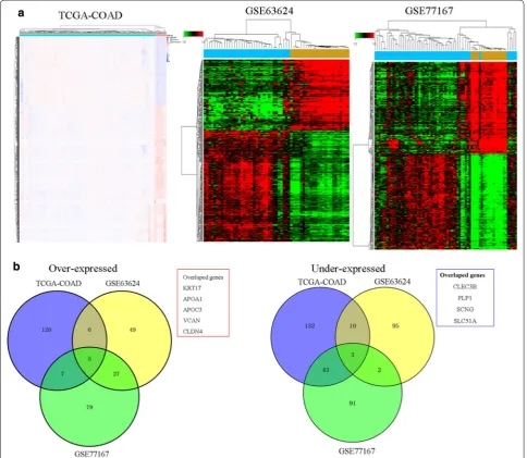
To identify DEGs that are played key role in colon tum-origenesis, we used an integrative analysis of TCGA colon adenocarcinoma (TCGA-COAD) and RNA-seq data and colon cancer gene expression data includin-ging GSE63624, and GSE77167 the publicly available GEO databases. We identified 175 genes deregulated in the TCGA data, 77 in GSE63624 datasets, and 57 in GSE77167 datasets under the condition of “Q < 0.001 and fold change > 4”. Total these DEGs are shown clustered
in Fig. 1a, then we founded only five genes consistently up-regulated and four down-regulated in all datasets (Fig. 1b).
GO functional and pathway enrichment analysis
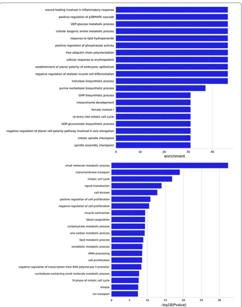
To determine significantly DEGs in human colon can-cer, gene ontology (GO) and pathway enrichment analysis were performed. We showed that the up/down-regulated DEGs were significantly enriched in wound healing involved in inflammatory response, positive regulation of phosphatase activity, cellular response to erythropoietin, negative regulation of skeletal muscle
cell differentiation, mitotic cell cycle, and cell division. Important genes and pathways involved in this process are shown in Fig. 2.
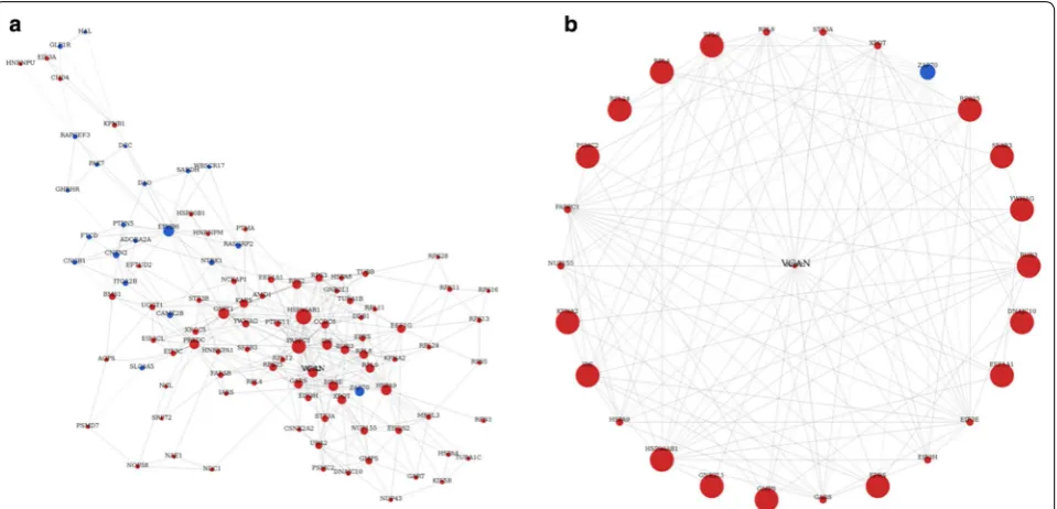
Co‑expression gene‑network analysis and candidate biomarker identification
To determine which gene or genes may play a pivotal role in the development of human colon cancer, we construct a gene–gene co-expression network. This co-expression network indicated HSP90AB1, VCAN, CLDN2, EPHB6, EIF3E, GSPT1, PRKDC, RPS2, GARS etc. play a key role in the progression of colon cancer (Fig. 3a). We selected VCAN over-expression genes because may be useful for early diagnosis biomarkers or therapeutic targets (Fig. 3b). We next further analysis the expression of in Colon adenocarcinoma (COAD), Adrenocortical carcinoma (ACC), Bladder Urothelial Carcinoma (BLCA), Breast invasive carcinoma (BRCA), Cervical squamous cell carcinoma and endocervical adenocarcinoma (CESC) using TCGA sequencing data and founded that is specifically upregulated in colon cancer (Fig. 4a).
VCAN expression is elevated in primary human colon cancer
To investigate the correlation between VCAN expres-sion and survival of colon cancer patient, we conducted
a Kaplan–Meier analysis with TCGA samples dichoto-mized into 2 groups with expression levels less than or equal to median and levels more than median of expres-sion. We fouded that patients in the high-risk group had significantly shorter median DFS than those in the low-risk group (Fig. 4b, c). qRT-PCR analysis were performed to detected the expression profile of VCAN in a panel of colon cancer cDNA arrays including 30 patients with colon cancer and 30 healthy controls. Result showed VCAN was significantly up-regulated at the mRNA level in colon cancer samples compared with normal colon tissues (Fig. 4d). We also founded that the expression level of VCAN mRNA was posi-tively correlated with clinical stage (P < 0.05), (Fig. 4e).
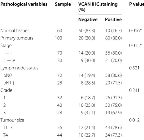
VCAN identified as a potential novel prognostic biomarker Immunohistochemical staining analysis in 100 human colon cancers and matched 60 adjacent tissue micro-arrays showed that VCAN was significantly over-expressed in colon cancer samples compared with adjacent tissue (Fig. 5a). Kaplan–Meier survival analysis showed that the overall survival and progression-free survival rates over 3 years for the high VCAN group were lower than those in the low VCAN group (Fig. 5b). Interestingly, we founded that VCAN expression lev-els significantly correlated with tumor size (P = 0.012) and clinical stage (P = 0.015) in colon cancer, but not
associated with other factors including pathological grading and lymph node status as shown in Table 1.
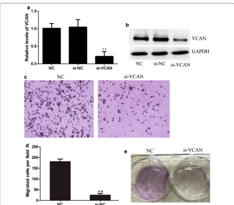
Silencing of VCAN inhibits HCT‑116 cell colony formation and migration
To determine the function of VCAN in regulating human colon cancer cell phenotype, we next performed knockdown of VCAN in HCT-116 cell line that with higher VCAN expression using small interfering RNA. Quantitative RT-PCR and Western blot analysis to quantitatively measure the effect of VCAN knockdown. Results that the VCAN expression was significantly decreased at both mRNA and protein levels in HCT116 cell lines (Fig. 6a, b). Transwell migration assays showed that knockdown of VCAN dramatically decreased cell migration (Fig. 6c, d). Furthermore, Colony formation assays showed that knockdown of VCAN inhibited cell proliferation in vitro.
Discussion
Treatments used for colon cancer may include some combination of surgery, radiation therapy, chemother-apy and targeted therchemother-apy [20–22]. Cancers that are con-fined within the wall of the colon may be curable with surgery while cancer that has spread widely are usually not curable, with management being directed towards improving quality of life and symptoms [23]. Five year survival rates in the United States are around 65%. This, however, depends on how advanced the cancer is, whether or not all the cancer can be removed with surgery, and the person’s overall health. Globally, colon cancer is the third most common type of cancer mak-ing up about 10% of all cases. In 2012, there were 1.4
Fig. 5 Expression of VCAN in colon cancer patient specimens. a Expression of VCAN in primary colon cancer and matched normal tissue (×100 or ×400). b Kaplan–Meier plots of VCAN expression in 37 cases of colon cancer patients. Overall survival rate was performed by log-rank test. (Immunoreactivity scores < 4 was ascribed to be low VCAN expression, immunoreactivity scores ≥ 4 was ascribed to be high VCAN expression)
Table 1 Correlation of VCAN protein expression with clinicopathological data (Fisher’s exact test)
*P value < 0.05 was consider significant
Pathological variables Sample VCAN IHC staining
(%) P value
Negative Positive
Normal tissues 60 50 (83.3) 10 (16.7) 0.016* Primary tumours 100 20 (20.0) 80 (80.0)
Stage 0.015*
I + II 70 14 (20.0) 56 (80.0) III + IV 30 9 (30.0) 21 (70.0)
Lymph node status 0.521
pN0 72 14 (19.4) 58 (80.6) pN1+ 28 8 (28.5) 20 (71.5)
Grade 0.241
1 32 6 (18.7) 26 (91.3)
2 40 10 (25.0) 30 (75.0)
3 28 9 (32.1) 19 (67.9)
Tumour size 0.012
million new cases and 694,000 deaths from the disease [6, 24].
Previous studies demonstrate VCAN is involved in cell adhesion, proliferation, migration and angiogen-esis and plays a central role in tissue morphogenangiogen-esis and maintenance. Zhao et al. reported miR-135a-5p could affect the proliferation, invasion and migration of thyroid carcinoma cells by targeting VCAN [18]. Sathyan et al. reported Versican plays an important role in extracellular matrix assembly and plays a major role in the pathogenesis of IA [17]. The linkage studies also indicated VCAN as a putative candidate gene for
IA in the 5q22-31 region. Chida et al. reported VCAN protein was detected exclusively in cancer stroma by immunohistochemistry, demonstrating a stepwise increase of stromal VCAN from normal tissues through stage 0 to stage IV tumors [25].
Our data demonstra that the expression level of VCAN mRNA was positively correlated with pathologic grade, clinical stage, VCAN were significantly over-expressed in metastasis samples compared with primary tumors. Immunohistochemical staining analysis in 100 human colon cancers and matched adjacent tissue microarray showed that VCAN was significantly over-expressed in
colon cancer samples compared with adjacent tissue. A loss-of-function study revealed that colony formation assays showed that knockdown of VCAN inhibited cell proliferation in vitro. Transwell migration assays showed that knockdown of VCAN dramatically decreased cell migration.
Conclusions
In conclusion, we demonstrated for the first time that VCAN is over-expressed in colorectal cancer and VCAN promotes colorectal cancer cell growth in vitro. These data suggest VCAN might serve as a potential target in the diagnosis and/or treatment in colorectal cancer.
Abbreviations
COAD: colon adenocarcinoma; ACC : adrenocortical carcinoma; BLCA: bladder urothelial carcinoma; BRCA : breast invasive carcinoma; CESC: cervical squa-mous cell carcinoma and endocervical adenocarcinoma; qRT-PCR: quantita-tive reverse transcription polymerase chain reaction; PBS: phosphate buffered saline; TCGA : The Cancer Genome Atlas; GEO: gene expression omnibus.
Authors’ contributions
We thanked XL for clinical information collection. Performed the experiments and collected data: XL, ZW. Analyzed the data: ZD. Conceived and designed the experiments: GL. Contributed reagents/materials: ZW. Wrote the paper: XL, ZW. Revised the paper: ZW. All authors read and approved the final manuscript.
Author details
1 Department of Gastroenterological Surgery, The First Affiliated Hospital of Chongqing Medical University, Chongqing Medical University, 1# Youyi Road, Chongqing 400016, People’s Republic of China. 2 Department of General Surgery, Mianyang Central Hospital, Mianyang 621000, Sichuan, People’s Republic of China.
Acknowledgements
We thanked Rong Jiang of Chongqing Medical University for clinical informa-tion collecinforma-tion.
Competing interests
The authors declare that they have no competing interests.
Availability of data and materials
The datasets used and/or analysed during the current study are available from the corresponding author on reasonable request.
Availability of data and materials
The datasets used and/or analysed during the current study are available from the corresponding author on reasonable request.
Consent for publication
The manuscript did not contain any individual person’s data in any form.
Ethics approval and consent to participate
The study has been approved by the ethical committee of Chongqing Medi-cal University.
Funding
This work was supported by National Natural Science Foundation of China (No. 81272753).
Publisher’s Note
Springer Nature remains neutral with regard to jurisdictional claims in pub-lished maps and institutional affiliations.
Received: 28 May 2018 Accepted: 10 September 2018
References
1. O’Keefe SJ. Diet, microorganisms and their metabolites, and colon cancer. Nat Rev Gastroenterol Hepatol. 2016;13(12):691–706.
2. Irrazabal T, Belcheva A, Girardin SE, Martin A, Philpott DJ. The mul-tifaceted role of the intestinal microbiota in colon cancer. Mol Cell. 2014;54(2):309–20.
3. Segal NH, Saltz LB. Evolving treatment of advanced colon cancer. Annu Rev Med. 2009;60:207–19.
4. Rustgi AK. The genetics of hereditary colon cancer. Genes Dev. 2007;21(20):2525–38.
5. Tauriello DVF, Palomo-Ponce S, Stork D, Berenguer-Llergo A, Badia-Ramentol J, Iglesias M, Sevillano M, Ibiza S, Canellas A, Hernando-Mom-blona X, et al. TGFbeta drives immune evasion in genetically reconsti-tuted colon cancer metastasis. Nature. 2018;554(7693):538–43. 6. Chung DC. Genetic testing and early onset colon cancer.
Gastroenterol-ogy. 2018;154(4):788–9.
7. Seidel DV, Azcarate-Peril MA, Chapkin RS, Turner ND. Shaping functional gut microbiota using dietary bioactives to reduce colon cancer risk. Semin Cancer Biol. 2017;46:191–204.
8. Viguier J, Morere JF, Brignoli-Guibaudet L, Lhomel C, Couraud S, Eisinger F. Colon cancer screening programs: impact of an organized screening strategy assessed by the EDIFICE surveys. Current oncology reports. 2018;20(Suppl 1):16.
9. Wu C. Systemic therapy for colon cancer. Surg Oncol Clin N Am. 2018;27(2):235–42.
10. Yu L, Zhou Y, Yang Y, Lu F, Fan Y. Efficacy and safety of compound kushen injection on patients with advanced colon cancer: a meta-analysis of randomized controlled trials. eCAM. 2017;2017:7102514.
11. Gkekas I, Novotny J, Pecen L, Strigard K, Palmqvist R, Gunnarsson U. Microsatellite instability as a prognostic factor in stage II colon cancer patients, a meta-analysis of published literature. Anticancer Res. 2017;37(12):6563–74.
12. Merlano MC, Granetto C, Fea E, Ricci V, Garrone O. Heterogeneity of colon cancer: from bench to bedside. ESMO open. 2017;2(3):e000218. 13. Franklin BR, McNally MP. Laparoscopy for colon cancer. Clin Colon
Rectal Surg. 2017;30(2):99–103.
14. Conde J, Oliva N, Zhang Y, Artzi N. Local triple-combination therapy results in tumour regression and prevents recurrence in a colon cancer model. Nat Mater. 2016;15(10):1128–38.
15. Staal FJ, van der Burg M, Wessels LF, Barendregt BH, Baert MR, van den Burg CM, van Huffel C, Langerak AW, van der Velden VH, Reinders MJ, et al. DNA microarrays for comparison of gene expression profiles between diagnosis and relapse in precursor-B acute lymphoblastic leukemia: choice of technique and purification influence the identifica-tion of potential diagnostic markers. Leukemia. 2003;17(7):1324–32. 16. Centeno BA, Enkemann SA, Coppola D, Huntsman S, Bloom G, Yeatman
TJ. Classification of human tumors using gene expression profiles obtained after microarray analysis of fine-needle aspiration biopsy samples. Cancer. 2005;105(2):101–9.
17. Sathyan S, Koshy LV, Balan S, Easwer HV, Premkumar S, Nair S, Bhat-tacharya RN, Alapatt JP, Banerjee M. Association of Versican (VCAN) gene polymorphisms rs251124 and rs2287926 (G428D), with intracra-nial aneurysm. Meta Gene. 2014;2:651–60.
18. Zhao X, Sun Z, Li H, Jiang F, Zhou J, Zhang L. MiR-135a-5p modulates biological functions of thyroid carcinoma cells via targeting VCAN 3′-UTR. Cancer Biomark. 2017;20(2):207–16.
•fast, convenient online submission
•
thorough peer review by experienced researchers in your field
• rapid publication on acceptance
• support for research data, including large and complex data types
•
gold Open Access which fosters wider collaboration and increased citations maximum visibility for your research: over 100M website views per year
•
At BMC, research is always in progress.
Learn more biomedcentral.com/submissions
Ready to submit your research? Choose BMC and benefit from: 20. Venook AP. Advances in adjuvant therapy for colon cancer: p value
or practical value. Journal of clinical oncology. 2018. https ://doi. org/10.1200/JCO.2018.77.8423.
21. Andre T, Vernerey D, Mineur L, Bennouna J, Desrame J, Faroux R, Fratte S, Hug de Larauze M, Paget-Bailly S, Chibaudel B, et al. 3 versus 6 months of oxaliplatin-based adjuvant chemotherapy for patients with stage III colon cancer: disease-free survival results from a randomized, open-label, international duration evaluation of adjuvant (IDEA) France, phase III trial. Journal of clinical oncology. 2018;36:1469–77.
22. Ilson DH. Adjuvant therapy in colon cancer: less is more. Lancet Oncol. 2018;19(4):442–3.
23. Park H, Chen B, Ciorba MA. Progress in PD-1-based Immunotherapy: new mechanistic insight may provide expanded hope for application to colon and gastrointestinal cancers. Gastroenterology. 2017;153(4):1162–3. 24. Fadelu T, Zhang S, Niedzwiecki D, Ye X, Saltz LB, Mayer RJ, Mowat RB,
Whittom R, Hantel A, Benson AB, et al. Nut consumption and survival in patients with stage III colon cancer: results from CALGB 89803 (Alliance). J Clin Oncol. 2018;36(11):1112–20.