Michał Moritz
1, A–F, Małgorzata Geszke-Moritz
2, A–FRecent Developments in the Application of Polymeric
Nanoparticles as Drug Carriers
1 Department of General and Analytical Chemistry, Faculty of Chemical Technology, Institute of Chemistry and Technical Electrochemistry, Poznan University of Technology, Poland
2 NanoBioMedical Centre, Adam Mickiewicz University, Poznań, Poland
A – research concept and design; B – collection and/or assembly of data; C – data analysis and interpretation; D – writing the article; E – critical revision of the article; F – final approval of article
Abstract
Nanotechnology is an interdisciplinary field of science offering interesting solutions for many branches of human life. Nanomaterials, defined as structures with at least one dimension below 100 nm, are the focus of increasing research attention as versatile tools for nanomedicine. Among the various nanostructures recently described in the literature, polymeric nanoparticles, characterized by satisfying biocompatibility, have aroused great interest as the carriers for various biologically active substances such as drugs, proteins and nucleic acids. These nanopar-ticles have already been reported as efficient vehicles for therapeutic agents in many disease entities. They can be delivered to the body via different administration routes. This review addresses recent advances in the usage of polymeric nanoparticles as drug carriers described in the years 2013 and 2014. The advantages of polymeric nanocarriers for medical application are highlighted, including their low toxicity, evaluated in vitro and in vivo. Moreover, the classification of polymeric nanoparticles is presented as well as various protocols of their synthesis (Adv Clin Exp Med 2015, 24, 5, 749–758).
Key words: biocompatibility, polymeric nanoparticles, drug carrier, nanomedicine.
EDITORIAL
Adv Clin Exp Med 2015, 24, 5, 749–758
DOI: 10.17219/acem/31802 © Copyright by Wroclaw Medical University ISSN 1899–5276
The enormous progress recently observed in the field of nanotechnology provides many inter-esting tools for medical sciences. Nanomaterials are structures with at least one dimension below 100 nm [1]. These materials possess unique fea-tures such as large surface-to-volume ratio result-ing from their small dimensions. The specific char-acteristics of nanostructures make them extremely attractive as valuable devices for nanomedicine. Among the various nanomaterials, quantum dots [2], graphene [3], carbon nanotubes [4], me-tallic and metal oxide nanoparticles [5] and poly-meric nanoparticles [6] have already been reported as promising candidates for medical applications. Particularly the latter have recently been investi-gated as carriers for biologically active substances and widely used for the potential treatment of ma-ny disease entities.
Polymeric nanoparticles may provide target-ed drug delivery, especially desirable for cancer therapy, markedly decreasing the systemic side
premature release and degradation before reach-ing the targeted site [18]. Polymer-based nanopar-ticles can be used as the carrier for numerous bi-ologically active molecules including drugs [18], proteins [7], monoclonal antibodies [13], nucleic acids [19, 20], biological extracts [16, 21] and oth-ers [22]. This review will be especially concerned with the application of polymer-based nanoparti-cles for the treatment of various disease entities in-cluding bacterial [23, 24], fungal [25] and parasit-ic [26] infections, ulcers [11], hypertension [12], angina [8], glaucoma [9], uveitis [27], asthma [22], cancer [15, 28], neurodegenerative disease [29] and many others [30, 31]. It deserves to be empha-sized that several polymers are characterized by satisfying biocompatibility and predictable biode-gradability [9, 22] while others are non-degradable or undergo slow degradation [22, 32]. Remarkably, polymer–based nanoparticles can be administered orally [20, 23], intravenously [18], percutaneous-ly [30], ophthalmically [27], pulmonarily [12, 25], transmucosally to nose and lungs [33] or delivered to the brain via inner ear administration [16]. It is noteworthy that selected polymers are approved by the Food and Drug Administration (FDA) to be used for therapeutic applications [27].
Classification and Properties
of Polymeric Nanoparticles
Recent advances in nanotechnology provide us with a wide range of polymers able to be for-mulated into nanoparticulate carriers for various active substances. Numerous synthetic and natu-ral polymeric materials, including biodegradable, biocompatible and non-biodegradable substanc-es, have already been used for nanoparticle prep-aration. Among the synthetic biodegradable poly-meric matrices, poly(lactide-co-glycolide) (PLGA) [26, 28, 34] and poly(D, L-lactide) (PLA) [35] are the most common. Polymeric materials such as poly(methylmethacrylate) (PMMA) [36] and poly(ethylene-co-vinyl acetate) (PEVA) [12] are characterized by satisfying biocompatibility. On the other hand, poly(ethylene glycol) (PEG) [15] and Eudragits® [10, 31] belong to the group ofnon-biodegradable synthetic polymeric materials. The -biodegradable natural polymers already used for the preparation of nanoparticulate DDSs include poly(L-glutamic acid) (PGA) produced by Bacillus subtilis [37], pullulan produced from starch by the fungus Aureobasidium pullulans [33], gelatin [6], alginate [9, 33], chitosan [15] and its derivatives including N-palmitoyl chitosan [14] or mannose-modified trimethyl chitosan-cysteine (MTC) con-jugate [20] and many others [19].
Intriguingly, the widespread application of PLGA results from its relatively inert composition, stable rate of degradation and known degradation products [22], namely lactic acid and glycolic ac-id [23]. PLGA-based nanoparticles are character-ized by good mechanical stability and narrow size distribution [38]. This polymer, used as a matrix during nanoparticulate carrier preparation, can protect the drugs from enzymatic degradation [27] after oral administration [23]. More relevantly, it has been reported that the drug release rate can be controlled by the PLGA molecular weight as well as by the glycolide to lactide ratio [23]. Polymer-based nanoparticles are also promising candidates for non-invasive transdermal delivery without re-moving the stratum corneum [30]. In this view, es-pecially biocompatible and biodegradable chitosan possessing mucoadhesive properties can signifi-cantly enhance penetration into the deeper skin lay-ers [30]. Furthermore, similarly to PLGA, it has the approval of the FDA for biomedical usage [15, 22]. Remarkably, PEG can be used as a stabilizer dur-ing polymeric nanoparticle preparation [15] or applied as a linker for conjugation of targeting li-gands to the surface of the nanoparticles [17]. Ad-ditionally, the modification of drug carriers with water-soluble PEG can contribute to a significant increase of the circulation half-life in vivo of as-prepared nanovehicles [18].
Biologically active substances can be encap-sulated in polymeric nanoparticles and consist of a so-called core-shell system [34]. In such a case, an active agent is protected from degradation by a polymeric coat able to be functionalized to specif-ically recognize the targeted site [28]. Other strate-gies consist of dispersion of the active substance in the polymeric matrix [24]. Recently, the polymeric form of a prodrug containing the molecules of the active substance coupled with the polymeric back-bone has been demonstrated to release the ther-apeutic agent upon hydrolytic degradation [39]. Curiously, drug-loaded polymeric nanoparticles incorporated into other matrices (e.g. synthetic polymers) can consist of a component of materials designed for biomedical applications [35].
Synthesis of Polymeric
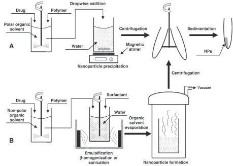
Nanoparticles
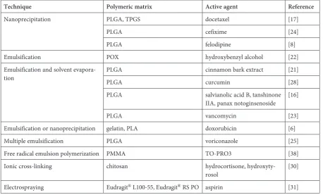
polymer-based nanoparticle preparation are nano-precipitation, emulsion/solvent evaporation and its modifications. These approaches of polymeric nanoparticle synthesis are schematically present-ed in Fig. 1. In the next paragraph, the strategies most recently described demonstrating fabrication of polymeric nanoparticles will be presented. Some examples are reviewed in the main text while oth-ers are summarized in Table 1.
Zhao and Feng [13] have described the prepa-ration of nanoparticles composed of poly(lactide)- -D-α-tocopheryl polyethylene glycol 1000 succi-nate copolymer (PLA-TPGS) and carboxyl group-terminated TPGS (TPGS-COOH) using the nano-precipitation method. The previously synthesized polymers, PLA-TPGS and TPGS-COOH, and the drug were dissolved in acetone and dropwise add-ed to water to form nanoparticles. The as-preparadd-ed carrier was loaded with docetaxel and chemically conjugated with a humanized monoclonal anti-body via a carboxyl terminal group present in the prepared polymeric matrix [13]. A curious instance of carvedilol-loaded nanoparticles composed of poly(ethylene-co-vinyl acetate) (PEVA) prepared
via the emulsion/solvent evaporation method has been described by Varshosaz et al. [12]. In this
work, the solution of the polymer in carbon tet-rachloride was mixed with a solution of the drug and PEG400 in dichloromethane. The as-pre-pared mixture was dispersed in the aqueous phase containing Tween 20 and agitated until complete evaporation of the organic phase [12]. Interest-ing studies have been also conducted by Alai and Lin [11] who have prepared lansoprazole-load-ed polymeric nanoparticles composlansoprazole-load-ed of PLGA. The nanoparticles were prepared using a double water-in-oil-in-water (W/O/W) emulsion/solvent evaporation method. The drug and polymer dis-solved in a mixture of dichloromethane and ace-tone were emulsified with an aqueous solution of NaHCO3. The as-prepared primary emulsion was
added to the aqueous solution of PVA and emul-sified. After stirring, the emulsion was evaporated and the nanoparticles were collected after centrif-ugation [11]. It is also worth mentioning that the group of Hazra et al. [36] have used an oil-in-wa-ter (O/W) modified atomized emulsification pro-cess to fabricate core/shell poly(methyl methac-rylate) (PMMA)/biosurfactant nanoparticles. The nanoparticles were loaded with ibuprofen, anthra-quinone and curcumin. As biosurfactants, rham-nolipid, surfactin and trehalose lipid have been
used. At the outset, the tenside was dissolved in water. Then, an aqueous solution of ammonium persulfate was added to the as-prepared solution to initiate the formation of free radicals. The mono-mer was sprayed over the surface of the biosurfac-tant solution to form nanoparticles [36]. As has been demonstrated by Katiyar et al. [9], the gela-tion method can be also employed for the prep-aration of polymeric nanoparticles. In this study, chitosan nanoparticles loaded with dorzolamide were formed by the dropwise addition of tripoly-phosphate to a dorzolamide-containing chito-san solution. Finally, the sedimented nanoparti-cles were suspended in a sodium alginate solution to obtain an in situ gel [9]. A curious instance has been described by Narayanan et al. [18] who have used the desolvation method for preparation of poly(ethylene glycol)-modified gelatin nanoparti-cles loaded with ibuprofen. High molecular weight aggregates of gelatin dispersed in water were mixed with a solution of ibuprofen in ethanol. The dropwise addition of acetone resulted in precipita-tion of the ibuprofen-loaded nanoparticles which were subsequently cross-linked by adding calcium chloride [18]. Another group has prepared pul-lulan-based nanoparticles using the polyelectro-lyte complexation method [33]. The negatively- -charged sulfated and positively-charged aminated derivatives of the polymer were firstly synthesized
by sulfonation and alkylation of the original poly-saccharide, respectively. The interactions of sulfat-ed pullulan-chitosan and aminatsulfat-ed pullulan-carra-geenan resulted in the formation of nanoparticles. The as-obtained nanocarriers were able to associ-ate with the model protein, bovine serum albumin (BSA), demonstrating the potential of the as-pre-pared system for protein delivery [33].
The group of Kwon et al. [39] has conducted compelling studies on the preparation of nanopar-ticles based on a polymeric prodrug of vanillin. In this work, the polymeric material was composed of poly(vanillin oxalate) (PVO) covalently coupled vanillin in its backbone. The as-synthesized PVO dissolved in dichloromethane was added to the so-lution of poly(vinyl alcohol) (PVA) to form oil- -in-water (O/W) emulsion. The nanoparticles were obtained by the evaporation of the solvent. The vanillin was released from the as-prepared na-no-carrier upon its hydrolytic degradation [39].
Toxicity and Bio-
-compatibility Concerns
The biocompatibility of polymeric nanoparti-cles used as a drug carrier is of crucial importance for development of drug delivery systems based on polymeric materials [33]. Hence, polymer-basedTable 1. Various strategies of polymeric nanoparticle fabrication
Technique Polymeric matrix Active agent Reference
Nanoprecipitation PLGA, TPGS docetaxel [17]
PLGA cefixime [24]
PLGA felodipine [8]
Emulsification POX hydroxybenzyl alcohol [22] Emulsification and solvent
evapora-tion PLGAPLGA cinnamon bark extractcurcumin [21][28]
PLGA salvianolic acid B, tanshinone IIA, panax notoginsenoside [16]
PLGA vancomycin [23]
Emulsification or nanoprecipitation gelatin, PLA doxorubicin [6] Multiple emulsification PLGA voriconazole [25] Free radical emulsion polymerization PMMA TO-PRO3 [38] Ionic cross-linking chitosan hydrocortisone,
hydroxyty-rosol [30]
Electrospraying Eudragit® L100-55, Eudragit® RS PO aspirin [31]
CA – cholic acid; PEG – poly(ethylene glycol); PLA – poly(lactide); PLGA – poly(lactide-co-glycolide);
nanoparticles should be examined through vari-ous in vitro and in vivo assays including cell via-bility, hemocompatibility and histological analy-sis [18]. One of the most common methods used to examine the in vitro cytotoxicity of polymer-ic nanopartpolymer-icles is the MTT test [13]. In the MTT (3-(4,5-dimethylthiazol-2-yl)-2,5-diphenyl tetra-zolium bromide) assay, the metabolic activity of cells is assessed relying on the evaluation of cel-lular enzyme activity [33]. In this test, the forma-tion of blue formazan dye is related to mitochon-drial dehydrogenase activity. High cell viability is ascribed to a high concentration of formazan dye, mirroring the high amount of metabolically active cells [33].
The group of Dionísio et al. [33] has exam-ined the in vitro cytotoxicity of pullulan-based nanoparticles using MTT assay. The experiment was performed on respiratory Calu-3 cells, which is an immortalized cell line derived from lung ade-nocarcinoma. It has been found that upon 24 h ex-posure to the polymeric nanoparticle formulation, cell viability was in the range from 74 to 94% inde-pendently of polymer modification, concentration or incubation time. Such results indicate the bio-logically acceptable viability of Calu-3 cells without disturbing their metabolic functions when in con-tact with pullulan-based nanoparticles [33]. The MTT assay was also employed for the evaluation the cytotoxicity of poly(vanillin oxalate) (PVO) nanoparticles [39]. The test, performed on RAW 264.7 cells, revealed dose-dependent cytotoxici-ty after 24 h of incubation with PVO nanoparti-cles. Interestingly, no or a negligible cytotoxic ef-fect was observed at nanoparticle doses less than 500 μg/mL, indicating a satisfying biocompatibil-ity of the prepared nanocarrier. Furthermore, tis-sue compatibility was examined using Balb/c mice intramuscularly injected with 100 μL of nanopar-ticles (1 mg/mL) suspended in phosphate buffer saline (PBS). The histological analysis of the mice, sacrificed at 7 days after injection, revealed min-imal inflammatory response as demonstrated by the very little formation of immune and inflamma-tory cells [39]. Another group [36] has examined the in vitro cytotoxicity of core/shell poly(methyl methacrylate)/biosurfactant nanoparticles upon incubation with peripheral blood mononuclear cells using MTT assay. Moreover, the nanoparti-cle samples were exposed to anticoagulated hu-man blood to assess the antihemolytic activity. The results of the MTT test revealed that cell sur-vival decreased with increasing concentrations of the nanoparticles. Meanwhile, a hemolysis as-say showed that nanoparticles coated with surfac-tin are less toxic than those surrounded by rham-nolipid and trehalose lipid biosurfactants [36].
Interestingly, Papa and co-workers [38] used a CellTiter96® Aqueous Non-Radioactive Cell
Pro-liferation Assay (3-(4,5-dimethylthiazol-2-yl)-5-(3- -carboxymethoxyphenyl)-2-(4-sulfophenyl)-2H-te- trazolium, inner salt, MTS) to determine the cell vi-ability of microglial cells treated with poly(methyl methacrylate) nanoparticles. Upon incubation with polymeric nanoparticles up to 6 days, the cel-lular viability was not affected [38].
It is worth noting that in cancer therapy the toxicity driven by a nanoparticulate carrier has a desirable effect. For example, Kulhari and co- -workers [7] employed the MTT test to assess the
in vitro cytotoxicity of PLGA nanoparticles load-ed with docetaxel and conjugatload-ed with bombe-sin peptide. The cellular toxicity studies ubombe-sing MDA-MB-231 breast cancer cells overexpress-ing gastrin releasoverexpress-ing peptide (GRP) receptor re-vealed 12-fold higher toxicity of the as-prepared nanoparticles as compared to pure docetaxel and Taxotere. Interestingly, the toxicity of bombesin peptide-conjugated nanoparticles was significant-ly improved as compared to the docetaxel-load-ed formulation in a dose-dependent manner from 6.25 to 100 ng/mL. The results demonstrated the potential of a nanocarrier in anticancer thera-py especially for the targeted therathera-py of cancer cells overexpressing GRP receptor [7]. Important
studies have been also conducted by Zhao and Feng [13] who have employed the MTT test for an examination of the in vitro cytotoxicity of Her-ceptin-functionalized nanoparticles composed of poly(lactide)-D-α-tocopheryl polyethylene glycol 1000 succinate copolymer (PLA-TPGS) and load-ed with doxorubicin. The viability studies were conducted using SK-BR-3 breast cancer cells up-on exposure to the drug-loaded nanocarrier with or without functionalization with herceptin. The experiment revealed dose-dependent toxicity and a reduction of cellular viability upon incubation with antibody-functionalized nanoparticles as compared to a non-conjugated polymeric carri-er. This may result from the intrinsic toxicity of Herceptin [13].
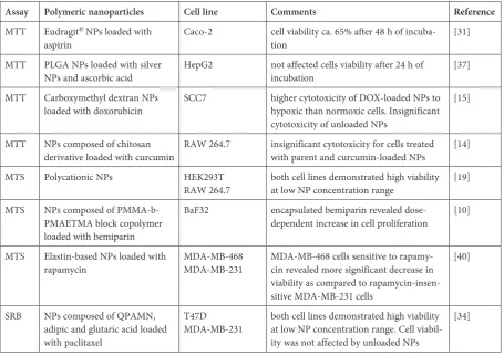
Other examples of cytotoxicity assays per-formed on cells exposed to polymer-based nanoparticles are summarized in Table 2.
Table 2. In vitro methods for evaluation of polymeric nanoparticle cytotoxicity
Assay Polymeric nanoparticles Cell line Comments Reference
MTT Eudragit®NPs loaded with
aspirin Caco-2 cell viability ca. 65% after 48 h of incuba-tion [31] MTT PLGA NPs loaded with silver
NPs and ascorbic acid HepG2 not affected cells viability after 24 h of incubation [37] MTT Carboxymethyl dextran NPs
loaded with doxorubicin SCC7 higher cytotoxicity of DOX-loaded NPs to hypoxic than normoxic cells. Insignificant cytotoxicity of unloaded NPs
[15]
MTT NPs composed of chitosan
derivative loaded with curcumin RAW 264.7 insignificant cytotoxicity for cells treated with parent and curcumin-loaded NPs [14] MTS Polycationic NPs HEK293T
RAW 264.7 both cell lines demonstrated high viability at low NP concentration range [19] MTS NPs composed of
PMMA-b-PMAETMA block copolymer loaded with bemiparin
BaF32 encapsulated bemiparin revealed
dose-dependent increase in cell proliferation [10]
MTS Elastin-based NPs loaded with
rapamycin MDA-MB-468MDA-MB-231 MDA-MB-468 cells sensitive to rapamy-cin revealed more significant decrease in viability as compared to rapamycin-insen-sitive MDA-MB-231 cells
[40]
SRB NPs composed of QPAMN, adipic and glutaric acid loaded with paclitaxel
T47D
MDA-MB-231 both cell lines demonstrated high viability at low NP concentration range. Cell viabil-ity was not affected by unloaded NPs
[34]
Polymeric Nanoparticles
as Carriers for Therapeutic
Agents
Polymer-based nanoparticles have been suc-cessfully used as carriers for antimicrobial agents. For instance, the group of de Carvalho et al. [26] has designed amphotericin-loaded polymeric nanocarriers decorated with maghemite nanopar-ticles to achieve a site-specific effect to treat cu-taneous leishmaniasis. The antibiotic was encap-sulated in poly(lactide-co-glycolide) (PLGA) and dimercaptosuccinic acid (DMSA) nanoparticles. The antimicrobial activity of the prepared system was evaluated on C57BL/G mice infected intra-dermally with promastigotes of Leishmania ama-zonensis. Remarkably, the controlled drug release from the prepared nanosystem was provided up-on exposure to an AC magnetic field resulting in a magnetohyperthermia effect. The nanocar-rier exhibited significantly greater reduction in
Leishmania amazonensis number and cell viabil-ity as compared to a free form of the antibiotic. Moreover, it allowed an effective reduction of the dose frequency required to obtain the same ther-apeutic effect [26]. Intriguingly, Chaudhary and Kumar [24] have demonstrated the antibacteri-al activity of Cefixime-loaded PLGA nanoparti-cles against the intracellular multidrug resistance (MDR) of Salmonella typhimurium. The results revealed a sustained release of the antibiotic from the prepared formulation and its better perme-ation across rat intestines as compared to the free drug [24]. A curious instance has been explored by Hill and co-workers [21], who demonstrated that PLGA nanoparticles loaded with the natural anti-microbial cinnamon bark extract are effective in-hibitors of Salmonella typhimurium and Listeria monocytogenes. The authors highlighted the poten-tial of the as-prepared system for the food industry to help prevent foodborne diseases [21]. Interest-ing studies have been conducted by Phan and co- -workers [35], who have examined the use of nata-mycin-loaded nanoparticles composed of dextran and poly(D, L-lactide) (Dex-b-PLA) for the sur-face modification of contact lenses fabricated from various polymeric materials. It has been found that extended drug release up to 12 h was observed for contact lens materials composed of N, N -dimeth-ylacrylamide or [tris(trimethylsiloxy)silyl]-propyl methacrylate. The authors suggested that natamy-cin-loaded Dex-b-PLA nanoparticles incorporated into contact lens materials have the potential to be used for targeted drug delivery directly to the cor-nea for the treatment of fungal infections [35]. An-other group [25] has prepared voriconazole-loaded
PLGA nanoparticles for pulmonary drug delivery. After administration to mice using an inhalation chamber, the sustained drug release was observed for 15 days [25].
Polymeric nanoparticles have also been used successfully as carriers for antihypertensive drugs. As an example, pulmonary delivery of the antihy-pertensive drug carvedilol was the subject of stud-ies performed by Varshosaz and co-workers [12]. The authors prepared drug-loaded poly(ethylene-co-vinyl acetate) (PEVA) nanoparticles coated with chitosan. The system revealed mucoadhe-sive properties and provided prolonged drug re-lease up to 8 h. The drug delivery systems based on these nanoparticles spray-dried in the pres-ence of mannitol revealed low density, satisfying flow ability, small aerodynamic diameter and fine powder fraction demonstrating potential as a suit-able inhaler powder for the pulmonary delivery of carvedilol [12]. Another group [8] has observed the enhancement of felodipine bioavailability from a formulation based on PLGA nanoparticles. Fur-thermore, in an ex vivo experiment performed on isolated rat stomach and intestinal segments, the nanovehicles demonstrated sustained release of the antihypertensive drug [8].
a better performance of triamcinolone-loaded nanoparticles as compared to microparticles of tri-amcinolone and prednisolone acetate. It has been found that polymeric nanoparticles can be used as an efficient triamcinolone carrier for topical treat-ment, providing better patient compliance [27]. Alai and Lin [11] have described the application of lansoprazole-loaded polymeric nanoparticles for the treatment of gastric ulcers. The drug en-capsulated in Eudragit® RS 100 nanoparticles and
PLGA nanoparticles revealed sustained release be-havior in vitro. After oral administration of enter-ic-coated capsules containing the nanoparticles, the sustained release of lansoprazole up to 24 h was observed in ulcer-induced Wistar rats [11]. Another group [20] has described application of mannose-modified trimethyl chitosan-cysteine conjugate nanoparticles for oral delivery of thera-peutic TNF-α siRNA to macrophages. The authors observed effective improvement of siRNA protec-tion in the physiological environment and its en-hanced permeation through intestinal epithelium. Furthermore, siRNA uptake by the macrophages was facilitated through clathrin-independent en-docytosis. In vitro experiments revealed that orally delivered nanoparticles inhibited TNF-α secretion
by macrophages, thus protecting mice with acute hepatic injury from inflammation-induced liver damage [20]. Enteric-coated polymeric nanopar-ticles have been the subject of studies performed by Hao and co-workers [31]. The authors synthe-sized core/shell Eudragit® L100-55/Eudragit® RS
nanoparticles loaded with aspirin. The external polymer coat provided a pH-sensitive drug release while polymeric core material played the role of the skeleton allowing sustained aspirin release [31].
Other examples demonstrating the application of polymeric nanoparticles as a carrier for various therapeutic agents are listed in Table 3.
Conclusion
The usage of polymeric nanoparticles as drug carriers for the treatment of many disease enti-ties opens encouraging possibilienti-ties in clinical ap-plication. The present-day progress achieved in adapting polymeric nanoparticles for drug deliv-ery makes the future of these nanovehicles bright and exciting. Employed as drug carriers, polymer-based nanoparticles offer numerous attractive fea-tures such as targeted drug delivery, sustained
Table 3. Use of polymeric nanoparticles as drug vehicles
Nanoparticle
components Active substance Functionalizing agent Cell culture/ /animal model Pharmacological effect Reference
PLA-TPGS,
TPGS-COOH docetaxel herceptin SK-BR-3 targeted delivery of anticancer drugs [13] PLGA docetaxel bombesin
pep-tide MDA-MB-231 targeted delivery of anticancer drugs. Sustained drug release [7] Carboxymethyl
dextran DOX 2-nitrozoimid-azole derivative SCC7 hypoxia-responsive and sustained drug release [15] PMMA,
biosur-factants (rham-nolipid, surfactin, trehalose)
ibuprofen, anthraqui-none, cur-cumin
– PMBCs PH-responsive and sustained drug release; antibacterial activity against
Bacillus subtilis and Pseudomonas aeru-ginosa by induction of oxidative stress
[36]
Chitosan dorzolamide – Goat cornea, male albino rabbit
reduced frequency of administration in the treatment of glaucoma [9]
POX
hydroxyben-zyl alcohol – RAW 264.7/Balb/c mice simultaneous antioxidant and anti-inflammatory effect [22]
PVO – – RAW 264.7/
Balb/c mice step-wise release of antioxidant and anti-inflammatory vanillin upon hydro-lytic degradation of polymeric matrix
[39]
DOX – doxorubicin; PLA-TPGS – poly(lactide)-D-α-tocopheryl polyethylene glycol 1000 succinate copolymer;
PLGA – poly(lactide-co-glycolide); PMMA – poly(methyl methacrylate); POX – polyoxalate; PVO – poly(vanillin oxalate); TPGS-COOH – carboxyl group-terminated D-α-tocopheryl polyethylene glycol 1000 succinate.
release or prolonged circulation half-life. Polymer-ic nanopartPolymer-icles can protect the drug from degra-dation in physiological conditions and improve its bioavailability. Polymeric nanoparticle-based drug delivery systems have been successfully employed as carriers for anticancer, anti-inflammatory and
antihypertensive agents, antioxidants, and nucle-ic acids, among others. In spite of the scientifnucle-ic ad-vances in the usage of polymeric nanoparticles as drug vehicles, many more efforts from scientists from a variety of disciplines are needed before they can be fully exploited for therapeutic applications.
References
[1] Moritz M, Geszke-Moritz M: Application of nanomaterials in medical sciences. Chemik 2012, 66, 219–226. [2] Geszke-Moritz M, Moritz M: Quantum dots as versatile probes in medical sciences: Synthesis, modification and
properties. Mater Sci Eng C 2013, 33, 1008–1021.
[3] Yang K, Feng L, Shi X, Liu Z: Nano-graphene in biomedicine: theranostic applications. Chem Soc Rev 2013, 42, 530–547.
[4] Battigelli A, Ménard-Moyon C, Ros TD, Prato M, Bianco A: Endowing carbon nanotubes with biological and biomedical properties by chemical modifications. Adv Drug Delivery Rev 2013, 65, 1899–1920.
[5] Moritz M, Geszke-Moritz M: The newest achievement in synthesis, immobilization and practical applications of antibacterial nanoparticles. Chem Eng J 2013, 228, 596–613.
[6] Han S, Li M, Liu X, Gao H, Wu Y: Construction of amphiphilic copolymer nanoparticles based on gelatin as drug carriers for doxorubicin delivery. Colloids Surf B 2013, 102, 833–841.
[7] Kulhari H, Pooja D, Shrivastava S, Naidu VGM, Sistla R: Peptide conjugated polymeric nanoparticles as a carrier for targeted delivery of docetaxel. Colloids Surf B 2014, 117, 166–173.
[8] Shah U, Joshi G, Sawant K: Improvement in antihypertensive and antianginal effects of felodipine by enhanced absorption from PLGA nanoparticles optimized by factorial design. Mater Sci Eng C 2014, 35, 153–163.
[9] Katiyar S, Pandit J, Mondal RS, Mishra AK, Chuttani K, Aqil M, Ali A, Sultana Y: In situ gelling dorzolamide loaded chitosan nanoparticles for the treatment of glaucoma. Carbohydr Polym 2014, 102, 117–124.
[10] Reyes-Ortega F, Rodríguez G, Aguilar MR, Lord M, Whitelock J, Stenzel MH, Román JS: Encapsulation of low molecular weight heparin (bemiparin) into polymeric nanoparticles obtained from cationic block copolymers: properties and cell activity. J Mater Chem B 2013, 1, 850–860.
[11] Alai M, Lin WJ: Novel lansoprazole-loaded nanoparticles for the treatment of gastric acid secretion-related ulcers: In vitro and in vivo pharmacokinetic pharmacodynamic evaluation. Amer Assoc Pharm Sci J 2014, DOI: 10.1208/ /s12248-014-9564-0.
[12] Varshosaz J, Taymouri S, Hamishehkar H: Fabrication of polymeric nanoparticles of poly(ethylene-co-vinyl acetate) coated with chitosan for pulmonary delivery of carvedilol. J Appl Polym Sci 2014, 131, 39694–39701. [13] Zhao J, Feng SS: Effects of PEG tethering chain length of vitamin E TPGS with a Herceptin-functionalized
nanoparticle formulation for targeted delivery of anticancer drugs. Biomaterials 2014, 35, 3340–3347.
[14] Pu HL, Chiang WL, Maiti B, Liao ZX, Ho YC, Shim MS, Chuang EY, Xia Y, Sung HW: Nanoparticles with dual responses to oxidative stress and reduced pH for drug release and anti-inflammatory applications. ACS Nano 2014, 8, 1213–1221.
[15] Thambi T, Deepagan VG, Yoon HY, Han HS, Kim SH, Son S, Jo DG, Ahn CH, Suh YD, Kim K, Kwon IC, Lee DS, Park JH: Hypoxia-responsive polymeric nanoparticles for tumor-targeted drug delivery. Biomaterials 2014, 35, 1735–1743.
[16] Zhang X, Chen G, Wen L, Yang F, Shao A, Li X, Long W, Mu L: Novel multiple agents loaded PLGA nanopar-ticles for brain delivery via inner ear administration: In vitro and in vivo evaluation. Eur J Pharm Sci 2013, 48, 595–603.
[17] Zhang X, Dong Y, Zeng X, Liang X, Li X, Tao W, Chen H, Jiang Y, Mei L, Feng SS: The effect of autophagy inhibitors on drug delivery using biodegradable polymer nanoparticles in cancer treatment. Biomaterials 2014, 35, 1932–1943.
[18] Narayanan D, Geena MG, Lakshmi H, Manzoor K, Shantikumar N, Menon D: Poly-(ethylene glycol) modified gelatin nanoparticles for sustained delivery of the anti-inflammatory drug ibuprofen-sodium: An in vitro and in vivo analysis. Nanomedicine: NBM 2013, 9, 818–828.
[19] Forbes DC, Peppas NA: Polycationic nanoparticles for siRNA delivery: Comparing ARGET ATRP and UV-initiated formulations. ACS Nano 2014, 8, 2908–2917.
[20] He C, Yin L, Tang C, Yin C: Multifunctional polymeric nanoparticles for oral delivery of TNF-α siRNA to mac-rophages. Biomaterials 2013, 34, 2843–2854.
[21] Hill LE, Taylor TM, Gomes C: Antimicrobial efficacy of poly(DL-lactide-co-glycolide) (PLGA) nanoparticles with entrapped cinnamon bark extract against Listeria monocytogenes and Salmonella typhimurium. J Food Sci 2013, 78, 626–632.
[22] Yoo D, Guk K, Kim H, Khang G, Wu D, Lee D: Antioxidant polymeric nanoparticles as novel therapeutics for airway inflammatory diseases. Int J Pharm 2013, 450, 87–94.
[23] Zakeri-Milani P, Loveymi BD, Jelvehgari M, Valizadeh H: The characteristics and improved intestinal perme-ability of vancomycin PLGA-nanoparticles as colloidal drug delivery system. Colloids Surf B 2013, 103, 174–181. [24] Chaudhary SH, Kumar V: Taguchi design for optimization and development of antibacterial drug-loaded PLGA
[25] Sinha B, Mukherjee B, Pattnaik G: Poly-lactide-co-glycolide nanoparticles containing voriconazole for pulmo-nary delivery: in vitro and in vivo study. Nanomedicine: NBM 2013, 9, 94–104.
[26] de Carvalho RF, Ribeiro IF, Miranda-Vilela AL, Filho J de Sousa, Martins OP, Cintra e Silva Dde O, Tedesco AC, Lacava ZGM, Báo SN, Sampaio RNR: Leishmanicidal activity of amphotericin B encapsulated in PLGA-DMSA nanoparticles to treat cutaneous leishmaniasis in C57BL/6 mice. Exp Parasitol 2013, 135, 217–222. [27] Sabzevari A, Adibkia K, Hashemi H, Hedayatfar A, Mohsenzadeh N, Atyabi F, Ghahremani MH, Dinarvand R:
Polymeric triamcinolone acetonide nanoparticles as a new alternative in the treatment of uveitis: In vitro and in vivo studies. Eur J Pharm Biopharm 2013, 84, 63–71.
[28] Palange AL, Mascolo DD, Carallo C, Gnasso A, Decuzzi P: Lipid-polymer nanoparticles encapsulating cur-cumin for modulating the vascular deposition of breast cancer cells. Nanomedicine: NBM 2014, DOI: 10.1016/j. nano.2014.02.004.
[29] Tiwari SK, Agarwal S, Seth B, Yadav A, Nair S, Bhatnagar P, Karmakar M, Kumari M, Chauhan LKS, Patel DK, Srivastava V, Singh D, Gupta SK, Tripathi A, Chaturvedi RK, Gupta KC: Curcumin-loaded nanopar-ticles potently induce adult neurogenesis and reverse cognitive deficits in Alzheimer’s disease model via canonical W nt/β-catenin pathway. ACS Nano 2014, 8, 76–103.
[30] Hussain Z, Katas H, Amin MCIM, Kumolosasi E, Buang F, Sahudin S: Self-assembled polymeric nanoparticles for percutaneous co-delivery of hydrocortisone/hydroxytyrosol: An ex vivo and in vivo study using an NC/Nga mouse model. Int J Pharm 2013, 444, 109–119.
[31] Hao S, Wang B, Wang Y, Xu Y: Enteric-coated sustained-release nanoparticles by coaxial electrospray: prepara-tion, characterizaprepara-tion, and in vitro evaluation. J Nanopart Res 2014, 16, 2204–2214.
[32] Jana S, Maji N, Nayak AK, Sen KK, Basu SK: Development of chitosan-based nanoparticles through inter-poly-meric complexation for oral drug delivery. Carbohydr Polym 2013, 98, 870–876.
[33] Dionísio M, Cordeiro C, Remuñán-López C, Seijo B, da Costa AMR, Grenha A: Pullulan-based nanoparticles as carriers for transmucosal protein delivery. Eur J Pharm Sci 2013, 50, 102–113.
[34] Bae J, Nael MA, Jiang L, Hwang PT, Mahdi F, Jun H-W, Elshamy WM, Zhou YD, Murthy SN, Doerksen RJ, Jo S: Quinone propionic acid-based redox-triggered polymer nanoparticles for drug delivery: Computational analysis and in vitro evaluation. J Appl Polym Sci 2014, 131, 40461–40470.
[35] Phan CM, Subbaraman L, Liu S, Gu F, Jones L: In vitro uptake and release of natamycin Dex-b-PLA nanopar-ticles from model contact lens materials. J Biomater Sci, Polym Ed 2014, 25, 18–31.
[36] Hazra C, Kundu D, Chatterjee A, Chaudhari A, Mishra S: Poly(methyl methacrylate) (core)-biosurfactant (shell) nanoparticles: Size controlled sub-100 nm synthesis, characterization, antibacterial activity, cytotoxicity and sus-tained drug release behavior. Colloids Surf A 2014, 449, 96–113.
[37] Stevanović M, Bračko I, Milenković M, Filipović N, Nunić J, Filipič M, Uskoković DP: Multifunctional PLGA particles containing poly(L-glutamic acid)-capped silver nanoparticles and ascorbic acid with simultaneous anti-oxidative and prolonged antimicrobial activity. Acta Biomater 2014, 10, 151–162.
[38] Papa S, Ferrari R, de Paola M, Rossi F, Mariani A, Caron I, Sammali E, Peviani M, Dell’Oro V, Colombo C, Morbidelli M, Forloni G, Perale G, Moscatelli D, Veglianese P: Polymeric nanoparticle system to target activated microglia/macrophages in spinal cord injury. J Controlled Release 2014, 174, 15–26.
[39] Kwon J, Kim J, Park S, Khang G, Kang PM, Lee D: Inflammation-responsive antioxidant nanoparticles based on a polymeric prodrug of vanillin. Macromolecules 2013, 14, 1618–1626.
[40] Shi P, Aluri S, Lin Y-A, Shah M, Edman M, Dhandhukia J, Cui H, MacKay JA: Elastin-based protein polymer nanoparticles carrying drug at both corona and core suppress tumor growth in vivo. J Controlled Release 2013, 171, 330–338.
Address for correspondence:
Michał Moritz
Faculty of Chemical Technology
Institute of Chemistry and Technical Electrochemistry Poznan University of Technology
Berdychowo 4 60-965 Poznań Poland
Tel. +48 61 665 23 16
E-mail: [email protected]
Conflict of interest: None declared