Address for correspondence
Barbara Choromańska
E-mail: [email protected]
Funding sources
None declared
Conflict of interest
None declared
Acknowledgements
Scholarship Project “I’m studying, research-ing, commercialize – UMB doctoral support program” co-financed by the European Union under the European Social Fund.
Received on May 3, 2015 Revised on Novemeber 13, 2015 Accepted on March 22, 2016
Abstract
Atherosclerosis is a progressive, chronic inflammation in artery walls. Oxidized low density lipoproteins (ox-LDL) play an important role in atherosclerotic plaque formation. ox-LDL are taken up by macrophages mainly through scavenger receptors, among which CD36 is considered to be the most important. Animal studies have shown that crossing atherogenic mice with a strain lacking the expression of CD36 prevent-ed the development of atherosclerosis despite a diet rich in saturatprevent-ed LCFA. In humans, autopsy studies performed in obese patients have demonstrated increased expression of CD36 receptor on macrophages, comprised within atherosclerotic plaques. Until recently it had been believed that CD36 is a major player in atherosclerosis progression in humans. However, recent studies challenge this conviction, showing in-creased incidence of coronary heart disease in the subgroup of patients with dein-creased expression of CD36. This article reviews the role of CD36 receptor in the development of atherosclerosis. The authors also discuss current possibilities to interfere with CD36, their potential benefits and hazards.
Key words: atherosclerosis, CD36 receptor, ox-LDL
DOI
10.17219/acem/62325
Copyright
© 2017 by Wroclaw Medical University This is an article distributed under the terms of the Creative Commons Attribution Non-Commercial License (http://creativecommons.org/licenses/by-nc-nd/4.0/)
The role of CD36 receptor in the pathogenesis of atherosclerosis
Barbara Choromańska
1, D, Piotr Myśliwiec
1, D, Katarzyna Choromańska
2, D, Jacek Dadan
1, E, Adrian Chabowski
3, F1 1st Department of General and Endocrine Surgery, Medical University of Bialystok, Poland
2 SKN Dental Biochemistry at the Department of Conservative Dentistry, Medical University of Bialystok, Poland 3 Department of Physiology, Medical University of Bialystok, Poland
A – research concept and design; B – collection and/or assembly of data; C – data analysis and interpretation; D – writing the article; E – critical revision of the article; F – final approval of article
Cardiovascular diseases are the main cause of death in Poland. It is often the process of atherosclerosis which leads to heart attack, stroke, heart failure or arrhythmias. Atherosclerosis is a multifactorial, progressive disease involving endothelial dysfunction and chronic inflam-mation of the arteries. Monocytes transformed into mac-rophages accumulate oxidized low-density lipoproteins (ox-LDL) in the vessel wall, forming foam cells. Mono-cytes and macrophages express plasmalemmal receptors participating in the uptake of ox-LDL. When overloaded with fatty acids, macrophages transform into foam cells. These cells are the most important in the development of atherosclerosis.
CD36 receptor, its structure and role
CD36 belongs to class B scavenger receptors, which chemically or oxidatively bind modified lipoproteins, polyanions and apoptotic cells.1,2 It is a membrane
glyco-protein with a mass of 88 kDa, composed of a single pro-tein chain.3,4 CD36 is present on the surface of a number
of cells, such as adipocytes, monocytes, macrophages, platelets, endothelial, cardiac, skeletal and smooth mus-cle cells, dendritic cells, retinal pigment epithelium and hematopoietic precursors of red cells (Table 1).1,2,5,6 It is
a multifunctional receptor with the independent ability to bind to three major classes of ligands (modified phospho-lipids, long chain fatty acids (LCFA) and proteins contain-ing structural domains of thrombospondin homologs) which include: ox-LDL, oxidized and negatively charged phospholipids, thrombospondin, collagen, apoptotic cells, hexarelin, red cells infected with plasmodium malariae or the outer segment of the retinal pigment epithelium.1,2,7
CD36 has 85% homology with FAT/CD36 protein – fatty acid translocase. It belongs to the transmembrane trans-porter proteins, facilitating the transport of LCFA.8
Pro-atherosclerotic effect
of CD36 receptor
Excessive uptake of LCFA results in their accumulation in the cells, contributing to lipotoxicity and secondarily to the development of metabolic diseases like obesity, atherosclerosis or diabetes, with their consequences such as coronary heart disease and ischemic stroke. CD36 binds ox-LDLs after recognizing them through lipid com-ponents, namely negatively charged phospholipids. Lack of the lipid component of ox-LDLs inhibits their uptake by CD36 receptor and, as a result, reduces lipoprotein ac-cumulation in macrophages.2 After entering monocytes
or macrophages, LCFA are bound by cytoplasmic fatty acid binding proteins (FABPc) and then, depending on the needs of the cell, they are targeted to suitable intra-cellular locations for rendering: oxidation in mitochon-dria and peroxisomes or storage (mainly esterified) in the endoplasmic reticulum and the cytoplasm.9
In vitro, macrophages devoid of CD36 receptor, incubat-ed with increasincubat-ed concentrations of ox-LDL, did not show increased intracellular transport of ox-LDL. These mac-rophages did not accumulate cholesterol esters and they did not undergo transformation into foam cells.10 CD36
facilitates translocation of LCFA in adipocytes, hepato-cytes, and heart and skeletal myohepato-cytes, where LCFA are important substrates for energy production.1 Thus CD36
receptor participates in lipid utilization within muscles, fat energy storage and fat absorption in the intestines.4
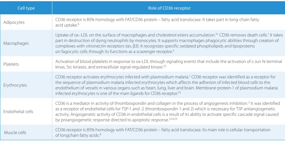
Table 1. The role of CD36 receptor in different cells
Cell type Role of CD36 receptor
Adipocytes CD36 receptor is 85% homology with FAT/CD36 protein – fatty acid translocase. It takes part in long-chain fatty acid uptake.8
Macrophages
Uptake of ox–LDL on the surface of macrophages and cholesterol esters accumulation.51 CD36 removes death cells.1 It takes
part in destruction of dying neutrophils by monocytes. It supports macrophages phagocytic abilities through creation of complexes with vitronectin receptors (αv, β3). It recognizes specific oxidated phospholipids and lipoproteins
on fagocytic cells through its functions as a scavenger receptor.3
Platelets Activation of blood platelets in response to ox-LDL through signaling events that include the activation of c-Jun N-terminal kinas, Src kinases, and extracellular signal-regulated kinase.52
Erythrocytes
CD36 receptor activates erythrocytes infected with plasmodium malaria.1 CD36 receptor was identified as a receptor for
the sequence of plasmodium malaria infected erythrocytes which affects the adhesion of infected blood cells to the endothelium of vessels in various organs such as heart, lung, liver and brain. Membrane protein-1 of plasmodium malaria infected erythrocytes is one of the main ligands for CD36 receptor.53
Endothelial cells
CD36 is a mediator in activity of thrombospondin and collagen in the process of angiogenesis inhibition.2 It was identified
as a receptor of endothelial cells for TSP-1 and -2 (thrombospondin 1 and 2) which is necessary for TSP antiangiogenetic activity. Angiogenetic activity of CD36 in endothelial cells is a result of its ability to activate specific cascade signal caused by proangiogenetic response directed to apoptotic response.3,54,55
in macrophages requires PAFR and CD36 recruitment into the same specific membrane microdomains (lipid rafts). oxLDL induced the expression of IL-10 mRNA only in HEK293T expressing both receptors CD36 and PAFR. In their experiments, they also observed a decrease of IL-10 production and oxLDL uptake caused by lipid raft disruption by treatment with methyl-β-cyclodextrin and an increase of oxLDL uptake when both receptors were present.16 By contrast, the blockage of CD36 reduced the
secretion of IL-1β, IL-6 and IL-8 and foam cell formation in human macrophages.17
Based on the results of the above studies, it can be con-cluded that reduction of CD36 receptor expression on macrophages prevents the development of atherosclero-sis. This has opened up the scope for further research on the regulation of CD36 expression to prevent atheroscle-rosis in humans.
However, Moore et al. showed that CD36 deficiency does not necessarily lead to a reduction in atherosclerotic lesions.18 They showed increased accumulation of foam
cells at the aortic sinus in CD36-ApoE double knockout mice compared to ApoE-KO mice.18 Further confusion
was caused by a Japanese study in 40 humans deficient in CD36 receptor. They were shown to have significantly higher incidence of coronary heart disease than the gen-eral population, suggesting that CD36 deficiency might in fact be atherogenic.19
Factors regulating the expression
of CD36 receptor
Expression of the CD36 gene is mainly controlled by the lipogenic transcription factor PPARγ, liver X receptor (LXR), nuclear pregnane X receptor (PXR) and testicular orphan nuclear receptor 4 (TR4).4,20
PPARγ (nuclear peroxisome proliferator-activated re-ceptor gamma) has ligands such as 15-deoksyΔ12,14-prostaglandin J2 and thiazolidinediones (drugs used to treat type 2 diabetes).PPARγ together with CD36 func-tion by positive feedback: the increased activity of PPARγ leads to increased expression of CD36 in macrophages.21
What is more, studies in recent years show that PPAR di-rectly affects CD36 expression and mediate oxLDL upta-ke in monocytes, which promotes foam cell formation.17
It is known that HDL has anti-atherogenic properties, ho-wever, the mechanism has not been fully elucidated. HDL probably reduces the accumulation of lipids in adipocytes by phosphorylation of PPAR.22 In contrast, Zhong et al.
suggest that HDL may increase oxLDL uptake in inflam-matory adipocytes stimulated by endotoxin lipopolysac-charide, which causes an increase in the expression of PPAR and CD36.23
Another factor regulating the expression of CD36 re-ceptor is HDL (high density lipoproteins). At higher con-centrations, HDL decrease the expression of CD36 by Upon interaction with ox-LDL, CD36 initiates a
sig-naling cascade, leading to ox-LDL uptake and foam cell formation. Cytoskeletal changes and inhibition of cell migration contribute to the trapping of foam cells within the atherosclerotic plaque. Interaction of ox-LDL with CD36 localized on macrophages triggers signals for the reaction, which is pro-inflammatory and pro-atheroscle-rotic. The signaling pathway involves activation of MAP kinases, Src family kinases and Vav family guanine nucle-otide exchange factors, leading to internalization of the ligand, foam cell formation and inhibition of migration.11
In vivo, the impact of CD36 on the development of atherosclerosis has been studied in mice with congenital deficiency of this receptor on macrophages. A significant reduction in atherosclerotic lesions (by 88%) was demon-strated.10 In order to assess the importance of CD36, an
atherogenic strain of mice lacking apolipoprotein E, was crossed with mice deficient in CD36. Mice deficient both in apolipoprotein E and CD36 showed no tendency to the development of atherosclerotic plaques, despite a diet rich in saturated LCFA. The extent of atherosclerotic lesions in the aorta was on average lower by 77% as compared to the mice lacking only apolipoprotein E.12
In addition to inhibition of lipid storage by macro-phages, mice deficient in CD36 receptor demonstrated improved insulin signaling.13 This implies the role of
CD36 receptor in insulin resistance development.
Animal studies have shown that CD36 participates in the inflammatory process by regulating membrane calcium influx. CD36 takes part in the activation of membrane calcium channels in response to endoplasmic reticulum stress. This results in phosphorylation of cyto-plasmic phospholipase A2 and its translocation to mem-branes in a CD36-dependent manner. The subsequent re-lease of arachidonic acid from phospholipids contributes to the generation of eicosanoids mediating the pleiotropic inflammatory effects. A major prostanoid metabolite of arachidonic acid formed with the participation of cyclo-oxygenases – prostaglandin E2 – is a potent proinflam-matory mediator.14 This study suggests that CD36 takes
part in the development of atherosclerosis by regulating the intracellular influx of calcium and synthesis of pros-taglandin E2 from arachidonic acid.
In humans, autopsy studies performed on atheroscle-rotic arteries in obese people with dyslipidemia showed increased expression of CD36 receptor on macrophages comprised within atherosclerotic plaques. In contrast, the expression of CD36 on macrophages present in the walls of arteries without atherosclerotic lesions was negligible.15
The stimulation of macrophages with oxLDL induced foam cell formation and cytokine secretion. Rios et al. showed that blocking CD36 and PAFR (Platelet Activat-ing Factor) decreases oxLDL uptake and IL-10 production by macrophages as well as PAFR and CD36 are colocal-ized in human atherosclerotic plaques.16 They found that
increasing the phosphorylation of PPARγ. This results in reduced transcription of CD36 mRNA.24
Expression of CD36 is enhanced by such pro-inflam-matory cytokines as IL-1 (interleukin-1), IL-4 and IL-18, as well as M-CSF (macrophage colony-stimulating factor) and GM-CSF (granulocyte-macrophage colony-stimu-lating factor).25–29 And increased concentration of
pro-inflammatory cytokines leads to increased expression of CD36 on the surface of macrophages and stimulates the accumulation of ox-LDL.21,24,30
CD36 expression is reduced by TGF-β (transforming growth factor beta) through phosphorylation of MAPK (mitogen-activated protein kinases), followed by phos-phorylation of PPARγ and reduced gene transcription of CD36 in macrophages.31 Also bacterial
lipopolysaccha-rides (LPS) and interferon gamma (IFN-γ) inhibit CD36 synthesis by reduction in mRNA expression in macro-phages.26,27,32 The reduced accumulation of cholesterol
in macrophages treated with IFN-γ and TNF-α, was also showed. This combined treatment of IFN-γ and TNF-α in mRNA reduced the CD36 expression on macrophages.33
Recent studies indicate that human neutrophil peptides (HNPs) take part in atherosclerosis development.34,35
Quinn et al.36 suggest that the HNP induces increase of
CD36 expression in the surface of macrophages and as a result contributes to the increased amount of foam cell formation.
Research on double knockout nuclear pregnane X re-ceptor (PXR) and ApoE mice showed the influence of PXR on the expression of CD36. Deficiency of PXR did not change the plasma levels of cholesterol and triglyc-eride in ApoE mice, but PXR and ApoE deficient mice had significantly decreased atherosclerotic lesions in the arteries.37 Moreover, expression of CD36, lipid
accumu-lation and CD36-mediated oxidized LDL uptake in the peritoneal macrophages of PXR and ApoE mice were re-duced by PXR deficiency. One of the human PXR antago-nists is bisphenol A (a chemical substance used in many consumer products). In PXR-humanized ApoE deficient mice, exposure to bisphenol A significantly increased the atherosclerotic lesion area in the aortic root and brachio-cephalic artery as well as increased CD36 expression and lipid accumulation in macrophages.38 This suggests that
deficiency of PXR reduces the risk of atherosclerosis de-velopment which may be due to decreased CD36 expres-sion and reduced lipid uptake in macrophages.37
Méndez-Barbero et al. found that CD36 expression also depends on RCAN1 (regulator of calcineurin 1 activity).39
RCAN1 genetic deletion decreased the expression of CD36 receptor on macrophages, reduced oxLDL uptake and inhibited macrophage migration, which contributes to reducing the size and severity of atherosclerosis in ApoEdeficient mice.39
Interestingly, AdipoR2 (adiponectin receptor 2) defi-ciency results in reduced atherosclerosis in the brachio-cephalic artery in ApoE deficient mice. The macrophages
from AdipoR2-/-ApoE-/- mice had lower expression of
CD36 compared to AdipoR2+/+ApoE-/- after incubation
with oxidized LDL.40
The authors have suggested that IRGM1 (a member of the immunity-related small GTPase family) plays a role in the regulation of oxLDL uptake by macrophages as a regulator for the CD36 receptor in the pathogenesis of atherosclerosis.41 IRGM1 regulates CD36 function by
controlling F-actin polymerization, and loss of IRGM/ IRMG1 (IRGM in humans) significantly decreases ox-LDL uptake in both mice and humans.41
Drugs modifying CD36 expression
Statins
Drugs lowering blood cholesterol concentration i.e., statins – indirectly decrease CD36 expression on mono-cytes and macrophages through reduction in the activ-ity of PPARγ. Statins also exert their anti-atherogenic action by inhibiting cholesterol synthesis and their anti-inflammatory and antithrombotic activity.42 The effect
of reducing the expression of CD36 by the statins is also confirmed by other researchers who have evaluated the expression of CD36 in monocytes isolated directly from the fresh anticoagulated blood of patients with acute cor-onary syndromes (ACS), and then repeated the test after 6 months of treatment with atorvastatin patients (ad-ministration of a statin). They showed significantly lower expression of CD36 after treatment with atorvastatin, which suggests that atorvastatin significantly reduces the expression of CD36. These studies also indicate the par-ticipation of CD36 in the development of both atheroscle-rosis and ACS in humans.43 Further studies have
provid-ed information that statins can activate PPARγ not only in macrophages but in other vascular cells. Fukuda et al. have observed that statins (fluvastatin and pitavastatin) can activate PPARγ in smooth muscle cells from the hu-man aorta, thereby confirming the anti-atherogenic ef-fects of this drug group.44
Thiazolidinediones
Thiazolidinediones are synthetic PPARγ ligands which increase the activity and expression of PPARγ and lead to enhanced expression of CD36 resulting finally in in-creased intracellular transport of ox-LDL.24,45,46 Despite
this, thiazolidinediones inhibit atherosclerosis, prob-ably by decreasing expression of other scavenger recep-tors, e.g. class A, so that the total uptake of ox-LDL does not change.47 Some clinical trials indicate that
thiazoli-dinediones decrease triglycerides and LDL-cholesterol in serum and at the same time increase serum levels of HDL-cholesterol in type 2 diabetes.48 These drugs
cholesterol from the cells with the involvement of apoli-poprotein A1. As a result, thiazolidinediones reduce the accumulation of cholesterol esters in macrophages and inhibit their transition into foam cells.49 These drugs also
inhibit the activation of macrophages by reducing the se-cretion of pro-inflammatory factors such as TNF-α (tu-mor necrosis factor alpha), IL-4 (interleukin-4) and IL-6 (interleukin-6) and the decreased activity of some tran-scription factors.45,47,49
Studies on testicular orphan nuclear receptor 4 (TR4) knockout mice indicate that it affects the expression of CD36. Transcription factor TR4 directly associated with CD36 regulates gene expression on macrophages. TR4-knockout mice showed reduced CD36 expression on macrophages. Thus TR4 can affect foam cell formation by modulating the expression of CD36. Moreover, it was shown that polyunsaturated fatty acids (PUFAs) such as omega-3 and -6 and thiazolidinediones (rosiglitazone) can enhance CD36 activation through TR4.24
Arginine
Arginine, whose supplementation protects endothelial function, also has an indirect effect on the development of atherosclerosis. Arginine contributes to the reduction of platelet aggregation and adhesion of mononuclear en-dothelial cells in hypercholesterolemia.7 The authors
ob-served an increase of CD36 protein expression in the aor-ta of fat diet rats compared to a control group of rats, and a decrease in the arginine group compared to the fat diet group of rats.50 Similar results were obtained in rat blood
mononuclear cell experiments. CD36 mRNA expression increased in blood mononuclear cells in fat diet rats com-pared to control rats and decreased in the arginine group compared to the fat diet rats. CD36 mRNA expression in rats in both the arginine and control groups was similar. Based on these results, the authors suggest that, indirec-tly, arginine may reduce the development of atheroscle-rosis by reducing CD36 expression in blood mononuclear cells and the aorta of fat diet rats.50
Conclusions
Atherosclerosis is a chronic progressive disease that be-gins in childhood. Knowledge about its exact pathomech-anism might produce a tool to prevent it and perhaps also to treat it. CD36 is a major protein involved in ox-LDL uptake into macrophages. Animal studies suggest an important role of CD36 receptor in the development of atherosclerosis. Lack of CD36 expression on the surface of macrophages has prevented the development of ath-erosclerosis in mice.4 However, recently, the methods of
these studies have been challenged and contradictory re-sults have been described. Therefore, it is important to discover the exact role of CD36 receptor in the
develop-ment of atherosclerosis on animal models as this knowl-edge will allow us to conduct human-targeted research and establish more effective methods of treatment. The incidence of coronary heart disease has been described to be higher in patients with decreased CD36 expression than in the general population. These findings define the urgent need to clarify the involvement of CD36 receptor in atherosclerosis initiation and progression in humans.
References:
1. Febbraio M, Hajjar DP, Silverstein RL. CD36: A class B scavenger receptor involved in angiogenesis, atherosclerosis, inflammation, and lipid metabolism. J Clin Invest. 2001;108:785–791.
2. Kuliczkowska-Płaksej J, Bednarek-Tupikowska G, Płaksej R, Filus A. Receptor CD36-występowanie, regulacja ekspresji oraz rola w pato-genezie miażdżycy. Część I. Postepy Hig Med Dosw. 2006;60:142–151. 3. Ohgami N, Nagai R, Ikemoto M, Arai H, Kuniyasu A, Horiuchi S,
Nakayama H. CD36, a member of the class B scavenger receptor family. As a receptor for advanced glycation end products. J Biol Chem. 2001;276(5):3195–3202.
4. Silverstein RL, Febbraio M. CD36, a scavenger receptor involved
in immunity, metabolism, angiogenesis and behavior. Sci Signal.
2009;2(72):re3.
5. Greenwalt DE, Lipsky RH, Ockenhouse CF, Ikeda H, Tandon NN, Jamieson GA. Membrane glycoprotein CD36: A review of its roles in adherence, signal transduction, and transfusion medicine. Blood.
1992;80:1105–1115.
6. Nicholson AC. Exspression of CD36 inmacrophages and atheroscle-rosis. The role of lipid regulation of PPARγ signaling. Trends Cardio-vasc Med. 2004;14:8–12.
7. Li W, Febbraio M, Reddy SP, Yu DY, Yamamoto M, Silverstein RL. CD36 participates in a signaling pathway that regulates ROS for-mation in Marine VSMCs. J Clin Invest. 2010;120(11):3996–4006. 8. Harasim E, Kalinowska A, Stępek T, Chabowski A. Udział
bia-łek transportujących (FAT/CD36, FABPpm, FATP) w
metaboli-zmie lipidów w mięśniach szkieletowych. Postepy Hig Med Dosw.
2008;62:433–441.
9. Kalinowska A, Harasim E, Łukaszuk B, Chabowski A. Białka trans-portujące kwasy tłuszczowe a metabolizm lipidów w mięśniu ser-cowym. Czynniki Ryzyka. 2009;4:43–49.
10. Febbraio M, Guy E, Silverstein RL. Stem cell transplantation reveals that absence of macrophage CD36 is protective against atheroscle-rosis. Arterioscler Thromb Vasc Biol. 2004;24:2333–2338.
11. Silverstein RL, Li W, Park YM, Rahaman SO. Mechanism of cell sig-naling by the scavenger receptor CD36: implications in atheroscle-rosis and thrombosis. Trans Am Clin Climatol Assoc. 2010;121:206-220.
12. Febbraio M, Podrez EA, Smith JD, et al. Targeted disruption of the class B scavenger receptor CD36 protects against atherosclerotic lesion development in mice. J Clin Invest. 2000;105:1049–1056. 13. Kennedy DJ, Kuchibhotla S, Westfall KM, Silverstein RL, Morton RE,
Febbraio M. A CD36-dependent pathway enhances macrophage and adipose tissue inflammation and impairs insulin signalling.
Cardiovasc Res. 2011;89:485–486.
14. Kuda O, Jenkins CM, Skinner JR, Moon SH, Su X, Gross RW, Abumrad NA. CD36 protein is involved in store-operated calcium flux, phos-pholipase A2 activation, and production of prostaglandin E2. J Biol Chem. 2011;286(20):17785–17795.
15. Nakata A, Nakagawa Y, Nishida M, et al. CD36, a novel receptor for oxidized lowdensity lipoproteins, is highly expressed on lipid-lad-en macrophages in human atherosclerotic aorta. Thromb Vasc Biol.
1999;19:1333–1339.
16. Rios FJ, Ferracini M, Pecenin M, Koga MM, Wang Y, Ketelhuth DF, Jancar S. Uptake of oxLDL and IL-10 production by macrophages requires PAFR and CD36 recruitment into the same lipid rafts. PLoS One. 2013ł8(10):e76893. doi: 10.1371.
18. Moore KJ, Kunjathoor VV, Koehn SL, et al. Loss of receptor-mediat-ed lipid uptake via scavenger receptor A or CD36 pathways does not ameliorate atherosclerosis in hyperlipidemic mice. J Clin Invest.
2005;115(8):2192–2201.
19. Yuasa-Kawase M, Masuda D, Yamashita T, et al. Patients with CD36 deficiency are associated with enhanced atherosclerotic cardiovas-cular diseases. J Atheroscler Thromb. 2012;19(3):263–275.
20. Xie S, Lee YF, Kim E, et al. TR4 nuclear receptor functions as afatty acid sensor to modulate CD36 expression and foam cell formation.
Proc Natl Nacad Sci USA. 2009;106(32):13353–13358.
21. Han J, Hajjar DP, Tauras JM, Nicholson AC. Cellular cholesterol reg-ulates expression of the macrophage type B scavenger receptor, CD36. J Lipid Res. 1999;40:830–838.
22. Zhao SP, Yang J, Li J, Dong SZ, Wu ZH. Effect of niacin on LXRalpha and PPARgamma expression and HDL-induced cholesterol efflux in adipo-cytes of hypercholesterolemic rabbits. Int J Cardiol. 2008;124:172–178. 23. Zhong Q, Zhao S, Yu B, et al. High-density lipoprotein increases the
uptake of oxidized low density lipoprotein via PPARγ/CD36 path-way in inflammatory adipocytes. Int J Biol Sci. 2015;11(3):256–265. 24. Han J, Hajjar DP, Zhou X, Gotto AMJr, Nicholson AC. Regulation
of peroxisome proliferator-activated receptor-gamma-mediated gene expression. A new mechanism of action for high density lipo-protein. J Biol Chem. 2002;277:23582–23586.
25. Chabowski A, Coort SL, Calles-Escandon J, et al. Insulin stimulates fatty acid transport by regulating expression of FAT/CD36 but not FABPpm. Am J Physiol Endocrinol Metab. 2004;287(4):E781-E789. 26. Kuliczkowska-Płaksej J, Bednarek-Tupikowska G, Płaksej R, Filus A.
Wpływ cukrzycy i insulinooporności na ekspresję receptora CD36. Część II. Udział receptora CD36 w patomechanizmie powikłań cukrzycy. Postepy Hig Med Dosw. 2006;60:152–162.
27. Yanai H, Chiba H, Morimoto M, Jamieson GA, Matsuno K. Type I CD36 deficiency in humans is not associated with insulin resis-tance syndrome. Thromb Haemost. 2000;83(5):786.
28. Bekkering S, Quintin J, Joosten LA, van der Meer JW, Netea MG, Rik-sen NP. Oxidized low-density lipoprotein induces long-term proin-flammatory cytokine production and foam cell formation via
epi-genetic reprogramming of monocytes. Arterioscler Thromb Vasc
Biol. 2014;34(8);1731–1738.
29. Sui Y, Park SH, Helsley RN, et al. Bisphenol A increases atheroscle-rosis in pregnane X receptor-humanized ApoE deficient mice. J Am Heart Assoc. 2014;3(2):e000492. doi: 10.1161/JAHA.113.000492. 30. Huang JT, Welch JS, Ricote M, et al. Interleukin-4-dependent
pro-duction of PPAR-g ligands in macrophages by 12/15-lipoxygenase.
Nature. 1999;400:378–382.
31. Han J, Hajjar DP, Tauras JM, Feng J, Gotto AM, Nicholson AC. Trans-forming growth factor-beta 1 (TGF-beta 1) and TGF-beta 2 decrease expression of CD36, the type B scavenger, through mitogen-acti-vated protein kinase phosphorylation of peroxisome proliferator-activated receptor-gamma. J Biol Chem. 2000;275:1241–1246. 32. Nakagawa T, Nozaki S, Nishida M, et al. Oxidized LDL increases
and interferongamma decreases expression of CD36 in human
monocyte-derived macrophages. Arterioscler Thromb Vasc Biol.
1998;18:1350–1357.
33. Chu EM, Tai DC, Beer JL, Hill JS. Macrophage heterogeneity and cho-lesterol homeostasis: Classically-activated macrophages are asso-ciated with reduced cholesterol accumulation following treatment with oxidized LDL. Biochim Biophys Acta. 2012;1831(2):378–386. 34. Zernecke A, Bot I, Djalali-Talab Y, et al. Protective role of CXC
recep-tor 4/CXC ligand 12 unveils the importance of neutrophils in ath-erosclerosis. Circ Res. 2008;102:209–217.
35. Baetta R, Corsini A. Role of polymorphonuclear neutrophils in ath-erosclerosis: current state and future perspectives. Atherosclerosis.
2010;210(1):1–13.
36. Quinn KL, Henriques M, Tabuchi A, et al. Human neutrophil pep-tides mediate endothelial-monocyte interaction, foam cell
for-mation, and platelet activation. Arterioscler Thromb Vasc Biol.
2011;31(9):2070–2079.
37. Sui Y, Xu J, Rios-Pilier J, Zhou C. Deficiency of PXR decreases athero-sclerosis in apoE-deficient mice. J Lipid Res. 2011;52(9):1652–1659. 38. Rios FJ, Ferracini M, Pecenin M, et al. Uptake of oxLDL and IL-10
pro-duction by macrophages requires PAFR and CD36 recruitment into the same lipid rafts. PLoS One. 2013;8(10):e76893. doi: 10.1371. 39. Méndez-Barbero N, Esteban V, Villahoz S, et al. A major role
for RCAN1 in atherosclerosis progression. EMBO Mol Med.
2013;5(12):1901–1917.
40. Lindgren A, Levin M, Rodrigo Blomqvist S, et al. Adiponectin receptor 2 deficiency results in reduced atherosclerosis in the bra-chiocephalic artery in apolipoprotein e deficient mice. PLoS One.
2013;8(11):e80330. doi: 10.1371.
41. Xia F, Li R, Wang C, et al. IRGM1 regulates oxidized LDL uptake by macrophage via actin-dependent receptor internalization during atherosclerosis. Sci Rep. 2013;3:1867. doi: 10.1038.
42. Han J, Zhou X, Yokoyama T, Hajjar DP, Gotto AM, Nicholson AC. Pitavastation downregulates expression of the macrophage type B scavenger receptor, CD36. Circulation. 2004;17:790–796.
43. Piechota M, Banaszewska A, Dudziak J, Slomczynski M, Plewa R. Highly upregulated expression of CD36 and MSR1 in circulating
monocytes of patients with acute coronary syndromes. Protein J.
2012;31(6):511–518.
44. Fukuda K, Matsumura T, Senokuchi T, et al. Statins meditate anti-atherosclerotic action in smooth muscle cells by peroxisome pro-liferator-activated receptor-γ activation. Biochem Biophys Res Com-mun. 2015;457(1):23–30.
45. Tontonoz P, Nagy L, Alvares JG, Thomazy VA, Evans RM. PPARγpromotes monocyte/macrophage differentiation and uptake of oxidized LDL. Cell. 1998;93:241–252.
46. Lehman JM, Moore LB, Smith-Oliver TA, Wilkison WO, Willson TM, Kliewer SA. An antidiabetic thiazolidinedione is a high affin-ity ligand for peroxisome proliferator-activated receptor gamma (PPAR gamma). J Biol Chem. 1995;270(22):12953-12956.
47. Ricote M, Li AC, Willson TM, Kelly CJ, Glass CK. The peroxisome pro-liferator-activated recptor-γ is a negative regulator of macrophage activation. Nature. 1998;391:79-82.
48. Komatsu A, Node K. Effects of PPARgamma agonist on dyslipid-emia and atherosclerosis. Nihon Rnsho. 2010;68(2):294–298. 49. Chinetti G, Lestavel S, Bocher V, et al. PPAR-α and PPAR-γ
activa-tors induce cholesterol removal from human macrophage foam cell through stimulation ABCA1 pathway. Nat Med. 2001, 7(1):53–58. 50. Xin Wang, Jianming Wei, Lijian Pan, Yijun Shi, Haihong Lin, Hui G.
The role of CD36 in the effect of arginine in atherosclerotic rats.
Med Sci Monit. 2015;21:1494–1499.
51. Nozaki S, Kashiwagi H, Yamashita S, et al. Reduced uptake of oxi-dized low density lipoproteins i monocyte – derived macrophages from CD36 – deficient subjects. J Clin Invest. 1995;96:1859–1865. 52. Magwenzi S, Woodward C, Wraith KS, et al. Oxidized LDL
acti-vates blood platelets through CD36/NOX2-mediated inhibition of the cGMP/protein kinase G signaling cascade. Blood. 2015;125(17): 2693–2703.
53. Areschoug T, Gordon S. Scavenger receptors: role in innate immu-nity and microbial pathogenesis. Cel Microbiol. 2009;11(8):1160–1169. 54. Dawson DW, Pearce SFA, Zhong R, Silverstein RL, Bouck NP. CD36
mediates the inhibitory effects of thromobospondin-1 on endo-thelial cells. J Cell Biol. 1997;138:707–717.