R E V I E W
Open Access
Potential therapeutic application of
mesenchymal stem cell-derived exosomes
in SARS-CoV-2 pneumonia
Ali Akbari and Jafar Rezaie
*Abstract
Background:
The outbreak of a new virus known as severe acute respiratory syndrome coronavirus 2 (SARS-CoV-2)
has now become the main health concern all over the world. Since effective antiviral treatments have not been
developed until now, SARS-CoV-2 is severely affecting countries and territories around the world.
Methods:
At the present review, articles in PubMed were searched with the following terms: mesenchymal stem
cells, exosomes, coronavirus, and SARS-CoV-2, either alone or in a combination form. The most relevant selected
functions were mesenchymal stem cell-derived exosomes and SARS-CoV-2 virus infection.
Results:
SARS-CoV-2 could damage pulmonary cells and induce secretion of different types of inflammatory
cytokines. In the following, these cytokines trigger inflammation that damages the lungs and results in lethal acute
respiratory distress syndrome (ARDS). The main characteristic of ARDS is the onset of inflammation in pulmonary,
hyaline formation, pulmonary fibrosis, and edema. Mesenchymal stem cell-derived exosomes (MSC-Exo) are
believed to have anti-inflammatory effects and immune-modulating capacity as well as the ability to induce tissue
regeneration, suggesting a significant therapeutic opportunity that could be used to SARS-CoV-2 pneumonia
treatment. Besides, exosomes may serve as a biomarker, drug delivery system, and vaccine for the management of
the patient with SARS-CoV-2.
Conclusion:
MSC-Exo may serve as a promising tool in the treatment of SARS-CoV-2 pneumonia. However, further
work needs to be carried out to confirm the efficacy of exosomes in the treatment of SARS-CoV-2 pneumonia.
Keywords:
Virus pneumonia, Mesenchymal stem cells, Exosomes, SARS-CoV-2
Background
The exploration of severe acute respiratory syndrome
coronavirus 2 (SARS-CoV-2) in the city of Wuhan in
China is done in December 2019. SARS-CoV-2 can
transmit human to human and spread rapidly to other
parts of China and then to other countries. The number
of individuals with SARS-CoV-2 has rapidly increased in
a few weeks; as of 8 July 2020, more than 11 million
cases have been reported across 213 countries and
terri-tories, resulting in more than 500,000 deaths, and more
than 4 million people have recovered [
1
]. The World
Health Organization (WHO) recognized it on January 12
and named it
“
new novel coronavirus 2019
(2019-nCoV)
”
; therefore, coronavirus 2019 (2019-nCoV) and
COVID-19 virus are stated as follows: 2019-nCoV is a
common name, and SARS-CoV-2 is a classification
name for this new emerging virus. Because of the
increasing afflicted people, concerns about reserve
restrictions, and emerging understanding on how to best
treat SARS-CoV-2, healthcare systems have established
© The Author(s). 2020Open AccessThis article is licensed under a Creative Commons Attribution 4.0 International License, which permits use, sharing, adaptation, distribution and reproduction in any medium or format, as long as you give appropriate credit to the original author(s) and the source, provide a link to the Creative Commons licence, and indicate if changes were made. The images or other third party material in this article are included in the article's Creative Commons licence, unless indicated otherwise in a credit line to the material. If material is not included in the article's Creative Commons licence and your intended use is not permitted by statutory regulation or exceeds the permitted use, you will need to obtain permission directly from the copyright holder. To view a copy of this licence, visithttp://creativecommons.org/licenses/by/4.0/. The Creative Commons Public Domain Dedication waiver (http://creativecommons.org/publicdomain/zero/1.0/) applies to the data made available in this article, unless otherwise stated in a credit line to the data.
* Correspondence:[email protected]
minimal therapeutic cares for both hospital admission
and mechanical ventilation [
2
]. Currently, there is a
sig-nificant lack of antiviral agents that could be used
specif-ically for the treatment of SARS-CoV-2 infection.
Although symptomatic and supportive cares are
recom-mended for severely infected individuals, those with
ad-vancing age and co-morbidities such as diabetes and
heart diseases remain to be at high risk for adverse
out-comes. Therefore, it is urgent to find a safe and effective
therapeutic approach to patients with severe
SARS-CoV-2 virus characterized by a severe acute respiratory
im-pairment [
3
]. It was demonstrated that mesenchymal
stem cells (MSCs) and their derivatives such as
exo-somes (MSC-Exo) considerably improved lung
inflam-mation and pathological damage resulting from different
types of lung injuries [
4
,
5
]. MSC-Exo have been shown
to harbor different proteins and RNAs that have
thera-peutic effects such as regenerative, anti-inflammatory,
pro-angiogenic, immunomodulatory, and anti-fibrotic
properties on damaged tissues [
6–8
]. Based on these
pieces of evidence, in this review, we aimed to discuss
the possible therapeutic application of exosomes in
SARS-CoV-2 virus infection.
Coronaviruses
Coronaviruses are enclosed, sphere-shaped, or
pleo-morphic viruses, containing single-strand positive-sense
RNA genome which is the longest among the RNA
viruses [
9
]. They belong to a large family of virus which
is the leading cause of common cold and severe
infec-tion such as Middle East respiratory syndrome (MERS)
and severe acute respiratory syndrome (SARS) [
10–12
].
Before the recent pandemic, six types of coronavirus
species are documented to induce human respiratory
diseases [
13
]. There are four different coronaviruses
types, namely NL63, human coronaviruses 229E, HKU1,
and OC43, which classically infect only the upper
respiratory system and cause fairly light symptoms [
14
],
whereas three other coronaviruses like SARS-CoV,
MERS-CoV, and more recently identified SARS-CoV-2
can infect the lower respiratory system and cause
pneu-monia, which can be lethal. SARS-CoV-2 is a member of
the betacoronavirus genus. It genetically resembles,
among human coronaviruses, SARS-CoV-2 with 79%
similarity [
15
], while among wholly recognized
corona-virus sequences, it has about 98% similarity to bat
coronavirus RaTG13 [
16
] and also share high similarity
to coronavirus sequences of pangolin [
17
].
Pathogenesis of SARS-CoV-2
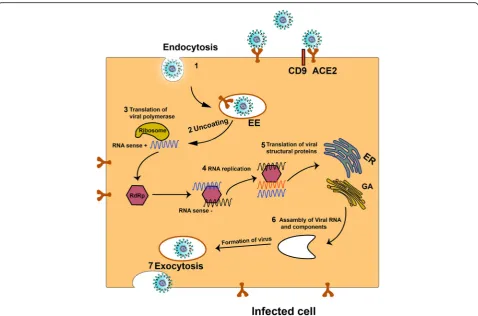
The affliction of coronaviruses is mediated by a trimeric
spike glycoprotein existing on the virion membrane.
Similar to the envelope of HIV or hemagglutinin of
influenza species, the spike proteins of coronavirus are
attracts several immune cells, especially T lymphocytes
and monocytes, but not neutrophils, from the circulatory
system into the infected tissue [
37
,
38
]. The entry of
im-mune cells into the lung tissue and the influx of
lympho-cytes into the airways may correlate with the high
neutrophil-to-lymphocyte ratio, increased T helper-to-T
regulatory cell ratio, and lymphopenia, which are present
in approximately 80% of individuals [
20
,
39
].
Further-more, SARS-CoV-2 virus, which is a cytopathic virus,
can induce injury and death in cells and tissues as a
con-sequence of the viral life cycle [
40
]. Viral infection and
replication in epithelial cells of the respiratory system
may induce pyroptosis associated with vascular leak, as
confirmed in people infected with SARS-CoV [
41
].
Pyr-optosis, a caspase-1-based cell death, is an inflammatory
type of programmed cell death that could be prompted
by cytopathic viruses [
42
] and activates inflammatory
re-sponse [
43
]. For example, IL-1
β
as a pyroptosis secretion
is increased during SARS-CoV-2 infection [
24
].
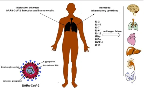
Collect-ively, in addition to the lesion on pulmonary cells, the
cytokine storm triggers inflammatory responses; thus, an
increasing effort is focused on the regeneration of
damaged cells as well as on the blocking or modulating
of inflammatory responses.
Extracellular vesicles
endosomes) located inside the cytoplasm via a complex
regularity system [
46
]. Once secreted, exosomes
distrib-ute into the extracellular matrix and bio-fluids and reach
the neighboring and distantly located target cells [
45
].
Exosomes bear different molecules both on their surface
and in the lumen. The density of sucrose (1.12
–
1.18 g/
mL) is equivalent to that of exosomes, and they appear
as cup-shaped vesicles under an electron microscopy
[
47
]. As exosomes contain active biomolecules from
parent cells, they can regulate function, fate, and shapes
of target cells, participating in different pathological and
physiological conditions [
45
,
48
].
Role of MSC-derived exosomes in SARS-CoV-2
treatment
MSCs, multipotent and self-renewal cells, are known as
adult stem cells that can be obtained from different
tis-sues such as the adipose tissue, bone marrow, umbilical
cord tissue, and amniotic fluid. These cells are capable
of differentiation into a variety of cells such as cartilage,
bone cells, neural cells, muscle cells, and skin cells [
49
].
MSCs are widely considered to be the most useful tool
in regenerative medicine because these cells can improve
damaged tissues and organs via differentiation into
dif-ferent cells and also producing many types of
chemo-kines, cytochemo-kines, growth factors, and EVs [
50
,
51
]. MSCs
may improve damaged lung tissue through homing
to-ward specific injuries of the lung to maintain
homeosta-sis as well as promote regeneration and secretion of
soluble factors and exosomes to moderate inflammation
and induce tissue regeneration [
52
,
53
]. More recently,
Leng et al. found that the transplantation of MSCs
con-siderably improved the pulmonary function of patients
with 2019-nCoV pneumonia over a period of 2 days
[
54
]. They administered about one million cells per
kilo-gram of weight once and declared that MSCs modulated
immune responses, participating in the improvement of
the outcome of SARS-CoV-2 pneumonia. Apart from
transdifferentiation into tissue cells, the other
mecha-nisms may involve in repairing tissue [
55
]; alternatively,
paracrine effects (such as exosomes) of stem cells are
the base of the beneficial effect of cell therapies [
56
].
administration, MSC-Exo can modulate immune
re-sponses like increased cytokine storms in related acute
lung injury and sepsis models [
59–64
]. Khatri and
co-workers found that the administration of MSC-Exo
attenuated influenza virus-induced acute lung injury
through suppressing influenza virus replication in a pig
model [
65
]. Similarly, a study conducted by Li and
colleagues showed that MSC treatment significantly
prevents avian H9N2-induced acute lung injury in mice
via decreasing the level of chemokines and
pro-inflammatory cytokines as well as reducing the
infiltra-tion of inflammatory cells onto the lungs [
66
]. In
addition, in
Escherichia coli
-induced pneumonia mouse
models, MSC-Exo had the ability to increase
phagocyt-osis of bacteria [
61
,
67
]. It seems that the hallmark of
the treatment of SARS-CoV-2 severe pneumonia is to
suppress
the
pro-inflammatory
immune
response
induced by the virus, thus reducing the damage of both
alveolar epithelial cells and capillary endothelial cells,
followed by the restoring function of lung cells and also
the recovery of the lung tissue which may be mediated
by MSC-Exo (Fig.
3
). Overall, as shown in Fig.
3
, the
possible therapeutic roles of MSC-Exo in SARS-CoV-2
virus
infection
may
comprise
inhibiting
pro-inflammatory cytokines, inducing M2 macrophages
through delivering PEG2, increasing secretion of
anti-inflammatory cytokines like IL-10, and regenerating
damaged tissue by producing KGF, VEGF, and HGF [
6
,
62
,
68–71
].
By June 2020, the public clinical trial database (https://
clinicaltrials.gov) prepared three studies (NCT04276987,
NCT0438938, and NCT04384445), and the Chinese
Clinical Trial Registry site (ChiCTR,
http://www.chictr.
org.cn
) records two clinical trials (ChiCTR2000030484
and ChiCTR2000030261) aiming to study the inhalation
of aerosol and intravenous administration of MSC-Exo
in the treatment of severe individuals with SARS-CoV-2
pneumonia. However, for SARS-CoV-2 pneumonia,
further studies, which take MSC-Exo into account, will
need to be undertaken, because various immune cells
re-spond differently against MSC-Exo and these exosomes
may modulate immune responses. For instance, Di
Tra-pani et al. found that MSC-Exo strongly inhibited the
function of B cells compared with the function of NK
and T cells [
72
]. Therefore, the immunological activity
of MSC-Exo toward different immune cells is dependent
on the type of target cell, the cellular maturity, cellular
status, and type of diseases among other factors [
73
,
74
].
However, different studies declared that MSC-Exo can
inhibit immune cell production and support an
immu-notolerant microenvironment.
Other potential therapeutic applications of
exosomes in 2019-nCoV treatment
This section provides some possible application of
exosomes in SARS-CoV-2 virus infection treatment;
however, there are some limitations regarding their
application, which are discussed in the later section. In
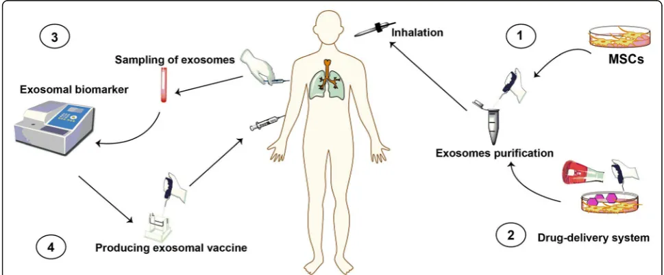
addition to exosome therapy, exosomes can serve as a
drug delivery system for the treatment of SARS-CoV-2
virus pneumonia (Fig.
3
). Exosomes exhibit superiority
to other nanocarriers as they are bi-phospholipid
vesi-cles, are of cell origin, safe, and have low
immunogen-icity and they can pass through the physiological barriers
[
75
]. In this regard, different strategies can be used to
construct optional exosomes such as (I) direct
engineer-ing method and (II) indirect engineerengineer-ing method [
76
].
By direct engineering methods, therapeutic agents
(bio-molecules and drugs) are directly loaded into
exosomes isolated from confident source cells, and then
these exosomes are delivered into the tissue of target. By
indirect engineering method, parent cells are co-cultured
with therapeutic agents or genetically modified to
pro-duce artificial/drug-loaded exosomes. Pascucci et al., for
instance, demonstrated the capacity of MSCs via
micro-vesicles to package and deliver anti-cancer drugs such as
paclitaxel, thus a potential tool for drug delivery.
Inter-estingly, paclitaxel-loaded exosomes exerted a strong
an-ticancer effect compared with exosomes isolated from
the control cells [
77
]. Another possible application of
exosomes is using them as a biomarker for SARS-CoV-2
virus infection. For other viruses, exosomes have been
shown to transfer viral components such as proteins and
miRNAs [
78
,
79
]. Although the detailed mechanism by
which SARS-CoV-2 viruses use exosomes for spreading
and infection has not been explained exactly, it is
reasonable that non-invasive sampling may be a new
av-enue to monitoring virus infection (Fig.
4
). Furthermore,
as mention above, exosomes from infected cells contain
viral components that make them ideal for vaccine
dis-covery. Although these exosomes could spread infection,
a growing body of evidence indicates that the mentioned
exosomes can induce immune responses [
80
]. It was
demonstrated that exosomes derived from serum
con-tain antigenic viral protein, suggesting the application of
these exosomes as a novel vaccine approach [
81
]. If viral
antigens are transferred on exosomes surface, it likely
seems these exosomes serve as a vaccine for the
treat-ment of SARS-CoV-2 virus infection (Fig.
4
). Recently,
exosomes have been shown to be inhibited both at the
biogenesis and uptake level [
82
,
83
]; therefore, inhibition
of the biogenesis and secretion of exosomes from
infected cells may reduce the speed and development of
infection; however, upcoming studies are essential for
confirmation. Collectively, in the case of MSC-Exo, the
therapeutic application of MSC-Exo in COVID-19
disease may comprise using them as a drug delivery
system as well as using them as therapeutic agents for
suppressing inflammatory responses and regeneration of
damaged tissues.
Challenges
Exosomes have attracted many attentions by both
aca-demic and medical industry communities. The existence
of different methods for EV isolation, purification, and
characterization not only makes big challenges in the
re-sult analysis but also causes the conclusions to be
un-clear in the use of exosomes in clinical trials. Some of
the previous studies did not use ISEV guidelines for the
characterization of the EVs and assessment of their
func-tion, since those experiments have been accomplished
prior to the 2014 and 2018 statements of ISEV
guide-lines of minimal experimental requirements for studies
dealt with EVs [
84
,
85
]. Although stem cell-based
ther-apies represent some benefits such as availability and
dif-ferentiation potential, they have several major hurdles
for therapy [
86
], which may include (i) determining and
targeting a reliable source of cells with phenotypic
stabil-ity, (ii) the immunological incompatibility that may
result in rejection, (iii) difficulty of handling and the
higher cost of expansion, and (iv) the potential of cancer
risks or formation of ectopic tissue. Exosome therapy
ex-hibits several advantages against cell therapy such as the
following: exosomes have a small size, which allows
them to pass through filter organs including the liver,
spleen, and lungs. Furthermore, as exosomes are
bi-phospholipid vesicles, their membrane-binding property
imparts excellent biocompatibility and biostability to the
loaded cargos and optional drugs, and also, exosomes
can pass through biological barriers like the brain-blood
barrier. As a hopeful therapeutic tool for novel cell-free
therapy, exosomes may serve as an alternative to stem
cells in the treatment of various diseases for
mainten-ance of the microenvironment for organ homeostasis
and tissue regeneration upon injury [
87
,
88
].
There are great developments in clinical
exosome-based studies (
https://clinicaltrials.gov
); however, until
now, no Food and Drug Administration (FDA)-approved
exosome products are available (
https://www.fda.gov/
).
In the case of using MSC-Exo, from a clinical
applica-tion point of view, a number of concerns should be
con-sidered before administering MSC-Exo to
SARS-CoV-2-infected people. These comprise the source of MSC-Exo.
MSCs, the heterogeneous cells, can be obtained from
various tissues, even though MSCs isolated from the
same sources may vary in entity and functionality [
89
,
90
]. For example, several donor-derived bone marrow
MSCs release exosomes with distinct cytokine profile
content [
91
]. Besides, young MSC-Exo as compared with
aged MSC-Exo improved LPS-induced acute lung injury
in a mouse model [
92
]. In addition to
immunomodula-tory properties, MSC-Exo actually also regulate other
biological processes, some with therapeutic functions
[
93
], and others that may induce unexpected
non-targeting effects. In order to inhibit the exosome
biogen-esis and secretion, future attempts should focus on
silen-cing or eliminating exosomes that selectively encourage
diseases which are not useful exosomes. Some research
groups tried to use exosome inhibitors as versatile tools
for exosome biology investigation; however, others have
evaluated the inhibitory potential of such drugs in
vari-ous disease models [
82
,
94
]. Most of the experiments
were carried out preclinical; therefore, clinical trials
must be done to confirm their validity. However, the
non-targeting effects of exosome inhibitors could be
considered as the main issue on exosome biogenesis of
healthy cells.
Another fascinating approach that exosomes can be
used as a therapeutic agent is the drug delivery potential
of them [
95–97
]. Exosomes from a safe source such as
MSCs may be loaded with optional compounds or
gen-etically engineered for targeting infected tissues/cells,
suggesting the exosome-based nanocarriers for the
treat-ment of infectious diseases. The advent of safe
nanocar-riers
with
high
efficiency
is
the
core
goal
of
nanomedicine. However, most of the studies are
designed in vitro and animal models; therefore, from a
clinical trial application point of view, there are still
more questions in the specificity, safety, and proficiency
of this method. Moreover, despite various available
de-livery strategies for therapeutic anti-viral vaccines,
thera-peutic
anti-viral
vaccines
in
humans
have
been
challenging to implement [
98
]. In another word, these
vaccines have not been a strong success so far, and it is
certainly valuable to consider more effective adjuvant
methods for refining vaccine efficiency. Furthermore,
an-other important thing is the source of
immunostimula-tory exosomes for human antiviral vaccines which needs
more considerations [
81
]. In a nutshell, researches and
explorations in the field of exosomes are in their early
stages; therefore, to implement many of the
aforemen-tioned ideas, the biology of EVs (especially exosomes)
from infected cells must be explored in-depth.
Conclusion
Although the exploration of novel specific vaccine for
the treatment of SARS-CoV-2 virus pneumonia is
critical, it seems to take a long time. Therefore, further
research into preventing and controlling the virus is
essential as soon as possible. Exosomes from MSCs may
be a useful tool for the treatment of SARS-CoV-2 virus
pneumonia due to their pivotal roles in the suppression
of inflammatory responses and the regeneration of
damaged tissues. Furthermore, exosomes may serve as a
biomarker, nanocarrier, and vaccine for the treatment of
SARS-CoV-2 virus. However, for a better outcome,
upcoming efforts are vital to carefully weigh the risk in
the use of MSC-Exo for SARS-CoV-2 virus pneumonia
against available pre-clinical findings in applicable
in vivo models.
Abbreviations
ARDS:Acute respiratory distress syndrome; EVs: Extracellular vesicle; FDA: Food and Drug Administration; IP10: Induced protein 10; MCP-1: Monocyte chemoattractant protein-1; MERS: Middle East respiratory syndrome; MSC-Exo: Mesenchymal stem cell-derived exosomes; SARS-CoV-2: Severe acute respiratory syndrome coronavirus 2; WHO: World Health Organization
Acknowledgements Not applicable.
Authors’contributions
J.R. and A.A. collected the data and edited the manuscript and with equal conceptualization. All authors read and approved the final manuscript.
Availability of data and materials
The datasets used and/or analyzed during the current study are available from the corresponding author on reasonable request.
Ethics approval and consent to participate Not applicable.
Consent for publication Not applicable.
Competing interests
The authors have no competing interests to declare.
Received: 17 June 2020 Revised: 9 July 2020 Accepted: 30 July 2020
References
1. WHO Coronavirus Disease (COVID-19) Dashboard.https://covid19.who.int/. Accessed 8 July 2020.
2. Gattinoni L, Coppola S, Cressoni M, Busana M, Rossi S, Chiumello D. Covid-19 does not lead to a“typical”acute respiratory distress syndrome. Am J Respir Crit Care Med. 2020;201:1299–300.
3. HuiDS IAE, Madani T, Ntoumi F, Koch R, Dar O. The continuing 2019-nCoV epidemic threat of novel coronaviruses to global health: the latest 2019 novel coronavirus outbreak in Wuhan, China. Int J Infect Dis. 2020;91:264–6. 4. Abraham A, Krasnodembskaya A. Mesenchymal stem cell-derived
extracellular vesicles for the treatment of acute respiratory distress syndrome. Stem Cells Transl Med. 2020;9:28–38.
5. Shah TG, Predescu D, Predescu S. Mesenchymal stem cells-derived extracellular vesicles in acute respiratory distress syndrome: a review of current literature and potential future treatment options. Clin Transl Med. 2019;8:25.
6. Jabbari N, Nawaz M, Rezaie J. Ionizing radiation increases the activity of exosomal secretory pathway in MCF-7 human breast cancer cells: a possible way to communicate resistance against radiotherapy. Int J Mol Sci. 2019;20: 3649.
7. Martin-Rufino JD, Espinosa-Lara N, Osugui L, Sanchez-Guijo F. Targeting the immune system with mesenchymal stromal cell-derived extracellular vesicles: what is the cargo’s mechanism of action? Front Bioeng Biotechnol. 2019;7:308.
8. Joo HS, Suh JH, Lee HJ, Bang ES, Lee JM. Current knowledge and future perspectives on mesenchymal stem cell-derived exosomes as a new therapeutic agent. Int J Mol Sci. 2020;21:727.
9. Belouzard S, Millet JK, Licitra BN, Whittaker GR. Mechanisms of coronavirus cell entry mediated by the viral spike protein. Viruses.2012;4:1011–33. 10. Kuiken T, Fouchier RA, Schutten M, Rimmelzwaan GF, Van Amerongen G,
van Riel D, Laman JD, de Jong T, van Doornum G, Lim W. Newly discovered coronavirus as the primary cause of severe acute respiratory syndrome. Lancet. 2003;362:263–70.
11. Drosten C, Günther S, Preiser W, Van Der Werf S, Brodt H-R, Becker S, Rabenau H, Panning M, Kolesnikova L, Fouchier RA. Identification of a novel coronavirus in patients with severe acute respiratory syndrome. N Engl J Med. 2003;348:1967–76.
12. de Groot RJ, Baker SC, Baric RS, Brown CS, Drosten C, Enjuanes L, Fouchier RA, Galiano M, Gorbalenya AE, Memish ZA. Commentary: Middle East respiratory syndrome coronavirus (MERS-CoV): announcement of the Coronavirus Study Group. J Virol. 2013;87:7790–2.
13. Su S, Wong G, Shi W, Liu J, Lai AC, Zhou J, Liu W, Bi Y, Gao GF. Epidemiology, genetic recombination, and pathogenesis of coronaviruses. Trends Microbiol. 2016;24:490–502.
14. Fehr AR, Perlman S: Coronaviruses: an overview of their replication and pathogenesis. New York: In Coronaviruses Springer; 2015. p. 1–23. 15. of the International CSG. The species severe acute respiratory
syndrome-related coronavirus: classifying 2019-nCoV and naming it SARS-CoV-2. Nat Microbiol. 2020;5:536.
16. Zhou P, Yang X-L, Wang X-G, Hu B, Zhang L, Zhang W, Si H-R, Zhu Y, Li B, Huang C-L. A pneumonia outbreak associated with a new coronavirus of probable bat origin. Nature. 2020;579:270–3.
17. Andersen KG, Rambaut A, Lipkin WI, Holmes EC, Garry RF. The proximal origin of SARS-CoV-2. Nat Med. 2020;26:450–2.
18. Whittaker GR, Millet JK: Biochemical characterization of Middle East respiratory syndrome coronavirus spike protein proteolytic processing. New York: In MERS Coronavirus Springer; 2020. p. 21–37.
19. La A, Alzoughool F, Atoum M. The human coronavirus disease COVID-19: its origin, characteristics, and insights into potential drugs and its mechanisms. Pathogens. 2020;9:331.
20. W-J G, Z-Y N, Hu Y, Liang W-H, Ou C-Q, He J-X, Liu L, Shan H, Lei C-L, Hui DS. Clinical characteristics of coronavirus disease 2019 in China. N Engl J Med. 2020;382:1708–20.
21. Pung R, Chiew CJ, Young BE, Chin S, Chen MI, Clapham HE, Cook AR, Maurer-Stroh S, Toh MP, Poh C. Investigation of three clusters of COVID-19 in Singapore: implications for surveillance and response measures. Lancet. 2020;395:1039–46.
22. Lauer SA, Grantz KH, Bi Q, Jones FK, Zheng Q, Meredith HR, Azman AS, Reich NG, Lessler J. The incubation period of coronavirus disease 2019 (COVID-19) from publicly reported confirmed cases: estimation and application. Ann Intern Med. 2020;172:577–82.
23. Chan JF-W, Yuan S, Kok K-H, To KK-W, Chu H, Yang J, Xing F, Liu J, Yip CC-Y, Poon RW-S. A familial cluster of pneumonia associated with the 2019 novel coronavirus indicating person-to-person transmission: a study of a family cluster. Lancet. 2020;395:514–23.
24. Huang C, Wang Y, Li X, Ren L, Zhao J, Hu Y, Zhang L, Fan G, Xu J, Gu X. Clinical features of patients infected with 2019 novel coronavirus in Wuhan, China. Lancet. 2020;395:497–506.
25. Liu Y, Yang Y, Zhang C, Huang F, Wang F, Yuan J, Wang Z, Li J, Li J, Feng C. Clinical and biochemical indexes from 2019-nCoV infected patients linked to viral loads and lung injury. Sci China Life Sci. 2020;63:364–74. 26. Chen N, Zhou M, Dong X, Qu J, Gong F, Han Y, Qiu Y, Wang J, Liu Y, Wei Y.
Epidemiological and clinical characteristics of 99 cases of 2019 novel coronavirus pneumonia in Wuhan, China: a descriptive study. Lancet. 2020;395:507–13. 27. Pan Y, Zhang D, Yang P, Poon LL, Wang Q. Viral load of SARS-CoV-2 in
clinical samples. Lancet Infect Dis. 2020;20:411–2.
28. Kim JY, Ko J-H, Kim Y, Kim Y-J, Kim J-M, Chung Y-S, Kim HM, Han M-G, Kim SY, Chin BS. Viral load kinetics of SARS-CoV-2 infection in first two patients in Korea. J Korean Med Sci. 2019;35:e86.
29. Zou L, Ruan F, Huang M, Liang L, Huang H, Hong Z, Yu J, Kang M, Song Y, Xia J. SARS-CoV-2 viral load in upper respiratory specimens of infected patients. N Engl J Med. 2020;382:1177–9.
30. Wang D, Hu B, Hu C, Zhu F, Liu X, Zhang J, Wang B, Xiang H, Cheng Z, Xiong Y. Clinical characteristics of 138 hospitalized patients with 2019 novel coronavirus–infected pneumonia in Wuhan, China. JAMA. 2020;323:1061–9. 31. Zhang B, Zhou X, Qiu Y, Feng F, Feng J, Jia Y, Zhu H, Hu K, Liu J, Liu Z. Clinical characteristics of 82 death cases with COVID-19. MedRxiv.2020;15:e0235458. 32. Adhikari SP, Meng S, Wu Y-J, Mao Y-P, Ye R-X, Wang Q-Z, Sun C, Sylvia S,
Rozelle S, Raat H. Epidemiology, causes, clinical manifestation and diagnosis, prevention and control of coronavirus disease (COVID-19) during the early outbreak period: a scoping review. Infect Dis Poverty. 2020;9:1–12. 33. Murthy S, Gomersall CD, Fowler RA. Care for critically ill patients with
COVID-19. JAMA.2020;323:1499–500.
34. Chen G, Wu D, Guo W, Cao Y, Huang D, Wang H, Wang T, Zhang X, Chen H, Yu H. Clinical and immunological features of severe and moderate coronavirus disease 2019. J Clin Invest. 2020;130:2620–9.
35. Diao B, Wang C, Tan Y, Chen X, Liu Y, Ning L, Chen L, Li M, Liu Y, Wang G. Reduction and functional exhaustion of T cells in patients with coronavirus disease 2019 (COVID-19). Front Immunol. 2020;11:827.
36. Yang Y, Shen C, Li J, Yuan J, Wei J, Huang F, Wang F, Li G, Li Y, Xing L, Peng L. Plasma IP-10 and MCP-3 levels are highly associated with disease severity and predict the progression of COVID-19. J Allergy Clin Immunol 2020.
https://doi.org/10.1016/j.jaci.2020.04.027.
37. Tian S, Hu W, Niu L, Liu H, Xu H, Xiao S-Y. Pulmonary pathology of early phase 2019 novel coronavirus (COVID-19) pneumonia in two patients with lung cancer. J Thorac Oncol. 2020;15:700–4.
38. Xu Z, Shi L, Wang Y, Zhang J, Huang L, Zhang C, Liu S, Zhao P, Liu H, Zhu L. Pathological findings of COVID-19 associated with acute respiratory distress syndrome. Lancet Respir Med. 2020;8:420–2.
39. Qin C, Zhou L, Hu Z, Zhang S, Yang S, Tao Y, Xie C, Ma K, Shang K, Wang W: Dysregulation of immune response in patients with COVID-19 in Wuhan, China. Clin Infect Dis.2020;71:762–8.
41. Zhang H, Zhou P, Wei Y, Yue H, Wang Y, Hu M, Zhang S, Cao T, Yang C, Li M. Histopathologic changes and SARS-CoV-2 immunostaining in the lung of a patient with COVID-19. Ann Intern Med. 2020;172:629–32.
42. Fink SL, Cookson BT. Apoptosis, pyroptosis, and necrosis: mechanistic description of dead and dying eukaryotic cells. Infect Immun. 2005;73: 1907–16.
43. Yang M. Cell pyroptosis, a potential pathogenic mechanism of 2019-nCoV infection. Available at SSRN 3527420; 2020.
44. Rezaie J, Rahbarghazi R, Pezeshki M, Mazhar M, Yekani F, Khaksar M, Shokrollahi E, Amini H, Hashemzadeh S, Sokullu SE, Tokac M. Cardioprotective role of extracellular vesicles: a highlight on exosome beneficial effects in cardiovascular diseases. J Cell Physiol. 2019;234:21732–45.
45. Kowal J, Tkach M, Théry C. Biogenesis and secretion of exosomes. Curr Opin Cell Biol. 2014;29:116–25.
46. Jabbari N, Karimipour M, Khaksar M, Akbariazar E, Heidarzadeh M, Mojarad B, Aftab H, Rahbarghazi R, Rezaie J. Tumor-derived extracellular vesicles: insights into bystander effects of exosomes after irradiation. Lasers Med Sci. 2019;35:1–15.
47. Kesimer M, Scull M, Brighton B, DeMaria G, Burns K, O’Neal W, Pickles RJ, Sheehan JK. Characterization of exosome-like vesicles released from human tracheobronchial ciliated epithelium: a possible role in innate defense. FASEB J. 2009;23:1858–68.
48. Hannafon BN, Ding W-Q. Intercellular communication by exosome-derived microRNAs in cancer. Int J Mol Sci. 2013;14:14240–69.
49. Han Y, Li X, Zhang Y, Han Y, Chang F, Ding J. Mesenchymal stem cells for regenerative medicine. Cells.2019;8:886.
50. Zhao T, Sun F, Liu J, Ding T, She J, Mao F, Xu W, Qian H, Yan Y. Emerging role of mesenchymal stem cell-derived exosomes in regenerative medicine. Curr Stem Cell Res Ther. 2019;14:482–94.
51. Satija NK, Singh VK, Verma YK, Gupta P, Sharma S, Afrin F, Sharma M, Sharma P, Tripathi R, Gurudutta G. Mesenchymal stem cell-based therapy: a new paradigm in regenerative medicine. J Cell Mol Med. 2009;13:4385–402.
52. Ullah M, Liu DD, Thakor AS. Mesenchymal stromal cell homing: mechanisms and strategies for improvement. iScience. 2019;15:421.
53. Mohammadipoor A, Antebi B, Batchinsky AI, Cancio LC. Therapeutic potential of products derived from mesenchymal stem/stromal cells in pulmonary disease. Respir Res. 2018;19:218.
54. Leng Z, Zhu R, Hou W, Feng Y, Yang Y, Han Q, Shan G, Meng F, Du D, Wang S. Transplantation of ACE2-mesenchymal stem cells improves the outcome of patients with COVID-19 pneumonia. Aging Dis. 2020;11:216. 55. Rogers TB, Pati S, Gaa S, Riley D, Khakoo AY, Patel S, Wardlow RD II,
Frederick CA, Hall G, He L-P. Mesenchymal stem cells stimulate protective genetic reprogramming of injured cardiac ventricular myocytes. J Mol Cell Cardiol. 2011;50:346–56.
56. Uemura R, Xu M, Ahmad N, Ashraf M. Bone marrow stem cells prevent left ventricular remodeling of ischemic heart through paracrine signaling. Circ Res. 2006;98:1414–21.
57. Keshtkar S, Azarpira N, Ghahremani MH. Mesenchymal stem cell-derived extracellular vesicles: novel frontiers in regenerative medicine. Stem Cell Res Ther. 2018;9:63.
58. Baglio SR, Pegtel DM, Baldini N. Mesenchymal stem cell secreted vesicles provide novel opportunities in (stem) cell-free therapy. Front Physiol. 2012;3:359. 59. Khaksar M, Sayyari M, Rezaie J, Pouyafar A, Montazersaheb S, Rahbarghazi R.
High glucose condition limited the angiogenic/cardiogenic capacity of murine cardiac progenitor cells in in vitro and in vivo milieu. Cell Biochem Funct. 2018;36:346–56.
60. Mansouri N, Willis GR, Fernandez-Gonzalez A, Reis M, Nassiri S, Mitsialis SA, Kourembanas S. Mesenchymal stromal cell exosomes prevent and revert experimental pulmonary fibrosis through modulation of monocyte phenotypes. JCI insight. 2019;4:e128060.
61. Monsel A, Zhu Y-G, Gennai S, Hao Q, Hu S, Rouby J-J, Rosenzwajg M, Matthay MA, Lee JW. Therapeutic effects of human mesenchymal stem cell–derived microvesicles in severe pneumonia in mice. Am J Respir Crit Care Med. 2015;192:324–36.
62. Park K-S, Svennerholm K, Shelke GV, Bandeira E, Lässer C, Jang SC, Chandode R, Gribonika I, Lötvall J. Mesenchymal stromal cell-derived nanovesicles ameliorate bacterial outer membrane vesicle-induced sepsis via IL-10. Stem Cell Res Ther. 2019;10:231.
63. Varkouhi AK, Jerkic M, Ormesher L, Gagnon S, Goyal S, Rabani R, Masterson C, Spring C, Chen PZ, Gu FX. Extracellular vesicles from interferon-γ–primed
human umbilical cord mesenchymal stromal cells reduce Escherichia coli– induced acute lung injury in rats. Anesthesiology. 2019;130:778–90. 64. Willis GR, Fernandez-Gonzalez A, Anastas J, Vitali SH, Liu X, Ericsson M,
Kwong A, Mitsialis SA, Kourembanas S. Mesenchymal stromal cell exosomes ameliorate experimental bronchopulmonary dysplasia and restore lung function through macrophage immunomodulation. Am J Respir Crit Care Med. 2018;197:104–16.
65. Khatri M, Richardson LA, Meulia T. Mesenchymal stem cell-derived extracellular vesicles attenuate influenza virus-induced acute lung injury in a pig model. Stem Cell Res Ther. 2018;9:1–13.
66. Li Y, Xu J, Shi W, Chen C, Shao Y, Zhu L, Lu W, Han X. Mesenchymal stromal cell treatment prevents H9N2 avian influenza virus-induced acute lung injury in mice. Stem Cell Res Ther. 2016;7:159.
67. Hao Q, Gudapati V, Monsel A, Park JH, Hu S, Kato H, Lee JH, Zhou L, He H, Lee JW. Mesenchymal stem cell–derived extracellular vesicles decrease lung injury in mice. J Immunol. 2019;203:1961–72.
68. Zhou F, Yu T, Du R, Fan G, Liu Y, Liu Z, Xiang J, Wang Y, Song B, Gu X. Clinical course and risk factors for mortality of adult inpatients with COVID-19 in Wuhan, China: a retrospective cohort study. Lancet. 2020;395:1054– 1062.
69. Harrell CR, Sadikot R, Pascual J, Fellabaum C, Jankovic MG, Jovicic N, Djonov V, Arsenijevic N, Volarevic V. Mesenchymal stem cell-based therapy of inflammatory lung diseases: current understanding and future perspectives. Stem Cells Int. 2019;4236973.
70. Devaney J, Horie S, Masterson C, Elliman S, Barry F, O’Brien T, Curley GF, O’Toole D, Laffey JG. Human mesenchymal stromal cells decrease the severity of acute lung injury induced by E. coli in the rat. Thorax. 2015;70:625–35. 71. Vasandan AB, Jahnavi S, Shashank C, Prasad P, Kumar A, Prasanna SJ.
Human mesenchymal stem cells program macrophage plasticity by altering their metabolic status via a PGE 2-dependent mechanism. Sci Rep. 2016;6: 38308.
72. Di Trapani M, Bassi G, Midolo M, Gatti A, Kamga PT, Cassaro A, Carusone R, Adamo A, Krampera M. Differential and transferable modulatory effects of mesenchymal stromal cell-derived extracellular vesicles on T, B and NK cell functions. Sci Rep. 2016;6:24120.
73. Doeppner TR, Herz J, Görgens A, Schlechter J, Ludwig A-K, Radtke S, de Miroschedji K, Horn PA, Giebel B, Hermann DM. Extracellular vesicles improve post-stroke neuroregeneration and prevent postischemic immunosuppression. Stem Cells Transl Med. 2015;4:1131–43. 74. Xie M, Xiong W, She Z, Wen Z, Abdirahman AS, Wan W, Wen C.
Immunoregulatory effects of stem cell-derived extracellular vesicles on immune cells. Front Immunol. 2020;11:13.
75. Malhotra H, Sheokand N, Kumar S, Chauhan AS, Kumar M, Jakhar P, Boradia VM, Raje CI, Raje M. Exosomes: tunable nano vehicles for macromolecular delivery of transferrin and lactoferrin to specific intracellular compartment. J Biomed Nanotechnol. 2016;12:1101–14.
76. Bunggulawa EJ, Wang W, Yin T, Wang N, Durkan C, Wang Y, Wang G. Recent advancements in the use of exosomes as drug delivery systems. J Nanobiotechnology. 2018;16:81.
77. Pascucci L, Coccè V, Bonomi A, Ami D, Ceccarelli P, Ciusani E, Viganò L, Locatelli A, Sisto F, Doglia SM. Paclitaxel is incorporated by mesenchymal stromal cells and released in exosomes that inhibit in vitro tumor growth: a new approach for drug delivery. J Control Release. 2014;192:262–70. 78. Naqvi AR, Shango J, Seal A, Shukla D, Nares S. Herpesviruses and
microRNAs: new pathogenesis factors in oral infection and disease? Front Immunol. 2018;9:2099.
79. Patters BJ, Kumar S. The role of exosomal transport of viral agents in persistent HIV pathogenesis. Retrovirology.2018;15:1–13.
80. Gunasekaran M, Xu Z, Nayak DK, Sharma M, Hachem R, Walia R, Bremner RM, Smith MA, Mohanakumar T. Donor-derived exosomes with lung self-antigens in human lung allograft rejection. Am J Transplant. 2017;17:474–84. 81. Devhare PB, Ray RB. A novel role of exosomes in the vaccination approach.
Ann Transl Med.2017;5:23.
82. Zhou X, Zhang W, Yao Q, Zhang H, Dong G, Zhang M, Liu Y, Chen J-K, Dong Z. Exosome production and its regulation of EGFR during wound healing in renal tubular cells. Am J Physiol Renal Physiol. 2017;312:F963–70. 83. Zheng Y, Tu C, Zhang J, Wang J. Inhibition of multiple myeloma-derived
exosomes uptake suppresses the functional response in bone marrow stromal cell. Int J Oncol. 2019;54:1061–70.
requirements for definition of extracellular vesicles and their functions: a position statement from the International Society for Extracellular Vesicles: Taylor & Francis. J Extracell Vesicles. 2014;3:26913.
85. Théry C, Witwer KW, Aikawa E, Alcaraz MJ, Anderson JD, Andriantsitohaina R, Antoniou A, Arab T, Archer F, Atkin-Smith GK. Minimal information for studies of extracellular vesicles 2018 (MISEV2018): a position statement of the International Society for Extracellular Vesicles and update of the MISE V2014 guidelines. J Extracell Vesicles. 2018;7:1535750.
86. Yu B, Zhang X, Li X. Exosomes derived from mesenchymal stem cells. Int J Mol Sci. 2014;15:4142–57.
87. Teixeira FG, Carvalho MM, Sousa N, Salgado AJ. Mesenchymal stem cells secretome: a new paradigm for central nervous system regeneration? Cell Mol Life Sci. 2013;70:3871–82.
88. Zhang ZG, Buller B, Chopp M. Exosomes—beyond stem cells for restorative therapy in stroke and neurological injury. Nat Rev Neurol. 2019;15:193–203. 89. Phinney DG. Functional heterogeneity of mesenchymal stem cells:
implications for cell therapy. J Cell Biochem. 2012;113:2806–12. 90. Radtke S, Görgens A, Liu B, Horn PA, Giebel B. Human mesenchymal and
murine stromal cells support human lympho-myeloid progenitor expansion but not maintenance of multipotent haematopoietic stem and progenitor cells. Cell Cycle. 2016;15:540–5.
91. Kordelas L, Rebmann V, Ludwig A, Radtke S, Ruesing J, Doeppner T, Epple M, Horn P, Beelen D, Giebel B. MSC-derived exosomes: a novel tool to treat therapy-refractory graft-versus-host disease. Leukemia.2014;28:970–3. 92. Huang R, Qin C, Wang J, Hu Y, Zheng G, Qiu G, Ge M, Tao H, Shu Q, Xu J.
Differential effects of extracellular vesicles from aging and young mesenchymal stem cells in acute lung injury. Aging (Albany). 2019;11:7996. 93. Arslan F, Lai RC, Smeets MB, Akeroyd L, Choo A, Aguor EN, Timmers L, van Rijen HV, Doevendans PA, Pasterkamp G. Mesenchymal stem cell-derived exosomes increase ATP levels, decrease oxidative stress and activate PI3K/ Akt pathway to enhance myocardial viability and prevent adverse remodeling after myocardial ischemia/reperfusion injury. Stem Cell Res. 2013;10:301–12.
94. Catalano M, O’Driscoll L. Inhibiting extracellular vesicles formation and release: a review of EV inhibitors. J Extracell Vesicles. 2020;9:1703244. 95. Lv L-H, Wan Y-L, Lin Y, Zhang W, Yang M, Li G-L, Lin H-M, Shang C-Z, Chen
Y-J, Min J. Anticancer drugs cause release of exosomes with heat shock proteins from human hepatocellular carcinoma cells that elicit effective natural killer cell antitumor responses in vitro. J Biol Chem. 2012;287:15874– 85.
96. Gnecchi M, He H, Noiseux N, Liang OD, Zhang L, Morello F, Mu H, Melo LG, Pratt RE, Ingwall JS. Evidence supporting paracrine hypothesis for Akt-modified mesenchymal stem cell-mediated cardiac protection and functional improvement. FASEB J. 2006;20:661–9.
97. Lamichhane TN, Jeyaram A, Patel DB, Parajuli B, Livingston NK, Arumugasaamy N, Schardt JS, Jay SM. Oncogene knockdown via active loading of small RNAs into extracellular vesicles by sonication. Cell Mol Bioeng. 2016;9:315–24.
98. Hung C-F, Ma B, Monie A, Tsen S-W, Wu TC. Therapeutic human papillomavirus vaccines: current clinical trials and future directions. Expert Opin Biol Ther. 2008;8:421–39.
Publisher
’
s Note