RESEARCH ARTICLE
Investigating the effect of extended high-frequency hearing loss
on duration pattern sequence test
Athareh Farahani1, Saeid Farahani1*, Nematollah Rouhbakhsh1, Farzaneh Zamiri Abdollahi1, Masoud Bolandi2
1- Department of Audiology, School of Rehabilitation, Tehran University of Medical Sciences, Tehran, Iran 2- Department of Audiology, University of Social Welfare and Rehabilitation Sciences, Tehran, Iran
Received: 2 Feb 2019, Revised: 11 Mar 2019, Accepted: 15 Apr 2019, Published: 15 Jul 2019
Abstract
Background and Aim: Temporal processing is affected in people exposed to occupational noi-se. The primary goal of this study was to eva-luate the temporal processing of people exposed to occupational noise of more than 85 dB A but have not experienced clinically significant chan-ges at hearing thresholds at conventional frequ-encies.
Methods: A comparison between groups were designed using individuals exposed to occupa-tional noise (n = 15 as the case group) and non-exposed individuals (n = 16 as the control gro-up). Two groups were age-matched (p < 0.05). The extended high-frequency audiometric thre-sholds and temporal processing system were evaluated through a duration pattern sequence test. Finally, the correlation between the exten-ded high-frequency hearing thresholds and the duration pattern test scores was investigated. Results: The case group had significantly high-er hearing thresholds than the control group at 14, 15, and 16 kHz (p < 0.05). Although in other frequencies, the mean hearing thresholds in the case group was higher than the control group, the difference was not significant. Also, the case
group had significantly lower duration pattern sequence scores than the control group in the right (p = 0.02) and the left ears (p = 0.03). There was no correlation between extended high-frequency hearing thresholds and duration pattern sequence test scores.
Conclusion: In people exposed to occupational noise, both extended high-frequency thresholds and temporal processing in lower frequency ran-ges (with normal hearing thresholds) are inter-rupted.
Keywords: Occupational noise; extended high-frequency hearing; duration pattern sequence test; temporal processing
Citation: Farahani A, Farahani S, Rouhbakhsh N, Zamiri Abdollahi F, Bolandi M.
Investigating the effect of extended high-frequency hearing loss on duration pattern sequence test. Aud Vestib Res. 2019;28(3):190-197.
Introduction
Excessive noise is the most common risk factor in work environments that causes hearing imp-airment [1]. Noise-induced hearing loss (NIHL) is usually an irreversible disorder and is a com-mon problem in industrial settings, especially where the noise level is harmful (more than 85 dB A) [2]. NIHL is the second most common
* Corresponding author: Department of Audiology,
form of acquired hearing loss after presbycusis, and it has long been known as a problem in professions that are too much associated with noise. Exposure to excessive noise can cause temporary threshold shift (TTS) or permanent threshold shift (PTS) [3]. However, NIHL can be a permanent and irreversible, but preventable disorder [4]. It has been suggested that frequ-encies higher than 8000 Hz may be more sen-sitive to noise, acoustic trauma, and ototoxic substances than lower frequencies. Therefore, hearing loss caused by noise at these frequ-encies can predict NIHL before it appears in lower frequencies, especially speech frequencies [5]. Türkkahraman et al. found that frequencies of 4000, 6000, 14000, and 16000 Hz were more exposed to noise. Therefore, they suggested that the extended high-frequency (EHF) hearing threshold with conventional audiometry should be used to identify and monitor people at risk of hearing loss [6].
The destructive effects of excessive noise are progressive and extensive, which are not fully detectable by conventional audiometric tests. Evidence suggests that in subjects with a history of noise exposure and normal auditory sensi-tivity, the temporal processing ability dramati-cally decreases [7]. Temporal auditory proce-ssing is one of the tasks of the central auditory nervous system that provides sound perception or sound changes in a given period. Also, tem-poral processing is an important aspect of audi-tory performance essential for a wide range of daily hearing activities, including speech and musical perception. Its defect can hinder the acquisition of speech, language, and reading [8]. According to the American Speech-Language-Hearing Association, evaluation of this aspect of auditory performance should be included in the auditory processing test batteries [9]. Therefore, temporal auditory processing is one of the cri-tical abilities of the auditory processing system, which includes temporal resolution, temporal sequencing or ordering, temporal integration or summation, and temporal masking [10]. Dura-tion pattern sequence test (DPST) is one of the auditory processing tests that evaluate skills such as temporal sequencing and duration
discrimination [11]. The temporal sequencing of acoustic stimuli is one of the most basic and essential skills of the central nervous system, which allows a person to recognize the sounds based on the sequence of an auditory stimulus [11]. Also, correct judgment on the temporal ordering and sequencing in the presence of the minimum interval between different sounds is necessary for the accurate perception of speech [10]. Excessive noise can cause significant dis-tortions in the processing of supra threshold temporal cues, which may add to difficulties in hearing in adverse listening conditions [7]. In areas of the cochlea where the hearing loss occurs, auditory processing of the signals is affected. This defect of auditory processing is associated with both hearing loss [12] and wea-ker supra threshold processing of the auditory system. However, processing defects may not be limited to frequencies where cochlear hea-ring loss is present. It may also extend to the surrounding area. The off-channel impact of cochlear lesions on signal processing has been indicated in both intensity and frequency coding [13,14]. It has recently been noted that in people with high-frequency hearing loss, temporal res-olution decreases in lower-frequency areas with almost normal-hearing sensitivity [15]. Accor-dingly, the purpose of this study was to inves-tigate the effect of extended high-frequencies hearing loss on the ability of temporal sequen-cing in people working in industrial environ-ments but have normal hearing in the frequency range of conventional audiometry.
Methods
temporal processing. The study subjects had the following criteria: right-handedness, normal hearing at conventional audiometric frequencies (250‒8000 Hz), lack of any otologic and neuro-logic disorder, not under any ototoxic medica-tions, and no exposure to organic solvents. Sub-jects in the case group were working in the car body production line with exposure to the more than 85 dB A noise. They had more than 16 hours break from noise exposure before the test [5] to eliminate any TTS. Informed consent was obtained from all participants. The noise level was measured by the health care team at the factory, and it was recorded in workers’ files. The following tests were conducted on the case (working in the car body production line) and control (working in the office without noise exposure) groups:
1) Examination of the head, neck, and external ear via otoscopy to be ensured of the healthy external ear, tympanic membrane and middle ear,
2) Immittance audiometry for ruling out any conductive deficit by Damplex Tymp87 (Den-mark). Subjects with A type tympanogram were included.
3) Pure tone audiometry at octave frequencies from 250‒8000 Hz by using Madsen ITERA (Denmark). Thresholds ≤ 25 dB were consi-dered normal for the case and control groups [17].
4) Extended High Frequency Audiometry (EHFA) (10‒16 kHz) was conducted by Beltone 2000 (USA) audiometer and Sennheiser HDA 200 headphones with the ascending-descending method. The lowest sound level at which wor-kers could detect stimuli in 50% of times was considered the threshold.
5) DPST was performed by calibrated Dell Ins-piron laptop, Beltone 2000 (USA) audiometer and Sennheiser HDA200 headphone. DPST was performed with the Musiek et al. [18] method. The 1000 Hz pure tone was presented with two different durations (short: 250 ms and long: 500 ms). We used patterns of three tones, with sti-muli intervals of 250 ms and pattern intervals of 6 seconds. Finally, there were 6 final patterns (short-short-long, short-long-short,
long-short-short, long-long-long-short-short, long-short-long, and short-long-long).
After instruction and trial items, 30 test items were presented for each ear, and the subjects’ responses were scored. The participants had to repeat patterns verbally. The number of correct repetitions was multiplied by 3.33, and the per-centages of correct responses were calculated for each ear.
Shapiro–Wilk test was used to test data distri-bution. The results showed that the data for 10 and 11 kHz in both ears and data for 12 kHz in the right ear were not normally distributed. But data for rest of frequencies had normal bution. DPST data did not have a normal distri-bution. Therefore the independent t-test was used for comparing means of two groups for normally distributed data and Mann-Whitney U test for data without normal distribution. DPST scores lacked normal distribution in both ears, so the Spearman test was used for testing the correlation between EHF hearing threshold and DPST score. For comparing right and left ear, paired t-test was used for data with normal distribution and Wilcoxon test for data without normal distribution. The significance level for all the tests was < 0.05, and SPSS 23 was used for data analysis.
Results
The mean and standard deviation of age in the case group (36.38 ± 3.84 years) and the control group (36.33 ± 4.03 years) were obtained. Sta-tistical analysis showed no significant difference between case and control groups in age (p > 0.05). Also, DPST and EHFA test showed no significant difference between the left and right ear in both groups of cases and controls (p > 0.05).
Extended high-frequency audiometry
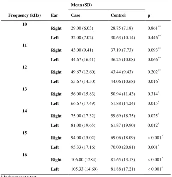
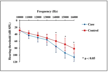
dB in the case group and 81.71 dB in the control group) (Fig. 1). The statistical analysis showed that at 3 frequencies of 14, 15, and 16 kHz, the case group significantly had worse hearing thresholds than the control group (Table 1). Although in other frequencies, the mean hearing thresholds of the case group were worse than the control group, this difference was not statistically significant.
Duration pattern sequence tests
In both groups, DPST scores did not have a normal distribution. In both ears, the case group had significantly lower DPST scores than the control group (p = 0.019 for the right ear) and p = 0.03 for the left ear).
Correlation between EHF hearing threshold and DPST scores
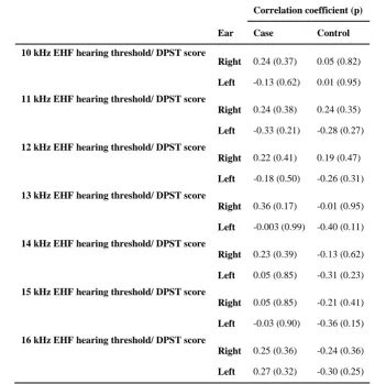
Considering that in the case group, the hearing thresholds of 14, 15, 16 kHz and DPST scores were significantly different with the control gro-up, the correlation between the hearing thresho-lds of EHF hearing threshold test and the DPST scores were examined in both ears (Table 2). The results indicated no linear correlation in all analyzed variables.
Discussion
Noise-induced hearing loss is one of the most common occupational and reversible disorders which happens in industrial environments. In the present study, ear effects were studied in Table 1. Descriptive and analytic data of extended high-frequency
audiometry test in case (n = 15) and control (n = 16) groups
Mean (SD)
Frequency (kHz) Ear Case Control p
10
Right 29.00 (6.03) 28.75 (7.18) 0.861**
Left 32.00 (7.02) 30.63 (10.14) 0.446** 11
Right 43.00 (9.41) 37.19 (7.73) 0.093**
Left 44.67 (16.41) 36.25 (10.08) 0.066** 12
Right 49.67 (12.60) 43.44 (9.43) 0.202**
Left 55.67 (14.50) 44.06 (10.68) 0.016* 13
Right 56.00 (15.83) 50.94 (11.43) 0.314*
Left 66.67 (17.49) 51.88 (14.24) 0.015* 14
Right 75.00 (17.32) 59.69 (18.75) 0.025*
Left 81.00 (19.65) 61.87 (19.90) 0.012*
15
Right 94.00 (15.02) 69.06 (18.09) < 0.001*
Left 95.33 (17.16) 70.00 (20.81) 0.001*
16
Right 106.00 (1284) 81.65 (13.13) < 0.001*
Left 105.33 (14.69) 81.88 (17.21) < 0.001*
DPST and EHFA, and there was not any sig-nificant difference between two ears. This fin-ding was in line with Mehrparvar et al. [5] and Balatsouras et al. [19] results. The lack of ear difference shows that two ears are affected by the same extent in industrial environments [20]. Tajik et al. studied temporal processing diffe-rences between normal and dyslexic children. The results showed no significant difference between the two ears [8]. Mustek et al. studied DPST in subjects with cortical lesions and reported impaired results for both ears without any significant inter-ear difference. They belie-ved that none of the hemispheres alone were capable of temporal pattern processing, and low scores was usually observed bilaterally [21]. There was no significant age difference between case and control groups in the present study because based on Somma et al. [22] findings, age has greater effects on extended high frequ-encies (9‒18 kHz) than lower frequfrequ-encies (less than 8 kHz). The results of the present study showed that auditory thresholds increase with test frequency, and the worst threshold was found at 16 kHz. This finding is in agreement
with Türkkahraman et al. [6] finding. Some stu-dies have shown that EHF hearing thresholds are considerably higher than conventional audi-ometric frequencies [6,23-25]. According to Mehrparvar et al. [5], most studies maintain that 16, 18, and 20 kHz frequencies are the most vulnerable frequencies to noise. Porto et al. stu-died conventional and extended high frequen-cies, and reported that the worst thresholds belonged to 6 and 14 kHz [25], but in the pre-sent study and da Rocha et al. study [26], only subjects with a normal hearing level at conven-tional frequencies were included. In general, it is demonstrated that noise exposure can affect EHF hearing threshold without any impact on conventional frequencies. This finding is in agr-eement with Wang et al. [27] results. They rep-orted that the effects of the noise exposure on extended high frequencies (10‒20 kHz) could be seen much earlier than lower frequencies (0.5‒6 kHz). Therefore, EHF hearing threshold can be beneficial in the early detection of NIHL. Recently some studies have conducted on the noise effects on central auditory processing des-pite normal hearing thresholds [7,28]. However,
0
20
40
60
80
100
120
10000 11000 12000 13000 14000 15000 16000
H
ear
ing
thres
hold
(dB
SP
L
)
Frequency (Hz)
Case
Control
*
* *
* p < 0.05
these studies have focused only on conventional frequencies (250‒8000 Hz). Temporal process-ing disorder and speech perception difficulty in noise in subjects with noise exposure might be secondary to extended high-frequency hearing loss. Therefore, Feng et al. studied temporal res-olution and speech understanding in the frequ-ency region with normal hearing in adults with sloping high frequency sensory neural hearing loss (at 4‒8 kHz). [15]. Temporal resolution was evaluated by amplitude modulation detection and gap detection tasks. Speech perception was evaluated via hearing in noise tests. The patients with high-tone loss showed poor performance in both tests. Test stimuli were limited to the frequencies in which patients had a normal hearing threshold, so they suggested that the
abnormality can be attributed to the extended high-frequency hearing loss. Therefore evalua-tion of the effects of EHF hearing thresholds on the central auditory processing in subjects with normal conventional audiometry can be helpful. The present discussion aimed at reviewing the ways temporal processing can be affected in subjects with exposure to the occupational noise without any hearing loss at conventional frequ-encies. By comparing the results of the case and control groups, it turned out that EHF hearing thresholds can affect temporal processing. This finding is in agreement with Kumar et al. [7] results. They reported that poor DPST perfor-mance in normal-hearing subjects with noise exposure might be due to alterations in the cen-tral auditory system secondary to the prolonged Table 2. Correlation between scores of duration pattern sequence
test and extended high-frequency hearing thresholds
Correlation coefficient (p)
Ear Case Control
10 kHz EHF hearing threshold/ DPST score
Right 0.24 (0.37) 0.05 (0.82)
Left -0.13 (0.62) 0.01 (0.95) 11 kHz EHF hearing threshold/ DPST score
Right 0.24 (0.38) 0.24 (0.35)
Left -0.33 (0.21) -0.28 (0.27) 12 kHz EHF hearing threshold/ DPST score
Right 0.22 (0.41) 0.19 (0.47)
Left -0.18 (0.50) -0.26 (0.31) 13 kHz EHF hearing threshold/ DPST score
Right 0.36 (0.17) -0.01 (0.95)
Left -0.003 (0.99) -0.40 (0.11) 14 kHz EHF hearing threshold/ DPST score
Right 0.23 (0.39) -0.13 (0.62)
Left 0.05 (0.85) -0.31 (0.23)
15 kHz EHF hearing threshold/ DPST score
Right 0.05 (0.85) -0.21 (0.41)
Left -0.03 (0.90) -0.36 (0.15)
16 kHz EHF hearing threshold/ DPST score
Right 0.25 (0.36) -0.24 (0.36)
Left 0.27 (0.32) -0.30 (0.25)
exposure to noise. Based on Kujawa and Liber-man reports, long duration of noise exposure can result in a fast and irreversible degeneration in spiral ganglion cells and a TTS so that neuron destructions may even persist in spite of hair cell and hearing sensitivity recovery. This red-uction in neuronal population might affect tem-poral processing [29]. Time, duration, and fre-quency information of the stimuli are encoded at lower levels of the auditory system. It seems that neurons responsible for transmission and coding of these characteristics are located at the level of the inferior colliculus (IC). There are neurons at IC that are tuned to signal duration [30].
Willott and LU studied IC in rats with noise exposure and found that excessive noise expo-sure would cause unpredictable changes in the temporal pattern of the action potentials. Conse-quently, NIHL would make some changes in neural functions and temporal coding [31]. In addition, the adverse effect of brain damages on auditory pattern sequence recognition is a pro-ven fact [21,32]. For that matter, DPST can be a suitable temporal processing test. Gold et al. tried to evaluate working memory in diabetic patients [33] and showed that grey matter in areas related to working memory (such as the hippocampus) had lower density. Seraji et al. studied the correlation between diabetes type I and DPST scores [34] and showed that these patients have lower DPST scores than the con-trol group. So DPST is related to working mem-ory function. Salame and Baddeley [35] repor-ted that noise exposure could interfere with short-term working memory and auditory atten-tion. As these two functions are vital for proper performance in DPST [7], noise exposure might lead to poor performance in DPST.
The present study failed to show any linear cor-relation between DPST and EHFA. This might be attributable to this fact that behavioral pure tone audiometry is simply a response of only a few inner and outer hair cells (IHCs and OHCs) and their related fibers [36]. Therefore normal hearing sensitivity in patients with noise expo-sure is not necessarily indicative of normal cochlear function. Animal studies show that it is
possible to have a normal hearing sensitivity accompanied by cochlear dysfunction [29]. Oto-acoustic emission (OAE) amplitude is the result of the cumulative activity of many OHCs, so OAE response is sensitive to cochlear defects and show changes accordingly; however, these changes might not be measurable by the aud-iometry [36]. Studying the correlation between OAE and DPST might address this issue, and it is highly recommended.
Conclusion
Noise exposure affects EHF hearing thresholds and temporal processing in spite of normal audi-ometric thresholds at the conventional frequen-cies. Therefore noise can distort supra threshold temporal cues considerably.
Acknowledgments
This study is extracted from the MSc thesis of A. Farahani with registration Ethic Code of IR.TUMS.FNM.REC.1397.155 at TUMS. The authors want to thank Mr. Farhad Alishahi, sup-ervisor of the Audiology Department of Iran Khodro Co., for his assistance in implementing the study project. We also would like to express our gratitude to all participants in the study.
Conflict of interest
The authors declared no conflicts of interest.
References
1. Kurmis AP, Apps SA. Occupationally-acquired noise-induced hearing loss: a senseless workplace hazard. Int J Occup Med Environ Health. 2007;20(2):127-36. doi:
10.2478/v10001-007-0016-2
2. Dunn DE, Robinowitz PM. Noise. In: Rosenstock L, Cullen MR, Brodkin CA, Redlich CA. editors. Textbook of clinical occupational and environmental medicine. 2nd ed. St Louis: Elsevier Saunders; 2005. p. 893-902. 3. Ryan AF, Kujawa SG, Hammill T, Le Prell C,
Kil J. Temporary and permanent noise-induced threshold shifts: a review of basic and clinical observations. Otol Neurotol. 2016;37(8):e271-5. doi:
10.1097/MAO.0000000000001071
4. Delphi M, Jarollahi F, Tahaie SA, Modarresi Y, Kamali MJBA-TUoMS. [Evaluating Mosleh monosylabic word lists in adults with noise-induced hearing loss]. Audiol. 2013;22(3):14-22. Persian.
402-6. doi: 10.4103/1463-1741.90295
6. Türkkahraman S, Gök U, Karlidağ T, Keleş E, Oztürk A. [Findings of standard and high-frequency audiometry in workers exposed to occupational noise for long durations]. Kulak Burun Bogaz Ihtis Derg. 2003;-10(4):137-42. Turkish.
7. Kumar UA, Ameenudin S, Sangamanatha AV. Tem-poral and speech processing skills in normal hearing individuals exposed to occupational. Noise Health. 2012;14(58):100-5. doi: 10.4103/1463-1741.97252
8. Tajik S, Adel Ghahraman M, Tahaie AA, Hajiabol-hassan F, Jalilvand Karimi L, Jalaie S. Deficit of auditory temporal processing in children with dyslexia-dysgraphia. Aud Vestib Res. 2012;21(4):76-83.
9. Zamyslowska-Szmytke E, Fuente A, Niebudek-Bogusz E, Sliwinska-Kowalska M. Temporal processing dis-order associated with styrene exposure. Audiol Neu-rootol. 2009;14(5):296-302. doi: 10.1159/000212108
10. Musiek FE, Chermak GD. editors. Handbook of central auditory processing disorder, volume I: Auditory neuroscience and diagnosis. 2nd ed. San Diego. Plural Publishing Inc; 2013.
11. Miranda ES, Pereira LD, Bommarito S, Silva TM. Auditory processing evaluation using nonverbal sounds in subjects with Parkinson's disease. Rev. Bras. Otorrinolaringol. 2004;70(4):534-9. doi: 10.1590/S0034-72992004000400015
12. Moore BC. Perceptual consequences of cochlear hearing loss and their implications for the design of hearing aids. Ear Hear. 1996;17(2):133-61.
13. Simon HJ, Yund EW. Frequency discrimination in listeners with sensorineural hearing loss. Ear Hear. 1993;14(3):190-201.
14. Schroder AC, Viemeister NF, Nelson DA. Intensity discrimination in normal-hearing and hearing-impaired listeners. J Acoust Soc Am. 1994;96(5 Pt 1):2683-93. 15. Feng Y, Yin S, Kiefte M, Wang J. Temporal resolution
in regions of normal hearing and speech perception in noise for adults with sloping high-frequency hearing loss. Ear Hear. 2010;31(1):115-25. doi:
10.1097/AUD.0b013e3181bb69be
16. Kumar AU, A V S. Temporal processing abilities across different age groups. J Am Acad Audiol. 2011;22(1):5-12. doi: 10.3766/jaaa.22.1.2
17. Cruickshanks KJ, Tweed TS, Wiley TL, Klein BE, Klein R, Chappell R, et al. The 5-year incidence and progression of hearing loss: the epidemiology of hearing loss study. Arch Otolaryngol Head Neck Surg. 2003;129(10):1041-6. doi:
10.1001/archotol.129.10.1041
18. Musiek FE, Baran JA, Pinheiro ML. Duration pattern recognition in normal subjects and patients with cerebral and cochlear lesions. Audiology. 1990;29(6):304-13. 19. Balatsouras DG, Homsioglou E, Danielidis V. Extended
high-frequency audiometry in patients with acoustic trauma. Clin Otolaryngol. 2005;30(3):249-54.
20. McGill TJ, Schuknecht HF. Human cochlear changes in noise induced hearing loss. Laryngoscope. 1976;86(9):1293-1302. doi: 10.1288/00005537-197609000-00001
21. Mustek FE, Baran JA, Pinheiro ML. Duration pattern
recognition in normal subjects and patients with cerebral and cochlear lesions. Audiology. 1990;29(6):304-13. 22. Somma G, Pietroiusti A, Magrini A, Coppeta L, Ancona
C, Gardi S, et al. Extended high-frequency audiometry and noise induced hearing loss in cement workers. Am J Ind Med. 2008;51(6):452-62. doi: 10.1002/ajim.20580
23. Singh R, Saxena R, Varshney SA. Early detection of noise induced hearing loss by using ultra high frequency audiometry. Int J Otorhinolaryngol. 2009;10(2):1-5. 24. Lopes AC, Otubo KA, Basso TC, Marinelli E, Lauris
JRPJAio. Occupational hearing loss: tonal audiometry x high frequencies audiometry. Intl. Arch. Otorhinolaryngol. 2009;13(3):293-9.
25. Porto MA, Gahyva DL, Lauris JR, Lopes AC. [Audiometric evaluation in extended high frequencies of individuals exposed to occupational noise]. Pro Fono. 2004;16(3):237-50. Portuguese.
26. Rocha RL, Atherino CC, Frota SM. High-frequency audiometry in normal hearing military firemen exposed to noise. Braz J Otorhinolaryngol. 2010;76(6):687-94. 27. Wang Y, Yang B, Li Y, Hou L, Hu Y, Han Y.
[Application of extended high frequency audiometry in the early diagnosis of noise--induced hearing loss]. Zhonghua Er Bi Yan Hou Ke Za Zhi. 2000;35(1):26-8. Chinese
28. Hope AJ, Luxon LM, Bamiou DE. Effects of chronic noise exposure on speech-in-noise perception in the presence of normal audiometry. J Laryngol Otol. 2013;127(3):233-8. doi: 10.1017/S002221511200299X
29. Kujawa SG, Liberman MC. Adding insult to injury: cochlear nerve degeneration after "temporary" noise-induced hearing loss. J Neurosci. 2009;29(45):14077-85. doi: 10.1523/JNEUROSCI.2845-09.2009
30. Johnson KL, Nicol TG, Zecker SG, Kraus N. Auditory brainstem correlates of perceptual timing deficits. J Cogn Neurosci. 2007;19(3):376-85. doi:
10.1162/jocn.2007.19.3.376
31. Willott JF, Lu SM. Noise-induced hearing loss can alter neural coding and increase excitability in the central nervous system. Science. 1982;216(4552):1331-4. 32. Mustek FE, Pinheiro ML. Frequency patterns in
cochlear, brainstem, and cerebral lesions: reconnaissance mélodique dans les lésions cochléaires, bulbaires et corticales. Audiology. 1987;26(2):79-88. doi: 10.3109/00206098709078409
33. Gold SM, Dziobek I, Sweat V, Tirsi A, Rogers K, Bruehl H, et al. Hippocampal damage and memory impairments as possible early brain complications of type 2 diabetes. Diabetologia. 2007;50(4):711-9. doi:
10.1007/s00125-007-0602-7
34. Seraji H, Mohamadkhani G, Nasli Esfahani E, Jalaei S. [Evaluation of temporal processing in patients with type1 diabetes in duration pattern sequence test]. Journal of Paramedical Sciences & Rehabilitation. 2018;7(3):17-25. Persian. doi: 10.22038/jpsr.2018.27557.1720
35. Salame P, Baddeley. Language. Disruption of short-term memory by unattended speech: Implications for the structure of working memory. 1982;21(2):150-64. doi:
10.1016/S0022-5371(82)90521-7