Implementation of Regional-CNN and SSD Machine Learning Object
Detection Architectures for the Real Time Analysis of Blood
Borne Pathogens in Dark Field Microscopy
Daniel Fleury,
John Marshall, USA
Angelica Fleury,
Universityof Minnesota,USA
The emerging use of visualization techniques in pathology and microbiol- ogy has been accelerated by machine learning (ML) approaches towards image preprocessing, classification, and feature extraction in an increasingly complex series of datasets. Modern Convolutional Neural Network (CNN) architectures have developed into an umbrella of vast image reinforcement and recognition methods, including a combined classification-localization of single/multi-object featured images. As a subtype neural network, CNN cre- ates a rapid order of complexity by initially detecting borderlines, edges, and colours in images for dataset construction, eventually capable in mapping intricate objects and conformities. This paper investigates the disparities between Tensorflow object detection APIs, exclusively, Single Shot Detector (SSD) Mobilenet V1 and the Faster RCNN Inception V2 model, to sample computational drawbacks in accuracy-precision vs. real time visualization capabilities. The situation of rapid ML medical image analysis is theoretically framed in regions with limited access to pathology and disease prevention departments (e.g. 3rd world and impoverished countries). Dark field mi- croscopy datasets of an initial 62 XML-JPG annotated training files were processed under Malaria and Syphilis classes. Model trainings were halted as soon as loss values were regularized and converged.
Additional Key Words and Phrases: Convolutional Neural Network,Single Shot Detector, Regional Convolutional Neural Network, Machine Learning, Visualization-Localization
Introduction
This medical object detection project explores and compares the benefits and costs in utilizing the SSD (Single Shot Detector) MobileNet and the Faster R-CNN Tensorflow Models as it pertains to detecting blood-borne pathogens- specifically Malaria and Syphilis in dark field microscopy images. Further, the paper will analyze the real-world practicality of utilizing both of these models in object detection challenges in addition to both models’ capacities in detecting sample slides from Malaria and Syphilis cultures. The authors propose a cost-benefit system to assess the performance of two distinct models in various settings. The Faster R-CNN model’s training process requires about half the time as needed to create a checkpoints and detect loss values in the images versus the SSD MobileNet architecture. In addition, Faster-RCNN marks a higher accuracy in detecting a greater number of cells as opposed to the SSD MobileNet. Although an assessment of precision and accuracy is required in both API models, the practical real time nature of the framework must be considered by observing the graphical compactness, efficiency, and lightness of each model in a limited environment (i.e. 3rd world setting). The communicability and variance of infectious diseases, such as Malaria and Syphilis, create a public health problem by making onsite medical intervention difficult. Further, the microheterogeneity of Malaria complicates the process of rapid human-controlled pathology diagnoses in local clinical laboratories.
CNN: Feed-forward artificial neural networks used for image visualization
R-CNN: Regional CNN deployed by Tensorflow for Object Detection
Single Shot Detector: Uses only one or a few training images for final detection.
The infeasibility of the“human eye” feature extraction alone is apparent as a full determination and processing of sample information can require up to 7 days in the case of unexpected complications in tissue-blood analysis. Although existing feature segmentation techniques facilitate the visualization of histology samples, there is a growing gap in diagnosis due to the lack of specificity and machine- confirmed confidence/percentile scores. In the scope of a limited 3rd world environment, the ideal of accessibility and functionality is questioned in models such as Faster RCNN Inception v2 and SSD Mobilenet v1 due to their inherent graphical capabilities in FPS rate and real time motion detection. Utilizing a deployable object detection model that can be integrated into common IoT (Internet of Things) or system architectures would minimize the accessibility gap for a multi diagnostic app. However, the concept of a Cognitive Domain in the pathology treatment process must be attained by preserving detection accuracy, specificity, and sensitivity.
GPU (Graphical Processing Unit) tests on the R-CNN and Mobilenet Tensorflow models reveal distinct parameters and characteristics: Faster R-CNN preserves suitable precision and accuracy, however, demands more processing time whilst operating at a low FPS rate, making real time object detection difficult. The SSD Mobilenet architecture (v1-v2) creates an opportunity for moderate accuracy and average precision coupled with high scalability, FPS rates, real time capabilities, and low processing times. The SSD Mobilenet versions-series offers high deployability on low-CPU/GPU graded devices, including smartphones, Raspberry pies, and other other low-performance motherboards and computers by offering minimal overconsumption in the image processing procedure. Common operating system platforms including Android and IOS function with the Mobilenet architecture due to high scalability and compactness in CPU consumption. Although the pretrained Faster R-CNN Inception model offers critical image detection accuracies, a combination of low scalability, inaccessibility, and graphics limitations creates an unpromising use of the model. The SSD Mobilnet architecture demands additional training to suffice the loss- accuracy values of the R-CNN model, however, offers practicality, scalability, and easy accessibility on smaller devices which reveals the SSD model as a promising candidate for further assessment.
Pathology Diagnosis: The Cognitive Domain
The Cognitive Domain establishes the analytical process in a pathology diagnosis through a walkthrough of various strategies and tools in the collection and manipulation of data and clinical- microscopy findings. The strategies allow a deduction of biological and clinical data to determine objective solutions. However, an overarching mechanism, known as metacognition, is exerted over these hypotheses and
is checked/evaluated in relation to the data collection. Metacognition is defined as a direct-active control over one’s thought process (Pena & Andrade-Fiho, 2009). Although the Cognitive Domain is a clinical laboratory ideal, human performance complications can cause missed
details during analysis and confirmation. More importantly, personal biases
during the pathology investigations can cause low specificity and false-positives or false-positives during the diagnostics process. Integrating an ML approach ensures that specificity and biases are continuously regulated in the training process by using constant
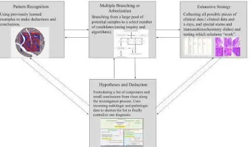
hyperparameters that prevent anomalies and complications during the machine evaluation. The traditional Cognitive Domain is demonstrated in figure 1.
Figure 1.0: The Cognitive Domain
The Cognitive Domain magnifies strategies that pathologists can use to draw preliminary hypotheses and deduction based on radiologic, microscopic, and clinical data. A default (given) procedure of pattern recognition is followed throughout the tissue-blood analysis by allowing the pathologists to extract features from the image and relate this to other biological samples. Multiple branching or arborization allows a breakdown of incoming data, while the exhaustive strategy is a rushed and uncoordinated approach towards massive data analysis.
The Cognitive Domain reveals a human oriented investigation in the pathology prognosis process, however, the efficacy of the system is not universal and is sacrificed due to natural human error. Moreover, the backbone of the Cognitive Domain takes advantage of metacognition to create an efficient toolkit and guideline of strategies to be utilized during the investigation. The Cognitive Domain uses a given premise that rationale and full-scale objectivity and understanding is maintained throughout the procedure, however, this is simply not the case: In a typical cancer diagnostic procedure, involving histologic analysis, nearly 1 million cases go undiagnosed in the process (American Cancer Society, 2018). Although this massive inflation in a lack of detection may seem non-unique in relation to pathology classification, continued complications and inconsistencies in histology practice exacerbate a trusting diagnostic process for the patient. The inconsistencies in cancer-histology detection represent a growing population pool of misdiagnosed diseases due to complex image features human simply cannot learn and apply.
Convolutional Neural Network (CNN) and Image Processing
Prior to considering Faster R-CNN, it is necessary to briefly introduce the basis of Convolutional Neural Networks since this model uses this neural network as a framework. Convolutional Neural Networks have been effective in image recognition and object detection. There are four essential components of the neural network: Convolutional Step, ReLU, Pooling and Fully Connected Layer. The Convolution Step extracts features from the input image and learns the image features using matrices of input data while allowing to
cultivate the spatial relationship of each feature within the image- this will produce a feature map. Non-Linearity(ReLU) is then introduced by replacing all negative pixel values in the feature map by zero in order to insert non-linearity. As a result, the network will learn to process inputs in a non-linear fashion that is similar to how data is conveyed in the real world
.
Pooling reduces the dimensionality of the feature map, however it retains the important information of each in addition to minimizing overfitting by reducing the number of parameters and computations in the network. The final stage is the input approaching the Fully Connected Layer, which is a Multi-Layer Perceptron which utilizes a softmax activation function in the output layer to ensure that the outputs are probabilities that add up to 1. The softmax function does this by taking a vector of arbitrarily valued scores and minimizing it to a vector of values explicitly between 0 and 1. As one of the classes approaches a value of 1, this would create a positive inference for the detection class, leaving all other negative values near 0 to indicate a fail. At this stage in the process, the output represents high-level features of the input image. The objective of the Fully Connected Layer is to utilize the features to classify the input image into different classes dependent upon the training data (Britz, 2015).
Figure 1.1: Typical feature extraction in a CNN passing through pooling layers, reaching the final neural network.
Tensorflow Object Detection API
Surfacing as a popular toolkit of machine learning technologies in early-mid 2017, the Tensorflow object detection API, released by Google, is an open source framework for object detection related tasks used for training both Single Shot Detector (SSD) and regional-Convolutional Neural Network (R-CNN) models from their model zoo (Mustamo 2018). The Tensorflow API was essentially purposed to offer scalability and potential for device deployment by Google. More importantly, Google prepared Tensorflow tools with necessary support for leading methods such as Multibox/SSD, and Fast/Faster R-CNN, which will be discussed further in this paper. The object detection API was created with an order/hierarchy of levels ranging from deployment to simple box operations. A low level API generally consists of box operations, Box representations, Target Assignment, and Visualization operations. A high level API is comprised of the heart-core structure of meta-architectures including SSD, Faster R-CNN, etc. Eventually, serving and deployment on technologies such as Jupyter Notebook and android is reached once training and visualization has been finalized. Figure
1.1 reveals an example output from the Jupyter Notebook object detection demo application.
.
Faster R-CNN Tensorflow Model
Building on the foundation of Convolutional Neural Networks, the Faster R-CNN essentially utilizes the CNN computed features and the Region Proposal Network half of the model by using the features collected to detect bounding boxes that have a probability of containing the object(s) of interest by obtaining bounding boxes, labels assigned to the boxes and probabilities (objectiveness score) for each label and box. The architecture for the model is as follows: Region Proposal Network, Anchors, Training/Loss, Region of Interest (RoI) Pooling, and Region Based CNN. Region Proposal Networks take an image as input and output rectangular object proposals, which are the regions believed to contain the object, with an objectiveness score. This is done by sliding a small network over the convolutional feature map output created by the last convolutional layer. The network then takes a sliding spatial window of the input convolutional feature map and the sliding window is then mapped to a lower-dimensional feature using ReLU. The newly created feature is then inputted into fully connected layers of a box- regression layer and a box-classification layer. This creates a single, unified network for object detection. Anchors are fixed sized reference bounding boxes within the window placed uniformly throughout the original image and the anchors are both Translational- Invariant, meaning that the functions and the proposals of that anchor are translative to varying locations of an object within the image
Multi-Scale anchors classify and regress bounding boxes with reference to anchor boxes of varying scales and aspect ratios to address multiple scales and sizes of the images.
In training the Region Proposal Network (RPN), a class label of either being an object or not is assigned to each anchor. A positive label is assigned to anchors with the highest Intersection-over- Union overlap with a ground truth box or with an overlap with a value >0.7 with any ground truth box, indicating an object has been detected. A negative label is assigned if the IoU ratio is <0.3 for all
ground-truth boxes, indicating there is no object detected. The RPN is able to be trained through backpropagation and stochastic gradient descent. To train the network, each mini-batch derives from a single image that contains many positive and negative example anchors in order to compute the loss function and to obtain a near 1:1 ratio (Ren et. al, 2016).
The Region of Interest Pooling (RoI Pooling) uses convolutional neural networks to detect multiple objects in an image by performing
max pooling on inputs of varying sizes to obtain a fixed-size feature maps of the image by dividing the region proposal into equal sized sections, finding the largest value of each section and copying the max values into the output buffer. (deepsense ai). This layer takes an input of a fixed-size feature map obtained from a deep convolutional network with several convolutions and max pooling layers. It also takes an input of a Region of Interests x 5 matrix that represents a list of regions of interests, where the first column represents the image index and the four are coordinates of the corners of the region. Figure 1.2reveals a simplified CNN-associated process diagram of the Faster R-CNN model:
The benefits of implementing a Faster RCNN Tensorflow model is that the training interval in which it takes in order to create a checkpoint file along with acceptable loss values is decreased by half the time as opposed to using the SSD MobileNet Tensorflow model. In addition, it also detects a higher amount of objects per image as well as increased accuracy for detection in comparison. However, the model is not able to be of use in real time object detection analysis (i.e. mobile smartphones) due to lacking the depthwise and pointwise convolutional layers, which also contributes to a slower detection rate.
SSD (Single Shot Detector) Mobilenet v1 Architecture
The Single Shot Detector Mobilenet architecture model is derived from depth wise separable convolutions, defined as a form of factorized convolutions which factor a standard convolution into a
The networking integrity of the Mobilenet system uses default mechanisms such as a batchnorm and ReLU nonlinearity operation (excluding the final fully connected layer possessing no non- linearity and utilizes a softmax layer for classification) for dataset processing. The compact nature of the SSD Mobilenet framework is associated with a reduction in both parameters and Mult-adds. Although the neural network structure of SSD Mobilenet is notorious for minimal accuracy in relation to the Faster R-CNN Inception models, an intensively trained Mobilenet model can equate accuracy and precision on output object detection. Nevertheless, preliminary average precision scores (mAP) on small background objects reveal reduced values in relation to the inception models. Table 1.0 exhibits the pretrained COCO- Tensorflow models and their relationships in speed (ms) and mAP(^1):
Coco Model Name Speed (ms) mAP^[1]
SSD Mobilenet V1 30 21
SSD Mobilenet V2 31 22
SSD Lite mobilenet v2 27 22
SSD inception v2 42 24
Faster RCNN Inception v2 58 28
Faster RCNN ResNet 50 89 30
Faster RCNN ResNet 50 Low Proposals 64 30
RFCN-ResNet 101 92 30
Faster RCNN ResNet 101 106 32
measures of average precision. The models of interest, SSD Mobilenet and Faster RCNN Inception, exhibit the most compatibility for future deployment with a coupling of efficiency and precision.
Prevalence of
Plasmodium Malariae
and
Treponema Pallidum
Particularly in 3rd world impoverished countries, the onset generation of devastating diseases has critically impacted communities by establishing communicability into waterways, sewers, and the common plumbing systems of undeveloped neighborhoods and towns. Although mobile medical departments have emerged in regions South of the African Sahara, immediate and handheld attention is scarce in households. Ultimately, individuals with ambiguous symptoms have minimal resource and awareness in detecting parasitic and onset disease types. More importantly, local pathology departments do not ensure full scale access to the broad community which translates into delayed diagnostic times and disruptions in quality medical intervention. Nearly 90% of present-day Malaria cases are rampant in Africa south of the Sahara. Further, in regions of free flowing Malaria transmission, both young children and pregnant women are vulnerable as a potential disease group being at risk of high morbidity and mortality. Children experiencing an initial Malaria infection experience behavioral-physical symptoms in their first year or two of life. The upsurge of Malaria in the young demographic is partly caused by inherent limitations in regional Obstetric departments and little mechanisms for on-birth diagnosis. Malaria creates cellular dysfunction and results in death by establishing an infection during pregnancy, an Acute Febrile illness, or Chronic repeated infection. An infection during pregnancy results in low birth weight during preterm delivery, whereas an Acute Febrile illness (a rapid onset of fevers, headaches, chills or muscle-joint pain) escalates Cerebral malaria causing a combination of respiratory distress and hypoglycaemia (World Health Organization, 2013). Chronic exposure and repeated infections result in severe anaemia leading to eventual death in children. According to the World Health Organization, outpatient clinical visits in Sub-Saharan-South Africa have been climbing the rungs by nearly 60% in regions including Malawi, UR Tanzania, Uganda, and Zambia since its emergence in 1985-2000. Increasing facilitation and global health initiative has been organized in regions resulting in the inhibited spread of Malaria across generations, however, little disruptive medical-technology implications have arose from 21st century clinical involvement in Africa. Faster RCNN ResNet 101 Low Proposals 82 32Faster RCNN Inception ResNet Atrous V2 620 37
Faster RCNN Inception ResNet Atrous v2 Low Proposals 241 37
+ 1833 43
Faster RCNN Nas Low Proposals 540 43
Table 1.0 (above) and Figure 1.4 (below): Relationships between typical object detection API models and their associated speeds and
Figure 1.5- Source: Routine health information system data,
ARFO,
for under-fives,
Against Malaria Foundation
The other hand of disease control is associated with Syphilis, or
Treponema Pallidum. Syphilis is underlined as an infectious disease
physiologic and psychologic map of Syphilis is characterized by periods of activity and latency, erratic systemic involvement, and a progression to acute complications in patients remaining untreated or having been inadequately treated. The diagnostic cycle for Syphilis is simply divided into both a recent case and a late case: A recent case is marked within a one year living period, whereas, following a one year bacterial presence, a late diagnosis is apparent. The current form of on point and personal diagnosis is a lookout for skin lesions, typically indicating primary Syphilis. However, skin lesions further delay the 1 year diagnostic stage by allowing the bacteria to continually replicate and eventually revealing skin lesions. Ultimately, superficial imaging techniques do not suffice immediate and real time diagnostic reports personally for both the patient and physicians in the region.
Dark Field Microscopy: Live Blood Analysis and
Accessibility
In broad terms, dark field microscopy is critical in the diagnosis of vital blood which is critical as an important holistic universal medical examination process carried out by clinics around the world. Although dark field microscopy reveals the internal environment (e.g the behavior of blood plasma, blood volume, and content), the macroscopic influences of small protein molecules in the human body from which microorganisms can arise are imaged on the sample. During dark field investigations, a drop of fresh blood is examined under the microscope without staining and the blood taken from the patient is put directly onto the slide and covered with a glass coverslip. Avoiding contamination and maintaining relative sterilization, the slide is examined immediately under 1200x magnification. The blood is ultimately exposed to severe stress in slide preparation through a lack of oxygen, non-circulation and exposure to intense light (Rau 2016). More importantly, dark field microscopy reveals the early and indirect signs and tendencies towards internal body conditions: By revealing the circulatory, and degenerative malign illnesses in the blood, doctors can use this as a precursor in a full case investigation/diagnosis. Due to the compact and non-staining requirement of dark field microscopes, immediate accessibility is maximized without strict laboratory preparation. Recent investigations in 2015 underlined the use of dark microscopy imaging with high power LED illumination, in contrast to traditional image staining techniques and halogen lamps. High power and energy efficient LED light bulbs have continually replaced halogen lamps, enhancing darkfield applications marking darkfield microscopy as an increasingly accessible imaging option. Figure 1.5 below reveals a high contrast imaging of Treponema Pallidum under
a traditional dark field microscope.
Figure 1.5: High contrast dark field imaging of Treponema Pallidum (Syphilis) blood sample. The image exhibits the potential for clear- sharp extraction of the specimen for diagnosis.
Disease Control in Impoverished 3rd World Countries
Health systems in developing countries typically have two objectives in mind to enhance social, economic, and medical advantages in the area: a.) improving the level and distribution of health outcome in the population and b.) to protect individuals from financial risks that are frequently very substantial and cause impoverished lifestyles (World Health Organization, 2000). Common financial risks are caused by illness-related loss of income and the drastic expenditures on treatment and care. However, much of this loss can be ameliorated by preventing illness or its progression by using new medical-financial architectures in the region. This paper centralizes both Malaria and Syphilis as a disease demographic, with a 90% prevalence in South Africa, however, also takes advantage of visualization techniques in a compact and easy diagnosis of human diseases. According to the Global Burden of Disease Study in 2010, diseases including Malaria and Syphilis, have been surprisingly masqueraded and unmentioned by local medical communities and departments in South African countries. This continued lack of awareness is partly rooted from the dense population pool (South- Sub Saharan Africa) experiencing this disease and the lack of regulation, assessment and modern diagnostic tools.
Common financial risks are caused by illness-related loss of income and the drastic expenditures on treatment and care. However, much of this loss can be ameliorated by preventing illness or its progression by using new medical-financial architectures in the region. This paper centralizes both Malaria and Syphilis as a disease demographic, with a 90% prevalence in South Africa, however, also takes advantage of
visualization techniques in a compact and easy diagnosis of human diseases. According to the Global Burden of Disease Study in 2010, diseases including Malaria and Syphilis, have been surprisingly masqueraded and unmentioned by local medical communities and departments in South African countries. This continued lack of awareness is partly rooted from the dense population pool (South- Sub Saharan Africa) experiencing this disease and the lack of regulation, assessment and modern diagnostic tools.
Proposal and Rationale: Real time Object Detection in
Disease Diagnostics
The objective in deploying use of object detection algorithm models in disease diagnostics is to utilize the Faster R-CNN model in order to detect Malaria and Syphilis in a hospital setting, due to the blood samples via dark field microscopy needing to be analyzed as frozen/still images. In addition, this model provides a significantly higher precision and accuracy rate when being able to detect every instance of the pathogens in each image. As seen on the figures and graphs of the Data Analysis portion
Methods and Protocols for Faster R-CNN Inception V2 COCO Model and SSD MobileNet V1
Before considering the methodology, a brief overview of the Tensorflow Object Detection API and data collection methods are overviewed:
1. Install TensorFlow-GPU v1.5, CUDA v9.0 and cuDNN v7
2. Download official Tensorflow Object Detection API repository from GitHub and setup the Tensorflow directory in the Windows C:/ drive
3. Download the Faster-RCNN-Inception-V2-COCO and SSD Mobilenet V1 models from the official Tensorflow model zoo and place inside of the \object_detection folder
4. Set up the Anaconda virtual environment and install packages: anaconda protobuf, pillow, lxml, Cython, jupyter, matplotlib, pandas, opencv-python
5. Configure PYTHONPATH environment variable to point to the correct directories in \models-masters
6. Compile Protobufs and run setup.py in the Anaconda
Command Prompt in order to create a name _pb2.py file from every name.proto file in the \protos folder
7. Test the Tensorflow Object Detection API to verify it has been set up successfully by launching the object_detection_tutorial.ipynb script with Jupyter
8. Gathered 31 images of Syphilis and 31 images of Malaria and saved into the images folder of the object detection directory
9. Open folder of images in the LabelImg software and create a new edited folder in LabelImg in order to hold the new .xml files once all of the verification boxes were saved in each image containing the every instance of the object of interest
10. Generate the training data by converting the .xml to .csv files in order to be read by the object detection classifier
11. Create a label map that tells the trainer what each object is defined by mapping class names (Malaria and Syphilis) to class ID numbers (Malaria:1, Syphilis: 2) and save the map as a .pbtxt file
12. Configure the object detection training pipeline. This is done by changing the number of classes to 2, changing the fine tune checkpoint to contain the Faster RCNN Inception V2 COCO model, changing the number of examples to 62 images and updating the eval_input_reader section, specifically the input_path and label_map_path to contain the directory of the test.record input path and the labelmap.pbtxt in the label map path.
13. Initiate training
14. Export the inference graph and modify the last saved checkpoint value post-training
15. Modify image path in python config file via idle to a newly saved image
16. Run command from Anaconda Prompt
Methodology
Implementing the Faster RCNN Inception V2 2018 and the SSD Mobilenet V1 2017 Models
Prior to initiating data collection and training, establishing a Tensorflow and Anaconda environment with default Python package imports (e.g. Numpy, Anaconda’s Protobuf, lxml, Cython, and Opencv) was necessary. Following package construction, the object detection model was then downloaded from the Tensorflow Model Zoo, a holding repository for all current models in both object detection and image classification. In this case, both the Faster- RCNN-Inception-V2-COCO and SSD Mobilent V1 2017 models were packaged and installed into the directories for testing.
Data Analysis: SSD Mobilenet V1 Training Loss Scalars
Figures 1.6-1.9:
Automated Tensorflow Object Detection API data was collected throughout the global steps as seen in the command prompt. Total loss parameters, as acknowledged in the CNN background, are ultimately summation functions of the biases and weights of the trained neural network (which result from the Softmax function and its counterparts). Although exponential loss decay is not comparable to the drastic decrease in the Faster RCNN Inception model, the SSD Mobilenet model maintained a high speed of ~30 ms for full image analysis. Based on the Single Shot approach of the neural mobile network, training and loss values did not converge at low values immediately. Loss value convergence occurred around 8,000 steps near a value of 4.00. Nevertheless, the SSD architecture allowed realtime analysis of images (e.g. an OpenCV webcam window which detected live images). Thus, although loss value, in proportion to precision, was lower for the SSD Mobilenet model, immediate image visualization was easier.Data Analysis: SSD Mobilenet V1 Training Loss Scalars
Figures 2.0-2.3:
The Faster RCNN Inception V2 model reveals exceptional loss values converging near minimum values of ~0.05 during the R-CNN training. Total loss values converged near steps ranging in 4,500-6,000, although, experienced naturalfluctuations. Evidently, the Faster RCNN model reveals higher scalability in terms of training parameters. In other words, loss values decreased drastically while precision inversely increases within a range of
10,000 global steps. However, the Faster RCNN model is notorious for overconsumption of GPU contained RAM. On NVIDIA Cuda cores, nearly all accelerators are utilized during the training process
which minimizes versatility of the model and the potential for deployment. The training model accurately localized smaller cells and borderline features on the image (i.e. not general-apparent details). Following the Python shell testing, it was apparent that the
is limited to ~2-4 frames per second (FPS), which eliminates potential for deployability on small device operating systems such as Android and IOS.
Object Detection API Deployment: Android OS Platforms
The rationale of this project-investigation is grounded in versatility and the universal nature of an pretrained object API neural network model. More importantly, after a review of disease burdens in regions and countries of South Africa, a standard of accessibility must be attained to allow rapid handheld usage. The levels of Tensorflow API usage are defined by the interactivity of the application itself. The project ultimately moved from a stage of moderate-low complexity (a Python shell console that required necessary path directory modifications) to a fully automated application that used a preloaded frozen_inference_graph.py to recognize novel images. The SSD Mobilenet V1 model was the sole candidate in performing real time
30 FPS rate object detection due to its compressed size and capacity. Figure 2.4 reveals the Android application detecting two Syphilis cells in a blood-specimen culture:
The android application was initially configured by utilizing Tensorflow’s open source android directory which enables Machine Learning deployment tools on mobile devices. Path definitions in the camera detection activity JAR files of the directory were modified to ultimately match the location of the SSD Mobilenet Frozen Inference graph.
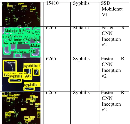
Object Detection Analysis: Faster RCNN v2 vs SSD
Mobilenet v1
15410
Syphilis SSD
Mobilenet
V1
6265
Malaria Faster R-
CNN
Inception
v2
6265
Syphilis Faster R-
CNN
Inception
v2
6265
Syphilis Faster R-
CNN
Inception
v2
Data Interpretation and Conclusions: SSD Mobilenet and
Faster R-CNN Comparisons
The combined approach of a Faster Regional Based Convolutional Neural Network and a Single Shot Detection Mobilenet architecture are apparent as they offer unique dimensions of accuracy, efficiency, and compatibility in immediate blood-data analysis. Although the Faster R-CNN model revealed minimal converging loss values, a standard of speed and immediate detection was sacrificed. In other words, detection time was extended and required demanding GPU performance to prevent crashing and processing overload. The SSD Mobilenet architecture provided moderate precision-accuracy while providing high speed, less demanding CPU-GPU consumption. With a modified batch size of ~5, the SSD Mobilenet model rapidly passed through global steps reporting decreasing loss values. More importantly SSD Mobilenet created an opportunity for scalability and accessible deployment by adapting to the architectures of small operating systems such as Android and IOS.
Future Applications
The broad constraints of a darkfield-based object detection application were sufficed in terms of moderate accuracy and compactibility in the
Open CV Image
Step
Count
Type API Model
SSD Mobilenet model. More importantly, the potential of Android-OSdevice deployment of the SSD Mobilenet architecture enables high accessibility and versatility on other cross-platforms. Moreover, the
15410 Syphilis SSD
Mobilenet
V1
15410
Malaria SSD
Mobilenet
V1
compressed nature of the SSD Mobilenet architecture allows precise object tracking and more rapid image analysis/processing time. In contrast to the Faster R-CNN model, single shot detection offers high general scalability. Primary future developments suggest that the SSD Mobilenet model may be integrated into a full scale application for multiple diagnostic disease detection. Additionally, to equate
Bibliography
An Intuitive Explanation of Convolutional Neural Networks. (2016, August 11). Retrieved July 3, 2018, from
https://ujjwalkarn.me/2016/08/11/intuitive-explanation-convnets/
American, C. (2015, March 30). Reasons for delays in getting your biopsy and cytology test results. Retrieved July 3, 2018, from https://www.cancer.org/treatment/understanding-your-
diagnosis/tests/testing-biopsy-and-cytology-specimens-for- cancer/how-long-does-testing-take.html
Dark field microscopy. (2011). Paracelsus Clinica,1-2. Retrieved July 3, 2018
Foundation, A. M. (2008). The burden of malaria in Africa. 1-7. Retrieved July 3, 2018.
Grel, T. (2017, February 28). Region of interest pooling explained. Retrieved July 3, 2018, from https://deepsense.ai/region-of-interest- pooling-explained/
Howard, A. G. (2017). MobileNets: Efficient Convolutional Neural Networks for Mobile Vision Applications. Arxiv: Computer Vision
and Pattern Recognition,1-9. Retrieved July 3, 2018.
J. (2018, January 18). Faster R-CNN: Down the rabbit hole of modern object detection. Retrieved July 3, 2018, from https://tryolabs.com/blog/2018/01/18/faster-r-cnn-down-the-rabbit- hole-of-modern-object-detection/
Jha, P. (2013). Disease Control Priorities in Developing Countries, 3rd Edition Working Paper #2. Economic Evaluation for Health,1- 66. Retrieved July 3, 2018.
Johnson, J. (n.d.). Convolution Neural Networks for Visual Recognition. Retrieved July 3, 2018, from http://cs231n.github.io/
Kennedy, E. J., Jr. (1996). DARKFIELD MICROSCOPY FOR THE DETECTION AND IDENTIFICATION OF TREPONEMA PALLIDUM. 1-18. Retrieved July 3, 2018.
Liu, W. (2015). SSD: Single Shot MultiBox Detector. Arxiv:
Computer Vision and Pattern Recognition,1-17.
doi:10.1007/978-3-
319-46448-0_2
Liu, W. (n.d.). SSD:Single Shot MultiBox Detector. Retrieved July 3,
2018, from
http://web.cs.ucdavis.edu/~yjlee/teaching/ecs289g- winter2018/SSD.pdf
Lu, Z. (n.d.). Tensorflow detection model zoo. Retrieved July 3, 2018, from
https://github.com/tensorflow/models/blob/master/research/object_d e tection/g3doc/detection_model_zoo.md
Mogeni, P., & Williams, T. (2017). Detecting Malaria Hotspots: A Comparison of Rapid Diagnostic Test, Microscopy, and Polymerase Chain Reaction. The Journal of Infectious Diseases,216(9), 27th ser.,
1091-1098. Retrieved July 3, 2018.
Mustamo, P. (2018). Object detection in sports: TensorFlow Object Detection API case study. University of Oulu: Faculty of Science,1- 43. Retrieved July 3, 2018.
Organization, W. H. (2016). Multiplexed Point-of-Care test for acute febrile illness (mPOCT). 1-2. Retrieved July 3, 2018. Pena, G. P., & Andrade-Filho, ;. (2009). How Does a Pathologist Make a Diagnosis? Archives of Pathology and Laboratory
Medicine and Archives of Pathology,1-9. Retrieved July 3, 2018.
Regazzi Avelleira, J., & Bottino, G. (2006). Syphilis: Diagnosis, treatment and control. Continuing Medical
Education,111-122. Retrieved July 3, 2018.
Ren, S., He, K., Girshick, R., & Sun, J. (2016). Faster R-CNN: Towards Real-Time Object Detection with Region Proposal Networks. 1-14. Retrieved July 3, 2018.
Xu, J. (2017). Deep Learning for Object Detection: A